NCERT Summary: Summary of Biology - 1 | Science & Technology for UPSC CSE PDF Download
| Table of contents |

|
| Components of Cell |

|
| Cell Division |

|
| Comparisons between Plant Cell and Animal Cell |

|
| Tissue |

|
Components of Cell
In living organisms, there are two types of cellular organizations. If we look at very simple organisms like bacteria and blue-green algae, We will discover cells that have no defined nucleus these are prokaryotes cells. The cells which have definite nucleus are known as a eukaryote.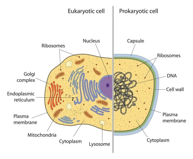 But the thing which both have in common is that there are compartments surrounded by some type of membrane. These are called cell membranes.
1. Cell Membranes
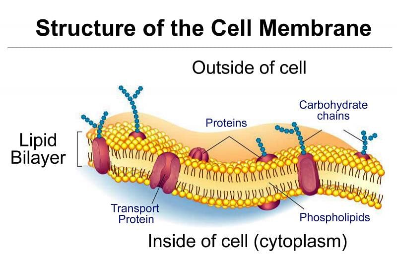
- It is like a plastic bag with some tiny holes that bag holds all of the cell pieces and fluids inside the cell and keeps foreign particles outside the cell. The holes are there to let some things move in and out of the cell.
- Compounds called proteins and phospholipids make up most of the cell membrane. The phospholipids make the basic bag. The proteins are found around the holes and help move molecules in and out of the cell.
- Substances like CO2 and O2 can move across the cell membranes by a process called diffusion.
- Diffusion is a process of movements of substance from a region of high concentration to a region where its concentration is low.
- Water also obeys the law of diffusion. The movement of water molecules is called osmosis.

2. Cytoplasm
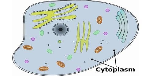
- It is the fluid that fills a cell. Scientists used to call the fluid protoplasm. Cytoplasm contains many specialized cells called organ cells. Each of these organ cells performs a specific function for the cell.

3. Cell Organelles
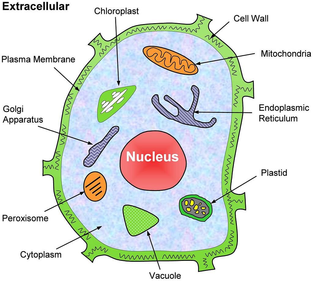
Organelles are living part of the cell have definite shape, structure and functions.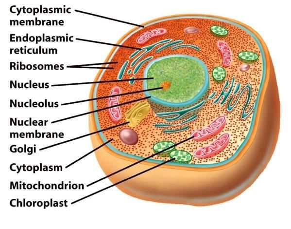 Cell OrganellesTo keep their function different from each other these organelles use membranes bound with little structure within themselves.
Some of the important organelles are:
(a) Endoplasmic Reticulum
- It is a network of tubular membranes connected at one end to the nucleus and on the other to the plasma membranes.
- Endoplasmic reticular (ER) are two types:
(i) Rough endoplasmic reticular (RER)
(ii) Smooth endoplasmic reticulum (SER) - Functions of Endoplasmic reticular (ER) are:
(i) It forms the supporting skeleton framework of the cell.
(ii) It provides a pathway for the distribution of nuclear material.
(iii) It provides surface for various enzymatic reactions.
(b) Ribosomes
Synthesizes protein, and ER sent this protein in various parts of the cell. Whereas SER helps in the manufacture of fats.
- Functions of these proteins and fats:
(i) Protein and fat (lipid) help in building the cell membranes. This process is known as membrane biogenesis.
(ii) Some other proteins and fat function as enzymes and hormones.
(iii) Smooth endoplasmic reticulum (SER) plays a crucial role in detoxifying many poisons and drugs.
(c) Golgi Apparatus
- It is found in most cells. It is another packaging organelle like the endoplasmic reticulum.
- It gathers simple molecules and combines them to make molecules that are more complex. It then takes those big molecules, packages them in vesicles and either stores them for faster use or sends them out of the cell.
- Other functions: Its functions include the storage modifications and packaging of products in vesicles. It is also the organelle that builds lysosomes (cells digestion machines).
(d) Lysosomes
- It is a kind of waste disposal system of the cell.
- It helps to keep the cell clean by digesting any foreign material.
- Old organs cell end up in the lysosomes. When the cell gets damaged, lysosomes may burst and the enzymes digest their own cell.
- Therefore lysosomes are also known as the “suicide bags” of the cell.
(e) Mitochondria
- It is known as the powerhouse of the cell.
- The energy required for various chemical activities headed for life is released by mitochondria in the form of ATP (adenosine triphosphate) molecules.
- ATP is known as the energy currency of the cell. The body uses energy is stored in ATP for making new chemical compounds and for mechanical work.
- Mitochondria are strange organelles in the sense that they have their own DNA and ribosomes, therefore mitochondria are able to make their own protein.
- Mitochondria is absent in bacteria and the red blood cells of mammals and higher animals.
(f) Centrioles
- It is a micro-tubular structure.
- Centrioles are concerned with cell division. It initiates cell division.
(g) Plastids
- These are present only in plant cells.
- There are two types of plastids:
(i) Chromoplasts (color plastids)
(ii) Leucoplast (white or colorless plastids) Plastid in Plant Cell
- Chromoplast imparts color to flowers and fruits.
- Leucoplasts are in which starch, oils and protein are stored.
- Plastids are self-replicating. i.e. they have the power to divide, as they contain DNA, RNA and ribosomes.
- Plastids contain the pigment chlorophyll that is known as chloroplast. It is the site for photosynthesis.
The above mentioned cell organelles are the living part of the cell but there are some non – living parts within the cell-like vacuoles and granules:
(h) Vacuoles
- It is a fluid-filled space enclosed by membranes.
- It is a storage sacs for solid or liquid contents. It stores excess water, minerals, food substance, pigments and waste products.
- Its size in animal is small and in plant it is big.
- Many substances of importance in the life of the plant cell are stored in vacuoles. These are amino acids sugars. It also Contains Various organic acids and some proteins.
(i) Granules
- It is not bounded by any membranes.
- It store fats, proteins and carbohydrates.
4. Cell Nucleus
- The cell nucleus acts like the brain of the cell.
- It helps control eating, movement and reproduction.
- Not all cells have a nucleus.
The Nucleus contain the following components:
(a) Nuclear Envelope
- It surrounds the nucleus and all of its contents nuclear envelope is a membrane similar to the cell membranes around the whole cell.
(b) Chromatin
- When the cell is in resting state there is something called chromatin in the nucleus.
- Chromatin is made up of DNA, RNA and nucleus protein. DNA and RNA are the nucleus acids inside the cell.
- When the cell is going to divide, the chromatin becomes very compact. It condenses when the chromatin comes together we can see the chromosomes.
(c) Chromosomes
- Chromosomes make organisms what they are. They carry all the information used to help a cell grow, thrive, and reproduce.
- Chromosomes are made up of DNA.
- Segments of DNA in specific patterns are called genes.
- In prokaryotes, DNA floats in the cytoplasm in an area called the nucleoid.
- Chromosomes are not always visible. They usually sit around uncoiled and as loose shards called chromatin.
- When it is time for all cells to reproduce, they condense and wrap up very tightly. The tightly round DNA in the chromosome.
- Chromosomes are usually found in pairs.
- Human Beings probably have 46 chromosomes (23 pairs).
- Peas only have 12, a dog has 78 chromosomes.
- The number of chromosomes is not related to the intelligence or complexity of the creature.
(d) Nucleolus
- It is a dense spherical granule contained within the nucleus, its size is related to the synthetic activity of the cell.
- Neurons cell have a comparatively larger nucleate than those cell have no synthetic activity.
- The nucleolus stores proteins.
Cell Division
Organisms grow and reduce through cell division. Plants continue to grow by cell division all their lives. But in most animals cells divide more slowly once the body takes shape.
There are two methods of replication mitosis and meiosis:
1. Mitosis
The main theme of this replication is that mitosis is the simple duplication of a cell and all of its parts. It duplicates its DNA and the two new cells (daughter cells) have the same pieces and generic code. Beyond the idea that two identical cells are created, there are five steps in this process. You should remember the term PMATI.
It breaks down to:
- Prophase
- Metaphase
- Anaphase
- Telophase
- Interphase
 Stages of Mitosis
➢ The Phases
- Prophase: A cell gets the idea that it is time to divide. First it has to get everything ready. Cell needs to duplicate DNA, get certain pieces in the right position (centrioles) and generally prepare the cell for the process of mitotic division.
- Metaphase: The DNA lines up along a central axis and then DNA condensed into chromosomes.
- Anaphase: Here the separation begins. Half of the chromosomes are pulled to one side of the cell half to go the other way.
- Telophase: Now the division is finishing up. We have now two separate cells each with half of the original DNA.
- Interphase: This is the normal state of the cell.
2. Meiosis
- It’s for sexual reproduction.
- The main theme of meiosis is that there are two cell division.
- Mitosis has one division and meiosis has two divisions in this process four cells are created where there was originally one.
- Meiosis happens when it's time to reproduce an organism.
- The steps of meiosis are very simple. When we break it down its just two PMATI’s in a row. The interphase that happens between the two processes is very short and the DNA is not duplicated.
- Meiosis is the great process that shuffles the cell’s gene-sis around. Instead of creating two new cells with equal number of chromosomes (like mitosis).
 Meiosis
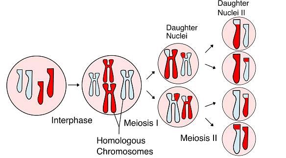
 - The cell does a second division soon after the first. This second division divides the number of chromosomes in half.
- Scientists call, this process as meiosis I and II, but its just two PMATI’s.
- Meiosis I
- This is basically live PMATI of regular mitosis.
- Pairs of chromosomes are lined up at the center of the cell and then pulled to each side.
- Meiosis is a bit different because there are something called crossing-over happens with the DNA.
- This crossing over is an exchange of genes. The genes are mixed up not resulting in a perfect duplicate like mitosis.
- The cell divides, having two new cells with a pair of chromosomes each. Since this is meiosis. There is very short interphase and division begins again.
- Meiosis II
- In this division the DNA that remains in the cell begins to condense and form short chromosomes and the center of the cell and the centrioles are in position for the duplication.
- Each one splits into two pieces. They don’t divide up the DNA between the cells. They split the DNA that exists.
- Each daughter cell will get one half of the DNA needed to make a functioning cell.
- When it’s all over we left with four haploid cells (means half the regular number) that are called gametes.
- The eventual purpose of the gametes will be to find other gametes with which they can combine.
➢ Some Important Facts Regarding Cells
- Nerve cells in animals are the longest cells.
- Smallest human cell is red blood cell.
- Largest human cell is female ovum.
- The single largest cell in the world is of an ostrich.
- The smallest cells are those of the mycoplasma.
- Every minute about 3 millions cells in our body die.
- Sieve tube in plants and the mature mammalian red blood cells do not have a nucleus.
- The red blood cell carries respiratory gases.
- Sieve cells in plants transport nutrients in plants.
- The lysosomal enzymes of the sperm cells digest the limiting membranes of the ovum (egg). Thus the sperm is able to enter the ovum.
- During the transformation of a tadpole into a frog. The embryonic tissues like gills and tail are digested by the lysosome.
- Mitochondria contain DNA, hence capable of replication.
- Matrix is a transparent, homogenous semi-fluid substance. In its active state. It remains saturated with water.
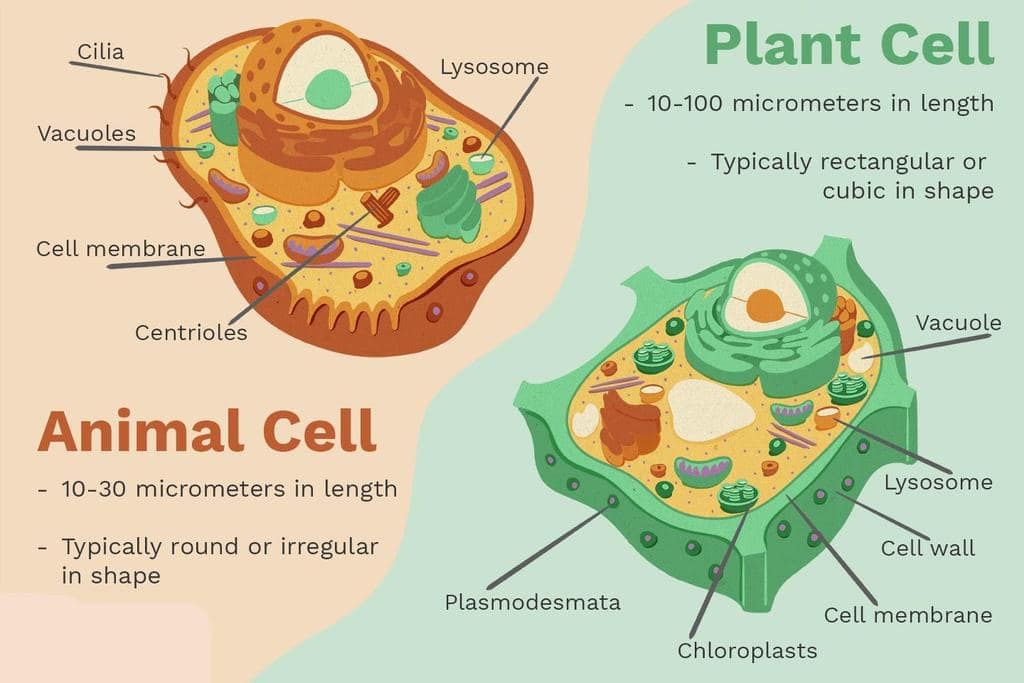
Comparisons between Plant Cell and Animal Cell
➢ Similarities
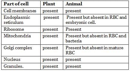
The nucleus is absent in mature mammalian red blood cells and sieve tubes in the phloem tissue of the vascular tube.
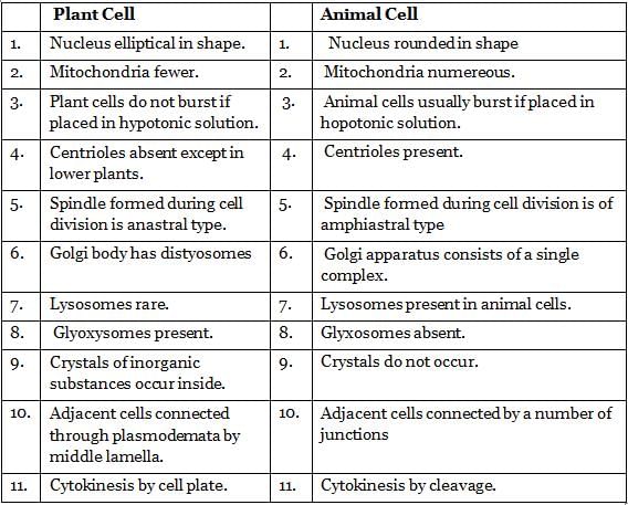
➢ Dissimilarities
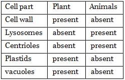
Tissue
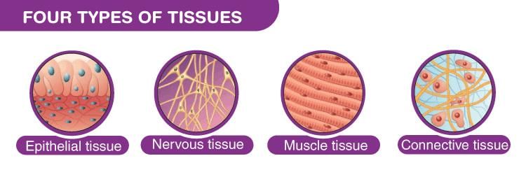
1. Epithelial Tissue
1. Epithelial Tissue
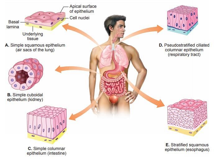
It is a tissue that is made up of tightly packed cells. Without much materials within these cells. The reasons for the tightly packed cells are to act as a barrier against mechanical injury, invading microorganisms, and fluid loss. We can define epithetical tissue by considering two points in mind one is the number of cell layers and two the shape of the cells.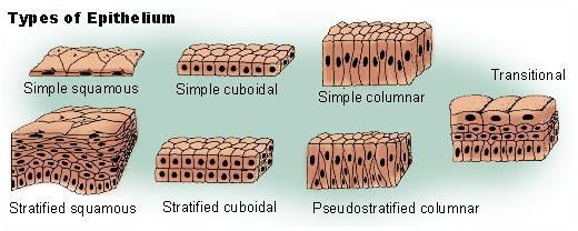
(a) On the basis of Cell layers
- When an epithelium has a single layer of cells it is called a simple epithelium.
- Whereas multiple tiers of cells are known as the stratified epithelium.
 Epithelial tissues in our body
(b) On the basis of the Simple Shape of Cells
- Cuboidal: Its occurrence is in kidney tubules, salivary glands, inner lining of the cheek. Its main function is to give mechanical strength.
- Columnar: Its occurrence is in sweat gland, tear gland, salivary gland its main function is to gives mechanical strength concerned with secretions.
- Squamous: When it forms a living as that of blood vessels, it is called the endothelium. Its main function is to protect the underlying parts from injury, entry of germs, etc.
- Connective tissue: Its main function is to bind and support other tissues. They have sparse populations of cells scattered through an extracellular matrix. This extracellular matrix is a web of fibers that is woven in a homogeneous ground substance that can be liquid, solid or jelly-like. There are a few types of connective tissue.
2. Connective Tissue

- Areolar tissue: It fills spaces inside organs found around muscles, blood vessels and nerves. Its main function is to join skin to muscles, support internal organs, help in the repair of tissues. Whereas tendon’s main function is to connect muscles to bones and ligament connects bones to each other.
- Adipose tissue: its occurrence is below skin, between internal organs and in the yellow bone Marrow. Its main function is to store fat and to conserve heat.
- Skeletal tissue: Bone & Cartilage cartilage occurrences is in nose pic, epiglottis, and in the intervertebral disc of mammals. Its main function is to provide support and flexibility to the body parts. Whereas bone protects internal delicate organs provides attachments for muscles, bone marrow makes blood cells.
- Fluid tissue: Blood & Lymph blood transport O2 nutrients, hormones to tissues and organs. Whereas leukocytes fight diseases and platelets help in the clotting of blood. Lymph transports nutrients into the heart and it also forms the defense system of the body.
3. Muscular Tissue
It is specialized for the ability to contract muscle cells. These are elongated and referred to as muscle fibers. When a stimulus is received at one end of a muscle cell, a wave of excitation is conducted through the entire cell so that all parts contract in harmony.
There were three types of muscle cells:
- Skeletal
- Cardiac
- Smooth muscles tissue
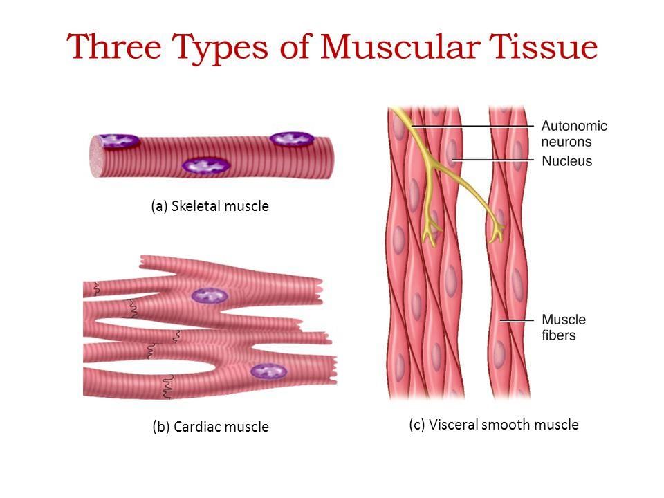
(a) Skeletal muscle: It attached primarily to bones. Its main function is to provide the force for locomotion and all other voluntary movements of the body.
(b) Cardiac muscle: It occurs only in the heart. The contraction and relaxation of the heart muscles help to pump the blood and distribute it to the various parts of the body.
(c) Smooth muscles: It can be found in the stomach, intestines, and blood vessels these muscles cause slow and prolonged contractions which are involuntary.
4. Nervous Tissue
This tissue is specialized with the capability to conduct electrical impulses and convey information from one area of the body to another. Most of the nervous tissue (98%) is located in the central nervous system. The brain and spinal cord.
There are two types of nervous tissue:
(i) Neurons
(ii) Neuroglia
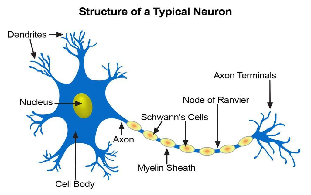
- Neurons: It actually transmits the impulses, receptor nerve ending of neurons react to various kind of stimuli and can transmit waves of excitation from the farthest point in the body to the central nervous system.
➢ Important facts regarding the Animal Tissue
- Muscles contain a special protein called contractile protein. Which contract and relax to cause movement.
- Fat storing adipose tissue is found below the skin and between internal organs.
- Two bones are connected to each other by a tissue called a ligament. This tissue is very elastic.
- The skin, the living of the mouth, the living blood vessels, kidney tubules are all made up of epithelial tissue.
- Voluntary muscles and cardiac muscles are richly supplied with water whereas involuntary muscles are poorly supplied with blood.
- Muscles tissue is composed of differentiated cells containing contractile protein.
|
146 videos|358 docs|249 tests
|
FAQs on NCERT Summary: Summary of Biology - 1 - Science & Technology for UPSC CSE
| 1. What are the components of a cell? |  |
| 2. How does cell division occur? |  |
| 3. What are the main differences between plant cells and animal cells? |  |
| 4. What is the importance of tissues in multicellular organisms? |  |
| 5. Can you provide a summary of the NCERT Biology - 1 textbook? |  |
|
146 videos|358 docs|249 tests
|

|
Explore Courses for UPSC exam
|

|
