NCERT Summary: Summary of Biology- 4 | Science & Technology for UPSC CSE PDF Download
| 1 Crore+ students have signed up on EduRev. Have you? Download the App |
Lymphocytes:
White blood cells known as lymphocytes arise from mitosis of stem cells in the bone marrow. Some lymphocytes migrate to the thymus and become T cells that circulate in the blood and are associated with the lymph nodes and spleen.
B cells remain in the bone marrow develop before moving into the circulatory and lymph systems. B cells produce antibodies.
1. Antibody-mediated (humoral) immunity is regulated by B cells and the antibodies they produce. Cellmediated immunity is controlled by T cells.
2. Antibody-mediated reactions defend against invading viruses and bacteria. Cell-mediated immunity concerns cells in the body that have been infected by viruses and bacteria, protect against parasites, fungi, and protozoans, and also kill cancerous body cells.
Antibody-mediated Immunity:
Stages in this process are:
(i) antigen detection
(ii) activation of helper T cells
(iii) antibody production by B cells
Each stage is directed by a specific cell type.
- Macrophages: Macrophages are white blood cells that continually search for foreign (nonself) antigenic molecules, viruses, or microbes. When found, the macrophages engulfs and destroys them. Small fragments of the antigen are displayed on the outer surface of the macrophage plasma membrane.
- Helper T Cells: Helper T cells are macrophages that become activated when they encounter the antigens now displayed on the macrophage surface. Activated T cells identify and activate B cells.
- B Cells: B cells divide, forming plasma cells and B memory cells. Plasma cells make and release between 2000 and 20,000 antibody molecules per second into the blood for the next four or five days. B memory cells live for months or years, and are part of the immune memory system.
- Antibodies: Antibodies bind to specific antigens in a lock-and-key fashion, forming an antigen-antibody complex. Antibodies are a type of protein molecule known as immunoglobulins. There are five classes of immunoglobulins: IgG, IgA, IgD, IgE, and IgM.
Antibodies are Y-shaped molecules composed of two identical long polypeptide (Heavy or H chains) and two identical short polypeptides (Light or L chains). Function of antibodies includes:
(i) Recognition and binding to antigens
(ii) Inactivation of the antigen
A unique antigenic determinant recognizes and binds to a site on the antigen, leading to the destruction of the antigen in several ways. The ends of the Y are the antigen-combining site that is different for each antigen.
Helper T cells activate B cells that produce antibodies. Supressor T cells slow down and stop the immune response of B and T cells, serving as an off switch for the immune system. Cytotoxic (or killer) T cells destroy body cells infected with a virus or bacteria. Memory T cells remain in the body awaiting the reintroduction of the antigen.
A cell infected with a virus will display viral antigens on its plasma membrane. Killer T cells recognize the viral antigens and attach to that cell’s plasma membrane. The T cells secrete proteins that punch holes in the infected cell’s plasma membrane. The infected cell’s cytoplasm leaks out, the cell dies, and is removed by phagocytes. Killer T cells may also bind to cells of transplanted organs. The immune system is the major component of this defense. Lymphocytes, monocytes, lymph organs, and lymph vessels make up the system.
The immune system is able to distinguish self from non-self. Antigens are chemicals on the surface of a cell. All cells have these. The immune system checks cells and identifies them as “self” or “nonself”. Antibodies are proteins produced by certain lymphocytes in response to a specific antigen. Blymphocytes and T-lymphocytes produce the antibodies. B-lymphocytes become plasma cells which then generate antibodies. T-lymphocytes attack cells which bear antigens they recognize. They also mediate the immune response.
Blood Types, Rh, and Antibodies
There are 30 or more known antigens on the surface of blood cells. These form the blood groups or blood types. In a transfusion, the blood groups of the recipient and donor should match.
If improperly matched, the recipient’s immune system will produce antibodies causing clotting of the transfused cells, blocking circulation through capillaries and producing serious or even fatal results. Individuals with blood type ‘A’ have the A antigen on the surface of their red blood cells, and antibodies to type B blood in their plasma. People with blood type ‘B’ have the B antigen on their blood cells and antibodies against type A in their plasma.
Individuals with type ‘AB’ blood produce have antigens for A and B on their cell surfaces and no antibodies for either blood type A or B in their plasma. Type O individuals have no antigens on their red blood cells but antigens of both A and B are in their plasma. People with type AB blood can receive blood of any type, So it is called as Universal Receptar.
Those with type O blood can donate to anyone. So it is called as Universal Donor. Hemolytic disease of the newborn (HDN) results from Rh incompatibility between an Rh- mother and Rh+ fetus. Rh+ blood from the fetus enters the mother’s system during birth, causing her to produce Rh antibodies. The first child is usually not affected, however subsequent Rh+ fetuses will cause a massive secondary reaction of the maternal immune system.
To prevent HDN, Rh- mothers are given an Rh antibody during the first pregnancy with an Rh+ fetus and all subsequent Rh+ fetuses.
Organ Transplants and Antibodies
Success of organ transplants and skin grafts requires a matching of histocompatibility antigens that occur on all cells in the body.
Chromosome 6 contains a cluster of genes known as the human leukocyte antigen complex (HLA) that are critical to the outcome of such procedures. The array of HLA alleles on either copy of our chromosome 6 is known as a haplotype.
The large number of alleles involved mean no two individuals, even in a family, will have the same identical haplotype.
Identical twins have a 100% HLA match. The best matches are going to occur within a family. The preference order for transplants is identical twin > sibling > parent > unrelated donor.
Chances of an unrelated donor matching the recipient range between 1 in 100,000-200,000. Matches across racial or ethnic lines are often more difficult. When HLA types are matched survival of transplanted organs dramatically increases.
Body Defences
The specialised cells which deal with germs and forcing particles by eating them up are called ‘phagocytes’ (phagein ‘to eat’; cyte ‘cell’). They are present in all tissues but are particularly concentrated in liver, spleen and bone marrow.
- Monocyter in the blood are the circulating counterparts of these cells.
- Specific acquired immunity can be categorised into two groups: humoral immunity and cellular immunity.
- Lymphoid organs produce lymphocytes. These organs include principally bone marrow, thymus, lymph, nodes, spleen and some ‘patches’ in the wall of the small intestine.
- The two types of lymphocytes — B lymphocytes concerned with humoral immunity, and T lymphocytes concerned with cellular immunity.
- Antibody production takes place in Numoral immunity. It is triggered by a protein called the antigen. It is the plasma cells which manufacture antibodies specific for the antigen presented.
- Theories which sring to explain the synthesis of specific antibodies—‘in structure’ and ‘selective’ theories. Instructive thrones postulate that all plasma cells are alike, it is the antigen that directs the plasma cells to manufacture a specific protein (antibody).
- Selective theories originally proposed by Busnet, assume that there are as many types of B cells as the antigens.
Antibodies are proteins belonging to a class called ‘gamma globulins’ or immunoglobulins.
Hepatitis Vaccine — Three doses are required: the interval between the first and second dose being one month, and that between the second and third being six months.
Oral typhoid vaccine is available in the form of capsule under the brand name ‘Typhoral’.
Blood: The Vital Fluid
Blood looks like a homogenous red fluid to the uncover edge. But when spread into a thin layer, it is found to be a suspension of different type of cells in a liquid called the ‘plasma’. Most of the cells are faint yellow and without a nucleus. A dense accumulation of these cells is responsible for the red colour of the blood. These cells are called ‘erythrocytes’ or red blood cells. These are also another two types of cells—the ‘leucocytes’ or white blood cells and ‘thrombocytes’ or platelets.
Plasma— is a straw coloured liquid, about 90 percent of which is water. The chief salt dissolved in plasma is sodium chloride, or common table salt. The salinity of plasma is onethird that of sea water.
- Fibrinogen is a protein which is essential for clotting of blood, another protein globulins aid in the defense mechanisms of the body.
- Red Blood Cells:– are the most numerous of the blood cells, they neither have a nucleus nor mitochondria, RBC are a reddish coloured protein containing iron.
- It is hemoglobin which makes it possible to deliver oxygen to tissue which need it.
The normal quantity of hemoglobin present in blood in 12-15 g in every 100 ml of blood. A decrease in this quantity is called ‘anemia’. - The nucleus membrane of the roof of the mouth (palate) is the best region to access the quantity of hemoglobin.
- The average life span of a red cell is about four months. They are produced in the hollow of the bones (bone marrow).
- White Blood Cells:– WBC are far less numerous than the RBC, the ratio being one white cell to every 600 red cells. They are slightly larger than the red cells, and differ in three aspects—first, they have nuclei, secondly, they do not contain hemoglobin, and are therefore nearly colourless, finally, some white cells can move and engulf particles or bacteria the process in called ‘phagocytosis’.
WBC are further subdivided in five groups.
(1) Neutroplis
(2) Eosinophils
(3) Basrophils
(4) Lymphocytes
(5) Monocytes
Platelets: are much smaller than red or white blood cells and are devoid of nuclei. They check the bleeding from an injury (harmostasis: haime ‘blood’; stages ‘standing’ Platelets contribute to this process of harmostasis by liberating a chemical called ‘sesolonies’.
- A, B, AB and O are the four blood groups. The classification is based on the type of substance present on the surface of red blood cells.
Lungs: The Life Linke
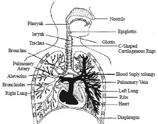
The bronchial tree consists of larynx, trachea, bronchus left lung, right lung.
Alveoli – is a cluster of thin walled air sacs which end in tiny air cells. It is covered with a tracery of capillaries. A men has about 600 million alveoli.
- Oxygen move from the alveoli into the blood and carbondioxide move out of the capillaries to entre the alveoli.
THE RESPIRATORY SYSTEM
Respiration in Single Cell Animals
Single-celled organisms exchange gases directly across their cell membrane. However, the slow diffusion rate of oxygen relative to carbon dioxide limits the size of single-celled organisms. Simple animals that lack specialized exchange surfaces have flattened, tubular, or thin shaped body plans, which are the most efficient for gas exchange. However, these simple animals are rather small in size.
Respiration in multicellular animals
Large animals cannot maintain gas exchange by diffusion across their outer surface. They developed a variety of respiratory surfaces that all increase the surface area for exchange, thus allowing for larger bodies. A respiratory surface is covered with thin, moist epithelial cells that allow oxygen and carbon dioxide to exchange. Those gases can only cross cell membranes when they are dissolved in water or an aqueous solution, thus respiratory surfaces must be moist.
Respiratory System Principles
1. Movement of an oxygen-containing medium so it contacts a moist membrane overlying blood vessels.

2. Diffusion of oxygen from the medium into the blood.
3. Transport of oxygen to the tissues and cells of the body.
4. Diffusion of oxygen from the blood into cells.
5. Carbon dioxide follows a reverse path
THE CIRCULATORY SYSTEM
Circulatory Systems in Single-celled Organisms
Single-celled organisms use their cell surface as a point of exchange with the outside environment. Sponges are the simplest animals, yet even they have a transport system. Seawater is the medium of transport and is propelled in and out of the sponge by ciliary action. Simple animals, such as the hydra and planaria lack specialized organs such as hearts and blood vessels, instead using their skin as an exchange point for materials. This, however, limits the size an animal can attain. To become larger, they need specialized organs and organ systems.
Circulatory Systems in Multicellular Organisms
Multicellular animals do not have most of their cells in contact with the external environment and so have developed circulatory systems to transport nutrients, oxygen, carbon dioxide and metabolic wastes. Components of the circulatory system include
i. Blood: a connective tissue of liquid plasma and cells
ii. Heart: a muscular pump to move the blood
iii. Blood vessels: arteries, capillaries and veins that deliver blood to all tissues
Vertebrate Cardiovascular System
The vertebrate cardiovascular system includes a heart, which is a muscular pump that contracts to propel blood out to the body through arteries, and a series of blood vessels.
The upper chamber of the heart, the atrium (pl. atria), is where the blood enters the heart. Passing through a valve, blood enters the lower chamber, the ventricle.
Contraction of the ventricle forces blood from the heart through an artery.
The heart muscle is composed of cardiac muscle cells.
Arteries are blood vessels that carry blood away from heart. Arterial walls are able to expand and contract. Arteries have three layers of thick walls. Smooth muscle fibers contract, another layer of connective tissue is quite elastic, allowing the arteries to carry blood under high pressure.
The aorta is the main artery leaving the heart.
The pulmonary artery is the only artery that carries oxygen-poor blood. The pulmonary artery carries deoxygenated blood to the lungs. In the lungs, gas exchange occurs, carbon dioxide diffuses out, oxygen diffuses in
Arterioles are small arteries that connect larger arteries with capillaries. Small arterioles branch into collections of capillaries known as capillary beds.
Capillaries, are thin-walled blood vessels in which gas exchange occurs.
In the capillary, the wall is only one cell layer thick.
Capillaries are concentrated into capillary beds. Some capillaries have small pores between the cells of the capillary wall, allowing materials to flow in and out of capillaries as well as the passage of white blood cells.
Changes in blood pressure also occur in the various vessels of the circulatory system.
Nutrients, wastes, and hormones are exchanged across the thin walls of capillaries.
Capillaries are microscopic in size, although blushing is one manifestation of blood flow into capillaries. Control of blood flow into capillary beds is done by nerve-controlled sphincters.
The circulatory system functions in the delivery of oxygen, nutrient molecules, and hormones and the removal of carbon dioxide, ammonia and other metabolic wastes. Capillaries are the points of exchange between the blood and surrounding tissues. Materials cross in and out of the capillaries by passing through or between the cells that line the capillary. The extensive network of capillaries in the human body is estimated at between 50,000 and 60,000 miles long. Thorough fare channels allow blood to bypass a capillary bed. These channels can open and close by the action of muscles that control blood flow through the channels.
Blood leaving the capillary beds flows into a progressively larger series of venules that in turn join to form veins. Veins carry blood from capillaries to the heart. With the exception of the pulmonary veins, blood in veins is oxygen-poor. The pulmonary veins carry oxygenated blood from lungs back to the heart. Venules are smaller veins that gather blood from capillary beds into veins. Pressure in veins is low, so veins depend on nearby muscular contractions to move blood along. The veins have valves that prevent backflow of blood
Blood pressure: Ventricular contraction propels blood into arteries under great pressure. Blood pressure is measured in mm of mercury; healthy young adults should have pressure of ventricular systole of 120mm, and 80 mm at ventricular diastole.
Higher pressures (human 120/80 as compared to a 12/1 in lobsters) mean the volume of blood circulates faster (20 seconds in humans, 8 minutes in lobsters).
As blood gets farther from the heart, the pressure likewise decreases. Each contraction of the ventricles sends pressure through the arteries. Elasticity of lungs helps keep pulmonary pressures low. Systemic pressure is sensed by receptors in the arteries and atria. Nerve messages from these sensors communicate conditions to the medulla in the brain. Signals from the medulla regulate blood pressure.
Diseases of the Heart and Cardiovascular System
Heart Attack: Cardiac muscle cells are serviced by a system of coronary arteries. During exercise the flow through these arteries is up to five times normal flow. Blocked flow in coronary arteries can result in death of heart muscle, leading to a heart attack. Blockage of coronary arteries. is usually the result of gradual buildup of lipids and cholesterol in the inner wall of the coronary artery. Occasional chest pain, angina pectoralis, can result during periods of stress or physical exertion. Angina indicates oxygen demands are greater than capacity to deliver it and that a heart attack may occur in the future. Heart muscle cells that die are not replaced since heart muscle cells do not divide. Heart disease and coronary artery disease are the leading causes of death today.
Hypertension, high blood pressure (the silent killer), occurs when blood pressure is consistently above 140/90. Causes in most cases are unknown, although stress, obesity, high salt intake, and smoking can add to a genetic predisposition. Luckily, when diagnosed, the condition is usually treatable with medicines and diet/exercise.
The Vascular System
Two main routes for circulation are the pulmonary (to and from the lungs) and the systemic (to and from the body). Pulmonary arteries carry blood from the heart to the lungs. In the lungs gas exchange occurs. Pulmonary veins carry blood from lungs to heart. The aorta is the main artery of systemic circuit. The vena cavae are the main veins of the systemic circuit. Coronary arteries deliver oxygenated blood, food, etc. to the heart.
Animals often have a portal system, which begins and ends in capillaries, such as between the digestive tract and the liver. Fish pump blood from the heart to their gills, where gas exchange occurs, and then on to the rest of the body. Mammals pump blood to the lungs for gas exchange, then back to the heart for pumping out to the systemic circulation. Blood flows in only one direction.
|
133 videos|399 docs|221 tests
|
FAQs on NCERT Summary: Summary of Biology- 4 - Science & Technology for UPSC CSE
| 1. What are the main topics covered in Biology-4? |  |
| 2. Can you give an example of a topic related to reproduction discussed in Biology-4? |  |
| 3. How does Biology-4 address the subject of genetics? |  |
| 4. Does Biology-4 cover the topic of evolution? |  |
| 5. Are there any sections in Biology-4 that focus on environmental issues? |  |
|
133 videos|399 docs|221 tests
|

|
Explore Courses for UPSC exam
|

|


