Body Fluids & Circulation Chapter Notes | Biology Class 11 - NEET PDF Download
| Table of contents |
|
| Introduction |
|
| Blood |
|
| Lymph (Tissue Fluid) |
|
| Circulatory Pathways |
|
| Double Circulation |
|
| Regulation of Cardiac Activity |
|
| Disorders of Circulatory System |
|

Introduction
The human body is made up of many cells, which are the basic building blocks of life. For these cells to stay healthy and work properly, they need a constant supply of essential things like nutrients, oxygen (O2), and other necessary substances. At the same time, the waste and harmful substances produced by these cells must be removed regularly. This process of bringing in what cells need and taking out what they don’t is called transport, and it is crucial for the proper functioning of tissues.
Transport Mechanisms in Different Organisms
Simple Organisms:
- Organisms like sponges and coelenterates are quite simple in their structure. They don’t have complex systems like higher animals. Instead, they circulate water from their surroundings through their body cavities. This movement of water helps their cells to exchange necessary substances with the environment.
Complex Organisms:
- More advanced organisms have developed specialized fluids within their bodies to carry out this transport. The most common body fluid used for this purpose is blood. Blood carries all the essential substances to the cells and helps remove waste products.
- Another fluid called lymph also plays a role in transporting certain substances, although it is not as widely used as blood.
In this chapter, we will explore in detail the composition and properties of blood and lymph, as well as the mechanism of blood circulation in the body. Understanding these processes is vital to grasp how our body maintains a healthy internal environment for all its cells.
Blood
Blood is a unique type of connective tissue composed of a liquid matrix called plasma and various formed elements.
Plasma
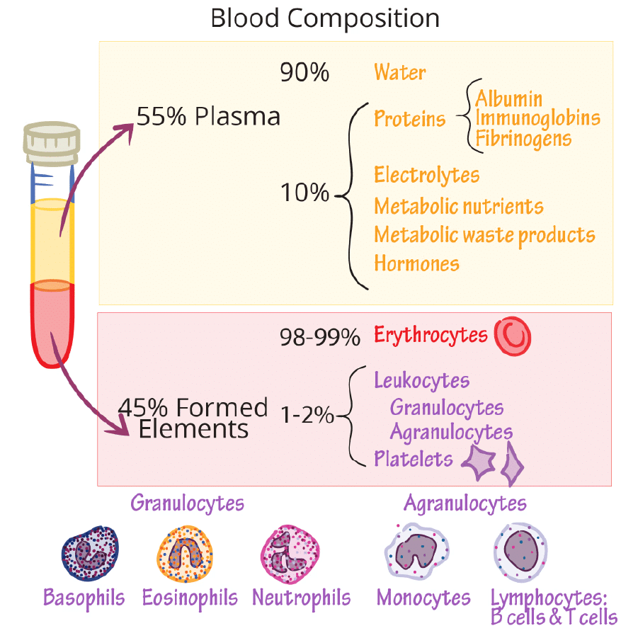
Plasma is a straw-colored, thick fluid that makes up nearly 55% of blood. It is 90-92% water, with proteins constituting 6-8% of its content. The main proteins in plasma are:
- Fibrinogen: Necessary for blood clotting.
- Globulins: Involved mainly in the body’s defense mechanisms.
- Albumins: Help maintain osmotic balance.
Plasma also contains small amounts of minerals such as sodium (Na+), calcium (Ca+2), magnesium (Mg+2), bicarbonate (HCO3 – ), and chloride (Cl– ). Additionally, substances like glucose, amino acids, and lipids are present in plasma as they are constantly in transit within the body. Coagulation factors are also present in plasma in an inactive form. When plasma is without these clotting factors, it is referred to as serum.
Formed Elements
Blood is made up of plasma and formed elements. The formed elements includes erythrocytes (RBCs), leukocytes (WBCs) and platelets. These formed elements constitute nearly 45 per cent of the blood.(i)
(i) Erythrocytes
- Erythrocytes are commonly known as red blood cells (RBCs) and are the most abundant cells in the blood.
- A healthy adult male typically has 5 to 5.5 million RBCs per cubic millimeter (mm³) of blood.
- In adults, RBCs are produced in the red bone marrow.
- Most mammalian RBCs lack a nucleus and have a biconcave shape.
- RBCs contain hemoglobin, a red, iron-containing protein that gives these cells their color and is crucial for transporting respiratory gases.
- A healthy individual has 12 to 16 grams of hemoglobin per 100 milliliters (ml) of blood.
- RBCs have an average lifespan of 120 days, after which they are destroyed in the spleen, often referred to as the "graveyard of RBCs".
(ii) Leucocytes
- Leucocytes, or white blood cells (WBCs), are colorless due to the absence of hemoglobin and are nucleated cells found in the blood.
- They are present in smaller numbers, with an average of 6,000 to 8,000 WBCs per cubic millimeter (mm³) of blood.
- Leucocytes have a shorter lifespan compared to RBCs and are classified into two main categories: granulocytes and agranulocytes.
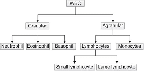
- Granulocytes include neutrophils, eosinophils, and basophils. Neutrophils make up the largest portion (60-65% ) of total WBCs, while basophils are the least common (0.5-1% ).
- Agranulocytes include lymphocytes and monocytes. Monocytes account for about 6-8% of total WBCs.
- Neutrophils and monocytes are phagocytic cells that help destroy foreign organisms entering the body.
- Basophils play a role in inflammatory reactions by secreting substances like histamine, serotonin, and heparin.
- Eosinophils (2-3% of WBCs) are involved in resisting infections and allergic reactions.
- Lymphocytes (20-25% of WBCs) are further divided into B and T lymphocytes, both of which are crucial for the body’s immune responses.
(iii) Platelets
- Platelets, also known as thrombocytes, are cell fragments derived from megakaryocytes, which are specialized cells in the bone marrow.
- Normal blood contains between 150,000 to 350,000 platelets per cubic millimeter (mm³).
- Platelets play a vital role in blood coagulation or clotting by releasing various substances involved in the clotting process.
- A reduction in platelet count can lead to clotting disorders, resulting in excessive blood loss from the body.
Blood Groups
Blood in human beings, while seemingly similar, actually differs in certain aspects. There are various types of blood grouping, but the two most widely used are the ABO and Rh groupings.
ABO Grouping
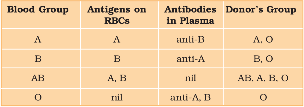
ABO grouping is determined by the presence or absence of two surface antigens, A and B, on the red blood cells (RBCs). The plasma of individuals contains natural antibodies against these antigens.
The distribution of antigens and antibodies in the four blood groups—A, B, AB, and O—plays a crucial role in blood transfusions.
- Blood Group A: Has A antigens on RBCs and anti-B antibodies in the plasma.
- Blood Group B: Has B antigens on RBCs and anti-A antibodies in the plasma.
- Blood Group AB: Has both A and B antigens on RBCs and no antibodies against A or B in the plasma.
- Blood Group O: Has no A or B antigens on RBCs and has both anti-A and anti-B antibodies in the plasma.
During blood transfusions, it is essential to match the donor's blood with the recipient's to prevent severe clumping and destruction of RBCs.
- Universal Donors: Blood group O can be donated to any other blood group.
- Universal Recipients: Blood group AB can accept blood from any other group.
 ABO Blood Grouping
Rh Grouping
The Rh grouping is based on the presence of the Rh antigen on the surface of RBCs. About 80% of humans have this antigen and are classified as Rh positive (Rh+ve), while those without it are Rh negative (Rh-ve).
- If an Rh-ve person is exposed to Rh+ve blood, they will produce antibodies against the Rh antigen.
- Therefore, it is important to match the Rh group before transfusions.
A specific case of Rh incompatibility occurs when an Rh-ve pregnant mother carries an Rh+ve fetus. During the first pregnancy, the maternal blood is not exposed to the Rh antigens of the fetus because their blood is separated by the placenta. However, during delivery, there is a risk of maternal exposure to small amounts of Rh+ve blood from the fetus, prompting the mother to produce antibodies against the Rh antigen.
In subsequent pregnancies, these Rh antibodies from the mother can cross into the blood of an Rh+ve fetus and destroy the fetal RBCs, leading to a condition called erythroblastosis foetalis. This condition can be fatal for the fetus or cause severe anemia and jaundice in the newborn.
To prevent erythroblastosis foetalis, anti-Rh antibodies can be administered to the mother immediately after the delivery of her first Rh+ve child.
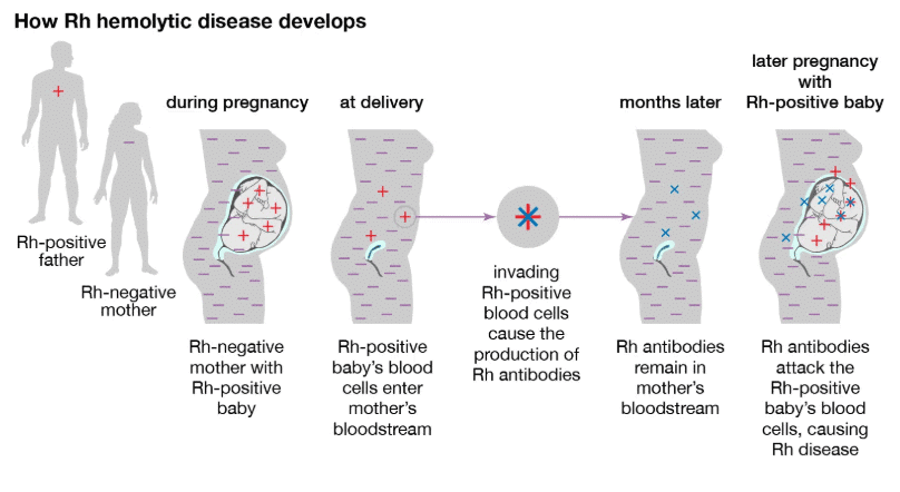 Erythroblastosis foetalis
Coagulation of Blood
- Coagulation is the process by which blood changes from a liquid to a gel, forming a blood clot. This is a crucial mechanism to prevent excessive blood loss from the body when there is an injury or trauma.
- When a blood vessel is damaged, the body initiates a series of reactions that lead to the formation of a clot. One of the key components in this process is fibrin, which is formed from fibrinogen, a protein present in the blood plasma.
- Fibrinogen is converted into fibrin by the enzyme thrombin. Thrombin is produced from prothrombin, another protein in the plasma, with the help of a complex called thrombokinase.
- Thrombokinase is formed through a cascade of enzymatic reactions involving various factors present in the plasma in an inactive state. When there is an injury, platelets in the blood release factors that activate the coagulation process, and tissue factors released at the injury site can also trigger this response.
- Calcium ions are essential for the clotting process, playing a vital role in the series of reactions that lead to the formation of a blood clot.
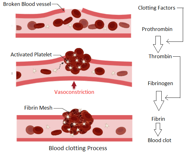 Clotting of Blood
Lymph (Tissue Fluid)
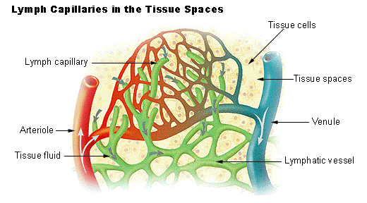
- When blood flows through the capillaries in tissues, some water and small water-soluble substances move out into the spaces between the cells, while larger proteins and most of the formed elements remain in the blood vessels. The fluid that is released is called interstitial fluid or tissue fluid. This fluid has a similar mineral distribution to that of plasma.
- The exchange of nutrients, gases, and other substances between the blood and cells occurs through this interstitial fluid.
- An intricate network of vessels known as the lymphatic system collects this fluid and drains it back into the major veins. The fluid present in the lymphatic system is called lymph.
- Lymph is a colourless fluid that contains specialized lymphocytes responsible for the body’s immune responses. Lymph also plays a crucial role in carrying nutrients, hormones, and other substances.
- Fats are absorbed through lymph in the lacteals located in the intestinal villi.
Circulatory Pathways
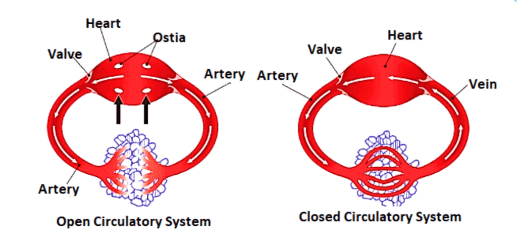
There are two types of circulatory patterns: open and closed.
Open Circulatory System
- Found in arthropods and molluscs.
- In this system, blood is pumped by the heart into large vessels that lead to open spaces or body cavities called sinuses.
Closed Circulatory System
- Present in annelids and chordates.
- Here, blood pumped by the heart is always circulated through a closed network of blood vessels.
- This system allows for more precise regulation of fluid flow and is considered more advantageous.
Heart Structure in Vertebrates
- All vertebrates have a muscular chambered heart.
- Fishes have a 2-chambered heart with one atrium and one ventricle.
- Amphibians and reptiles (except for crocodiles) have a 3-chambered heart with two atria and one ventricle.
- Crocodiles, birds, and mammals possess a 4-chambered heart with two atria and two ventricles.
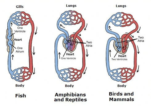 Heart Structures and Circulation in Different Vertebrates
Circulation in Different Vertebrates
- Fishes. Blood circulation is called single circulation. The heart pumps out deoxygenated blood, which is then oxygenated by the gills before being supplied to the body.
- Amphibians and reptiles. They also have single circulation, but the left atrium receives oxygenated blood from the gills, lungs, or skin, while the right atrium gets deoxygenated blood from the body. The blood mixes in the single ventricle before being pumped out.
- Birds and mammals. They have double circulation. The left and right atria receive oxygenated and deoxygenated blood, respectively, and the ventricles pump the blood out without mixing, creating two separate circulatory pathways.
Human Circulatory System
The human circulatory system, also known as the blood vascular system, comprises a muscular, chambered heart, a network of closed, branching blood vessels, and blood, which is the fluid being circulated.
The heart, which is derived from mesodermal tissue, is located in the thoracic cavity, between the two lungs and slightly tilted to the left. It is roughly the size of a clenched fist and is protected by a double-walled membranous bag called the pericardium, which contains a fluid known as pericardial fluid.
The heart consists of four chambers: two smaller upper chambers, known as atria, and two larger lower chambers, called ventricles.
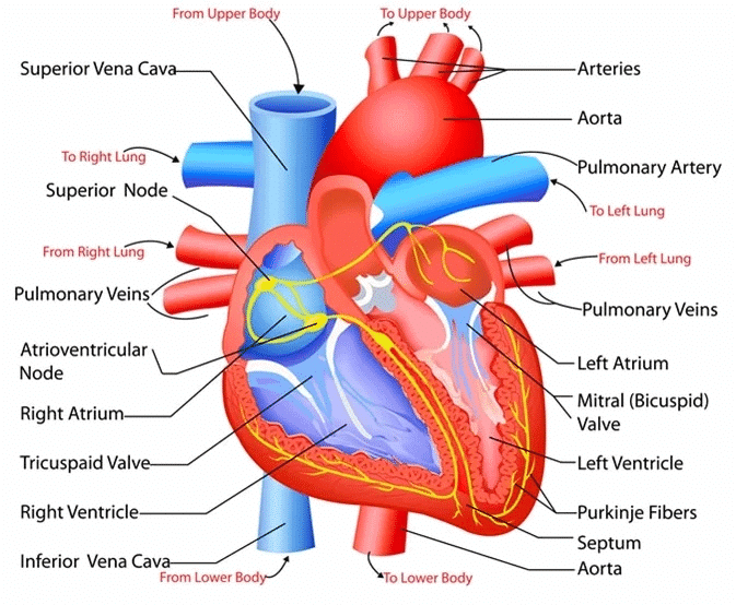 Structure of Heart
Structure of Heart
Heart Chambers
- Interatrial Septum:. thin muscular wall that separates the right and left atria.
- Interventricular Septum:. thick wall that separates the left and right ventricles.
Septa and Valves
- Atrioventricular Septum:. fibrous tissue that separates the atria from the ventricles on each side.
- Tricuspid Valve: Guards the opening between the right atrium and right ventricle.
- Bicuspid (Mitral) Valve: Guards the opening between the left atrium and left ventricle.
- Semilunar Valves: Located at the openings of the right and left ventricles into the pulmonary artery and aorta, respectively.
Functions of Heart
- Blood Flow Regulation: The valves ensure that blood flows in only one direction: from the atria to the ventricles and from the ventricles to the pulmonary artery or aorta, preventing any backward flow.
- Cardiac Muscle: The heart is made up of cardiac muscle, with the walls of the ventricles being thicker than those of the atria.
- Nodal Tissue: Specialized cardiac muscle tissue, known as nodal tissue, is distributed throughout the heart. This includes the sinoatrial node (SAN) in the right atrium, which initiates and maintains the heart's rhythmic activity, and the atrioventricular node (AVN), which helps coordinate the heart's contractions.
Heartbeat and Nodal System
- Atrioventricular Bundle: The bundle of nodal fibers that continues from the AVN, passing through the atrioventricular septum and dividing into right and left bundles on top of the interventricular septum.
- Purkinje Fibres: Minute fibres that spread throughout the ventricular muscle, ensuring coordinated contractions.
- Autoexcitability: The nodal tissue can generate action potentials without external stimuli, but different parts of the system have varying rates of generating these potentials. The SAN generates the most, at 70-75 per minute, and is therefore known as the pacemaker.
- Average Heart Rate: The normal heart rate is 70-75 beats per minute, with an average of 72.
Cardiac Cycle
The heart functions in a rhythmic cycle known as the cardiac cycle, which includes both systole (contraction) and diastole (relaxation) of the heart chambers. Let us understand how the heart works in detail.
Phases of Cardiac Cycle
1. Joint Diastole:
- Initially, all four chambers of the heart are relaxed in a phase called joint diastole.
- During this phase, the tricuspid and bicuspid valves are open. Blood flows from the pulmonary veins and vena cava into the left and right ventricles, respectively, through the left and right atria.
- The semilunar valves are closed at this stage.
2. Atrial Systole:
- The sinoatrial node (SAN) generates an action potential, stimulating both atria to contract simultaneously (atrial systole).
- This contraction increases blood flow into the ventricles by about 30 percent.
3. Ventricular Systole:
- The action potential is then conducted to the ventricles by the atrioventricular node (AVN) and the AV bundle, causing the ventricular muscles to contract (ventricular systole).
- As the ventricles contract, the pressure increases, causing the tricuspid and bicuspid valves to close, preventing backflow into the atria.
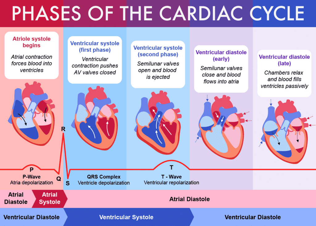
4. Blood Ejection:
- As ventricular pressure continues to rise, the semilunar valves guarding the pulmonary artery (right side) and the aorta (left side) are forced open.
- This allows blood to flow from the ventricles into the pulmonary artery and aorta, entering the circulatory pathways.
5. Ventricular Diastole:
- Following contraction, the ventricles relax (ventricular diastole), and the ventricular pressure falls.
- This decrease in pressure causes the semilunar valves to close, preventing backflow of blood into the ventricles.
- As ventricular pressure continues to decline, the tricuspid and bicuspid valves are pushed open by the pressure from blood in the atria, which is being emptied into them by the veins.
- Blood flows freely from the atria into the ventricles, and the cycle begins again.
This sequence of events is called the cardiac cycle, and it is repeated continuously. The heart typically beats 72 times per minute, indicating that the duration of a cardiac cycle is about 0.8 seconds.
Cardiac Output:
- During each cardiac cycle, each ventricle pumps out approximately 70 mL of blood, known as the stroke volume.
- The cardiac output is calculated by multiplying the stroke volume by the heart rate (number of beats per minute).
- It represents the volume of blood pumped out by each ventricle per minute and averages 5000 mL or 5 liters in a healthy individual.
Variation in Cardiac Output:
- The body can adjust both the stroke volume and heart rate, thereby altering the cardiac output.
- For example, an athlete has a significantly higher cardiac output compared to an average person due to their trained cardiovascular system.
Heart Sounds:
- During each cardiac cycle, two prominent sounds are produced, which can be easily heard with a stethoscope.
- The first heart sound (lub) is associated with the closure of the tricuspid and bicuspid valves.
- The second heart sound (dub) is linked to the closure of the semilunar valves.
- These sounds have clinical diagnostic significance and provide information about the heart's functioning.
Electrocardiograph (ECG)
Electrocardiograph (ECG) is a machine used to obtain an electrocardiogram (ECG), which is a graphical representation of the electrical activity of the heart during a cardiac cycle. To obtain a standard ECG, a patient is connected to the machine with three electrical leads that continuously monitor heart activity. The peaks in the ECG are identified with letters from P to T, representing specific electrical activities of the heart.
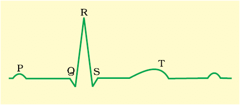
P-wave. Represents the electrical excitation (depolarization) of the atria, leading to their contraction.
QRS complex. Indicates the depolarization of the ventricles, initiating their contraction and marking the beginning of systole.
T-wave. Signifies the repolarization of the ventricles, returning them to their normal state, and marks the end of systole.
- By counting the number of QRS complexes in a given time period, the heart rate can be determined. ECGs from different individuals have similar shapes for a given lead configuration, so any deviation indicates a possible abnormality or disease, making ECGs clinically significant.
Double Circulation
Structure of Blood Vessels
- Blood flows through blood vessels in a fixed route.
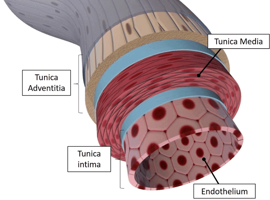
- Blood vessels consist of three layers:
- Inner Layer: Squamous endothelium, known as the tunica intima.
- Middle Layer: Smooth muscle and elastic fibers, called the tunica media. This layer is thinner in veins.
- Outer Layer: Fibrous connective tissue with collagen fibers, known as the tunica externa.
Circulation Process
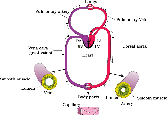
- Pulmonary Circulation: Blood flow from the heart to the lungs and back.
- Systemic Circulation: Blood flow from the heart to the body and back.
Pulmonary Circulation
- Blood pumped by the right ventricle enters the pulmonary artery.
- Deoxygenated blood is transported to the lungs.
- Oxygenated blood from the lungs is carried by the pulmonary veins into the left atrium of the heart.
Systemic Circulation
- Blood pumped by the left ventricle enters the aorta.
- Oxygenated blood is distributed to tissues through a network of arteries, arterioles, and capillaries.
- Deoxygenated blood is collected by venules and veins, returning to the heart via the vena cava and emptied into the right atrium.
Functions of Systemic Circulation
- Delivers nutrients, oxygen, and essential substances to tissues.
- Removes carbon dioxide and harmful substances for elimination.
Hepatic Portal System
- Unique vascular connection between the digestive tract and liver.
- The hepatic portal vein carries blood from the intestine to the liver before it enters systemic circulation.
Coronary System
- Special system of blood vessels for circulation to and from the heart muscle (cardiac musculature).
Regulation of Cardiac Activity
- Normal activities of the heart are regulated intrinsically, i.e., auto regulated by specialised muscles (nodal tissue), hence the heart is called myogenic.
- A special neural centre in the medulla oblangata can moderate the cardiac function through autonomic nervous system (ANS).
- Neural signals through the sympathetic nerves (part of ANS) can increase the rate of heart beat, the strength of ventricular contraction and thereby the cardiac output.
- On the other hand, parasympathetic neural signals (another component of ANS) decrease the rate of heart beat, speed of conduction of action potential and thereby the cardiac output.
- Adrenal medullary hormones can also increase the cardiac output.
Disorders of Circulatory System
1. High Blood Pressure (Hypertension)
- Hypertension refers to blood pressure that is higher than normal, specifically above 120/80 mm Hg.
- In this measurement, 120 mm Hg represents the systolic (pumping) pressure, while 80 mm Hg indicates the diastolic (resting) pressure.
- If an individual's blood pressure consistently measures 140/90 mm Hg or higher, it indicates hypertension.
- High blood pressure is a serious condition that can lead to heart diseases and negatively impact vital organs such as the brain and kidneys.
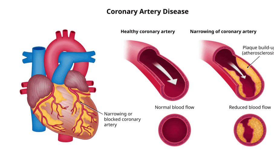
2. Coronary Artery Disease (CAD)
- Coronary Artery Disease, also known as atherosclerosis, affects the arteries that supply blood to the heart muscle.
- This condition is caused by the buildup of calcium, fat, cholesterol, and fibrous tissues in the arteries, which narrows their lumen (the inner space of the artery).
3. Angina
- Angina, or angina pectoris, is a symptom characterized by acute chest pain resulting from insufficient oxygen reaching the heart muscle.
- Angina can affect both men and women of any age, but it is more prevalent among middle-aged and elderly individuals.
- This condition occurs due to factors that impair blood flow to the heart.
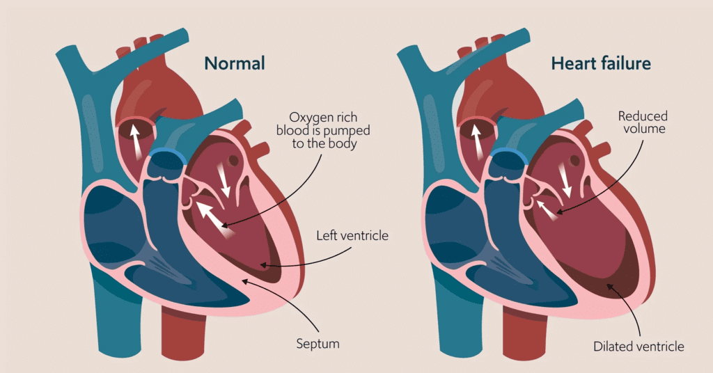
4. Heart Failure
- Heart failure refers to a condition where the heart is unable to pump blood effectively to meet the body's needs.
- It is sometimes called congestive heart failure because one of the main symptoms is lung congestion.
- It is important to note that heart failure is not the same as cardiac arrest (when the heart stops beating) or a heart attack (when the heart muscle is suddenly damaged due to inadequate blood supply).
|
150 videos|399 docs|136 tests
|
FAQs on Body Fluids & Circulation Chapter Notes - Biology Class 11 - NEET
| 1. What are the different formed elements present in blood? |  |
| 2. How does ABO grouping work in blood typing? | |
| 3. What is the function of lymph in the circulatory system? | |
| 4. How does coagulation of blood prevent excessive bleeding? | |
| 5. What is the cardiac cycle and how does it contribute to circulation in the human body? | |


Summary
,study material
,Sample Paper
,Body Fluids & Circulation Chapter Notes | Biology Class 11 - NEET
,Important questions
,Previous Year Questions with Solutions
,Body Fluids & Circulation Chapter Notes | Biology Class 11 - NEET
,practice quizzes
,Extra Questions
,Objective type Questions
,Viva Questions
,mock tests for examination
,past year papers
,shortcuts and tricks
,MCQs
,ppt
,video lectures
,Free
,Semester Notes
,Exam
,Body Fluids & Circulation Chapter Notes | Biology Class 11 - NEET
;


Chapter Notes: Body Fluids & Circulation Free PDF Download
Importance of Chapter Notes: Body Fluids & Circulation
Chapter Notes: Body Fluids & Circulation
Chapter Notes: Body Fluids & Circulation NEET Questions
Study Chapter Notes: Body Fluids & Circulation on the App
|
© EduRev
|
Education Revolution
|


|


