Cell: The Unit of Life Chapter Notes | Biology Class 11 - NEET PDF Download
| Table of contents |

|
| What is Cell? |

|
| What is Cell Theory? |

|
| Overview of Cells |

|
| Prokaryotic Cell |

|
| Eukaryotic Cells |

|
Our world is full of many different things. Some of these are living things like animals and plants that grow and survive. Others are not alive, like rocks and chairs, which don't have the qualities of living beings.
What is a difference Between living and non living organisms ?
The answer lies in the fundamental unit of life – the cell. Cells are the building blocks of all living organisms, and their presence is what sets living things apart from non-living things.
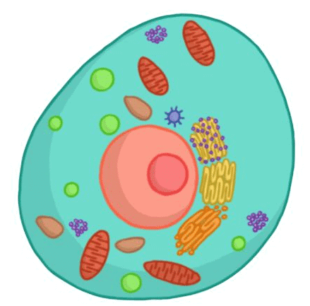 Fundamental Unit of Life - Cell
What is Cell?
A cell is the basic structural and functional unit of life in all living organisms. Cells are the smallest entities that can carry out the processes necessary for life, such as metabolism, growth, reproduction, and responding to environmental stimuli. They are often referred to as the building blocks of life.
Note: Cytology: (G.k. kyios = cell ; logas = study) is the branch of biology which comprises the study of cell structure and function. Cell is the structural and functional unit of all living beings.
What is Cell Theory?
Cell theory states that "All living organisms are composed of cells and their products".
Matthias Schleiden (1838) observed different plant cells forming plant tissues.
Theodore Schwann (1839) identified the plasma membrane in animal cells and recognized cell walls as unique to plant cells.
Schleiden and Schwann proposed that both animals and plants are made of cells and their products, However this theory lacked an explanation for the process of cell formation.
In 1855 Rudolf Virchow provided an explanation by stating that cells divide and give rise to new cells from pre-existing ones
Therefore cell theory states that:
(i) All living organisms are made up of cells and their products.
(ii) Cells can only arise from the division of pre-existing cells.
Note: We here at EduRev are also providing an interesting document of mnemonics for your better and easy understanding of the chapter .
If you click on the provided link, you'll be taken to the mnemonics document.
Overview of Cells
An overview of a typical cell involves understanding its fundamental components, structures, and functions. Cells are the basic building blocks of life and can vary in size, shape, and function depending on the organism and its specialized roles. Broadly, cells can be categorized into two main types: prokaryotic cells and eukaryotic cells.
(a) Prokaryotic Cell (b) Eukaryotic Cell
Cells are also classified on the basis of their shape and function they perform some the different types with different shapes are given below
Different Shapes of Cells
Cells can vary in shape and structure depending on their functions and the organisms they belong to. While the basic structural components of cells are similar, there can be variations.
- Spherical Cells: These cells have a round or spherical shape. Examples include many animal cells, such as blood cells (erythrocytes) and some bacterial cells.
- Cuboidal Cells: Cuboidal cells are cube-shaped, with all sides approximately equal in length. These cells are often found in epithelial tissues, such as those lining the kidney tubules.
- Columnar Cells: Columnar cells are elongated and have a tall, rectangular shape. They are commonly found in the lining of the intestines, where they aid in absorption.
- Squamous Cells: Squamous cells are flat and thin, resembling scales or fried eggs. They are found in tissues where a thin structure is needed, like the lining of blood vessels and the skin's outermost layer.
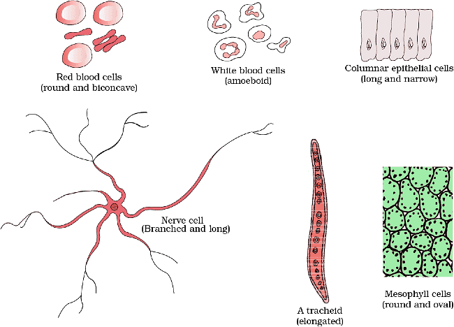 Shapes of Cell
Prokaryotic Cell
Prokaryotic cells exhibit a less complex structure when compared to eukaryotic cells, as they do not possess a genuine nucleus or membrane-bound organelles.
- These cells are primarily present in organisms like bacteria and archaea.
- They are typically smaller and less intricate than eukaryotic cells but have shown remarkable adaptability and have evolved to thrive in a wide variety of environments."
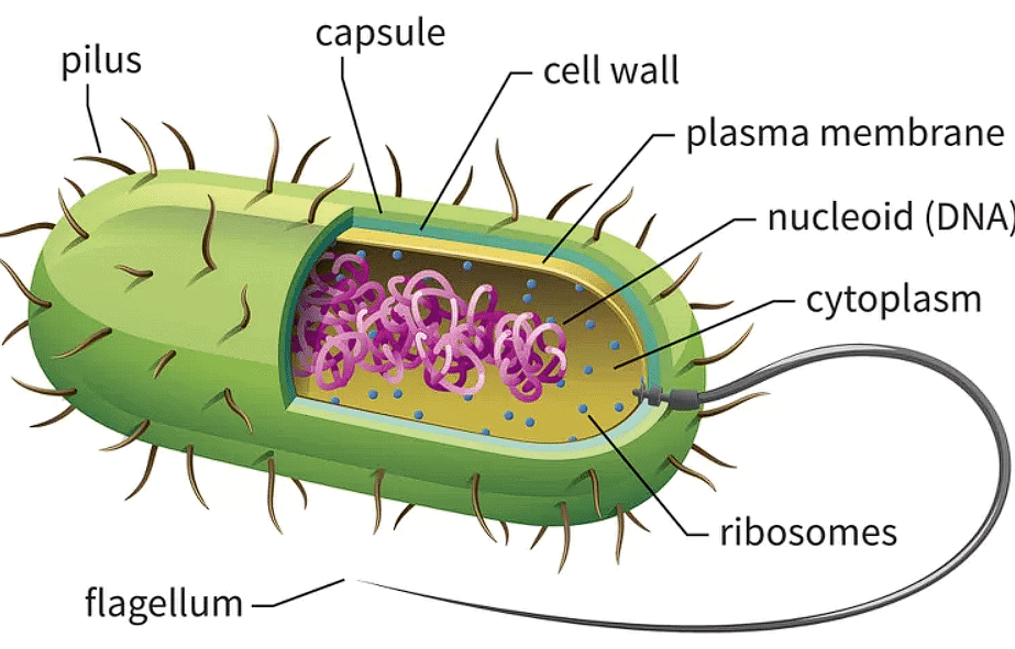 Prokaryotic Cell
Cell Envelope and its Modifications
(i) Prokaryotic cells, especially bacteria, feature a complex cell envelope comprising three tightly bound layers:
- Glycocalyx: The glycocalyx varies in composition and thickness, with some bacteria having a slime layer and others forming a capsule.
- Cell wall: The cell wall shapes the cell and provides structural support.
- Plasma membrane: Selectively permeable, interacts with the external environment and resembles that of eukaryotes.
- Mesosomes: mesosomes are formed by plasma membrane extensions, play roles in cell wall formation, DNA processes, respiration, and increasing membrane surface area.
(ii) These layers, while distinct, function together as a protective unit.
(iii) Bacteria are categorized as Gram-positive (retain Gram stain) or Gram-negative (do not retain Gram stain) based on their cell envelope differences.
Note: Bacteria are categorized as either Gram-positive or Gram-negative based on their cell envelope structure and response to a staining procedure called the Gram stain:
- Gram-Positive Bacteria: They have a thick peptidoglycan layer in their cell wall that retains the stain, making them appear purple when stained.
- Gram-Negative Bacteria: They have a thinner peptidoglycan layer and an outer membrane that does not retain the stain, causing them to appear pink or red after staining
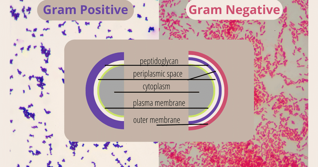 Gram-positive & Gram-negative
- Chromatophores: Pigment-containing structures found in various cells.
- Bacterial Cell Mobility: Bacterial cells can be either motile or non-motile.
- Flagella: Thin, filamentous extensions from the cell wall of motile bacteria.
- Composed of three parts: filament, hook, and basal body.
(i) Filament is the longest part and extends outside the cell.
(ii) Pili (Pilus, Singular): Elongated tubular structures made of a specialized protein.
(iii) Fimbriae: Small, bristle-like fibers on the surface of bacterial cells.
Ribosomes and Inclusion Bodies
Location: Prokaryotic ribosomes are found at the cell's plasma membrane.
Size: They are about 15 nm by 20 nm in size and consist of 50S and 30S subunits, forming 70S ribosomes.
Function: These ribosomes are where protein synthesis takes place, and multiple ribosomes can attach to one mRNA, forming polysomes.
Inclusion Bodies
Storage: Prokaryotic cells store reserve materials as inclusion bodies in the cytoplasm.Characteristics: Inclusion bodies are not enclosed by membranes and include materials like phosphate, cyanophycean, and glycogen granules.
(c) Gas Vacuoles
Presence: Gas vacuoles are found in specific photosynthetic bacteria, such as blue-green and purple-green varieties.
- Primary and Secondary Wall: The primary wall of a young plant cell is capable of growth, which gradually diminishes as the cell matures, and the secondary wall is formed on the inner (towards membrane) side of the cell.
Eukaryotic Cells
Complex cells with a true nucleus and membrane-bound organelles.
- Building blocks of multicellular organisms.
- Organized internal structure, including nucleus, endoplasmic reticulum, mitochondria, and other organelles.
- Involved in various cellular processes.
- Found in plants, animals, fungi, and some single-celled organisms.
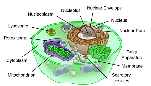 Eukaryotic Cell
Now, let's examine specific cellular organelles to gain insight into their structures and roles in the cells.
1. Cell Wall
The cell wall is a rigid, protective structure that surrounds the plasma membrane of plant cells, fungi, and some bacteria. It was first discovered by Robert Hooke in 1665.
The composition of cell walls varies depending on the type of organism. In plant cells, the primary component of the cell wall is cellulose, a complex carbohydrate made up of glucose molecules. Fungal cell walls contain chitin, while bacterial cell walls can be composed of peptidoglycan or other materials.
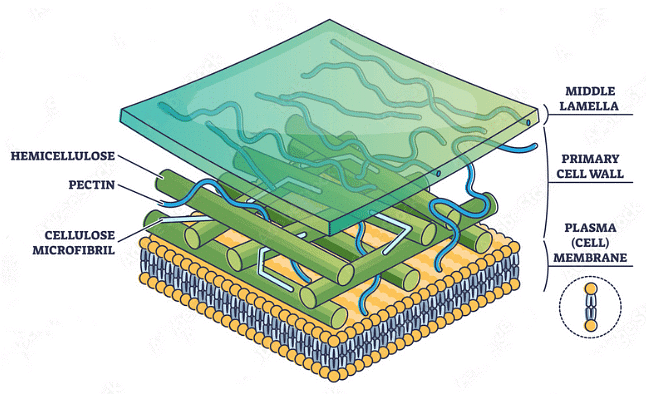 Components of Cell Wall
Components of Cell wall
- Cellulose is the primary structural component of the plant cell wall. It is a complex carbohydrate (polysaccharide) made up of glucose molecules linked together.
- Hemicellulose is another type of polysaccharide found in the plant cell wall. It helps in binding cellulose microfibrils together, contributing to the overall structural integrity of the cell wall.
- Pectin is a complex polysaccharide that is found in the middle lamella of plant cell walls. It acts as a cementing substance, holding adjacent cells together and helping to maintain the structure of tissues.
- Primary and Secondary Wall: The primary wall of a young plant cell is capable of growth, which gradually diminishes as the cell matures, and the secondary wall is formed on the inner (towards membrane) side of the cell.
- Middle Lamella: The middle lamella is a layer mainly of calcium pectate that holds or glues the different neighbouring cells together.
(i) Composition: The middle lamella primarily consists of pectin, a complex polysaccharide. Pectin molecules are rich in negatively charged ions and have the ability to form bonds with other pectin molecules. This adhesive quality of pectin makes the middle lamella effective in cementing adjacent cell walls.
(ii) Function: The primary function of the middle lamella is to hold plant cells together, especially in plant tissues. It ensures the structural integrity of the tissue by firmly joining the cell walls of neighboring cells.
(iii) Plasmodesmata: Plasmodesmata (singular: plasmodesma) are microscopic channels that traverse the cell wall and middle lamellae, connecting the cytoplasm of adjacent plant cells.
2. Cell Membrane
The cell membrane, also known as the plasma membrane, is a fundamental structural component of all cells in living organisms. It is a selectively permeable barrier that separates the interior of the cell from its external environment. The cell membrane plays several crucial roles in maintaining the integrity and function of the cell.
Every living cell is externally covered by a thin transparent electron microscopic, elastic regenerative and selective permeable membrane called plasma membrane.
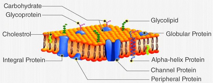 Cell Membrane
According to Singer and Nicolson, it resembles a "protein iceberg floating in a lipid sea."
- In plant cells, various microorganisms, some protists, and fungal cells, there exists an outer cell wall situated outside the plasma membrane.
- Membranes are also found within cells, and these are collectively referred to as biomembranes.
The term "cell membrane" was initially coined by C. Nageli and C. Cramer in 1855 to describe the outer membrane covering of the protoplast. However, the term "plasmalemma" or "plasma membrane" was introduced by Plowe in 1931 to replace "cell membrane."
Functions of Plasma Membrane
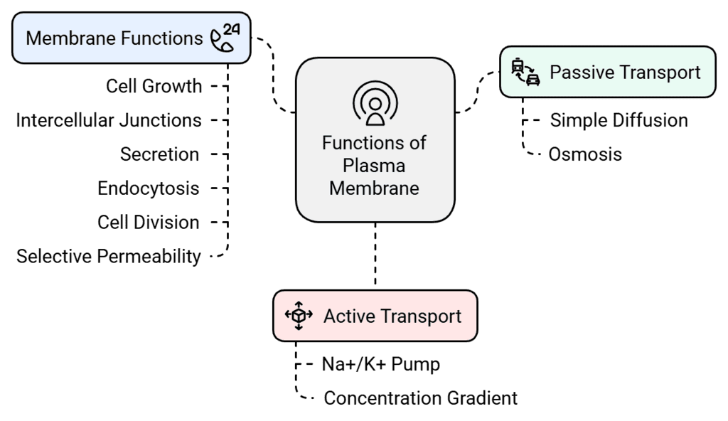 Functions of Plasma Membrane
(i) Involved in Membrane Functions: Supports cell growth, intercellular junctions, secretion, endocytosis, and cell division. Selectively permeable for molecules on both sides. passive transport takes place.
(ii) Transfer of Molecules without energy: Simple diffusion for neutral solutes (from high to low concentration). Osmosis for water (movement by diffusion).
(iii) Active Transport: Requires energy (typically ATP). Moves ions or molecules against their concentration gradient. Example: Na+/K+ Pump.
The Endomembrane System
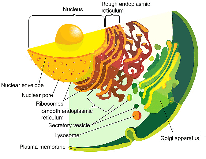
The Endomembrane system is a important component within eukaryotic cells, serving as a complex network of membranes and organelles that work together to perform various essential functions. It includes a range of organelles like the endoplasmic reticulum (ER), Golgi apparatus, lysosomes, and vacuoles. These organelles are interconnected by membranes and function together to carry out tasks such as protein synthesis, modification, and transport, lipid metabolism, and cellular waste management.
 The Endomembrane System
The endomembrane system encompasses organelles such as the endoplasmic reticulum (ER), Golgi complex, lysosomes, and vacuoles. These organelles work together to perform specific tasks.
Note: The mitochondria, chloroplasts, and peroxisomes do not operate in coordination with the components of the endomembrane system. Therefore, they are not classified as part of this system.
Let's study these organelles in detail:
1. Endoplasmic Reticulum
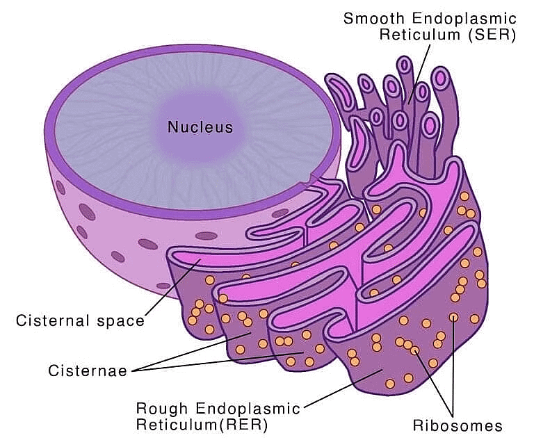
Electron microscopy studies conducted on eukaryotic cells reveal the presence of a network or reticulum made up of small tubular structures scattered throughout the cytoplasm. This network is known as the endoplasmic reticulum (ER).
 Endoplasmic Reticulum
The ER often shows ribosomes attached to their outer surface. The endoplasmic reticulum bearing ribosomes on their surface is called rough endoplasmic reticulum (RER). In the absence of ribosomes they appear smooth and are called smooth endoplasmic reticulum (SER).
- Rough endoplasmic reticulum: RER is frequently observed in cells actively engaged in protein synthesis and secretion. It is extensive and maintains a continuous connection with the outer membrane of the nucleus.
- Smooth endoplasmic reticulum: SER primarily responsible for lipid synthesis. In animal cells, SER is involved in the synthesis of lipid-like steroidal hormones.
2. Golgi Apparatus
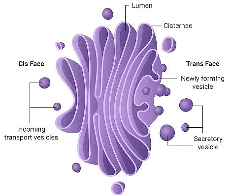
Camillo Golgi discovered reticular structures near the nucleus in 1898, and these structures were later named Golgi bodies after him. Golgi bodies consist of flat, disk-shaped sacs or cisternae, typically measuring 0.5µm to 1.0µm in diameter.
 Golgi Apparatus
These cisternae are stacked parallel to each other and can vary in number within a Golgi complex. Golgi cisternae are arranged concentrically near the nucleus, featuring a distinct convex cis face and a concave trans face. Despite their differences, the cis and trans faces of the Golgi apparatus are interconnected.
Functions:
- The primary function of the Golgi apparatus is to package materials, which can be directed either to intracellular destinations or secreted outside the cell.
- Materials to be packaged, in the form of vesicles from the endoplasmic reticulum (ER), merge with the cis face of the Golgi apparatus and move toward the maturing face.
- The Golgi apparatus remains closely associated with the endoplasmic reticulum due to this process.
- Many proteins synthesized by ribosomes on the endoplasmic reticulum undergo modifications within the Golgi cisternae before being released from its trans face.
- The Golgi apparatus plays a crucial role in the formation of glycoproteins and glycolipids.
3. Lysosomes
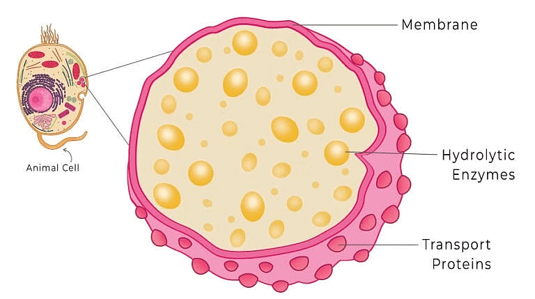
Lysosomes are vesicular structures enclosed by membranes, and they are created through the packaging process in the Golgi apparatus. These isolated lysosomal vesicles have been discovered to contain a wide range of hydrolytic enzymes (known as hydrolases), including lipases, proteases, and carbohydrases.
 Lysosomes
Function: These enzymes are most effective when the environment is acidic. They have the capacity to break down carbohydrates, proteins, lipids, and nucleic acids.
4. Vacuoles
Vacuoles are membrane-bound compartments present in the cytoplasm of cells. They contain water, sap, waste products, and other substances that aren't essential for the cell's functioning. A single membrane known as the tonoplast encloses the vacuole. In plant cells, vacuoles can occupy a significant portion, up to 90 percent, of the cell's volume.
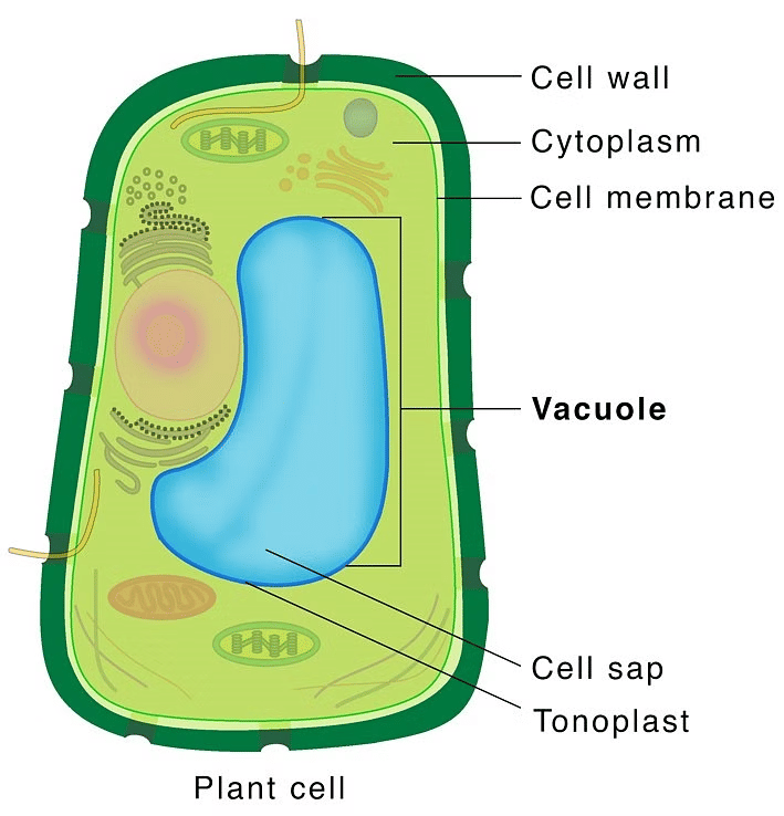 Vacuole - Plant Cell
Function:
- In plant cells, the tonoplast plays a crucial role in transporting various ions and substances into the vacuole against concentration gradients. As a result, these substances are found in much higher concentrations within the vacuole compared to the cytoplasm.
- In organisms like Amoeba, the contractile vacuole is important for regulating osmotic balance and eliminating waste. Additionally, in many cells, such as those in protists, food vacuoles are formed when food particles are engulfed by the cell.
5. Mitochondria
Mitochondria are semi autonomous having hollow sac like structures present in all eukaryotes except mature RBCs of mammals and sieve tubes of phloem. These are absent in all prokaryotes like bacteria and cyanobacteria. Mitochondria are organelles found in the cells that produce energy. They are not easily visible under the microscope and the number of mitochondria per cell varies depending on the cells' physiological activity.
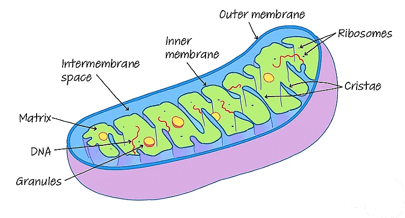 Mitochondria
- Mitochondria have a considerable degree of variability in shape and size. They are typically sausage-shaped or cylindrical in shape and are double membrane-bound structures.
- The outer membrane forms the continuous limiting boundary of the organelle, while the inner membrane divides the lumen into two aqueous compartments: the outer compartment and the inner compartment.
- The inner compartment is filled with a dense homogeneous substance called the matrix. The inner membrane also forms a number of infoldings called cristae that increase the surface area.
- Mitochondrial Division: Mitochondria divides by fission.
Functions of Mitochondria:
- Mitochondria are the sites of aerobic respiration and produce cellular energy in the form of ATP.
- They have their own specific enzymes associated with mitochondrial function.
- Additionally, the matrix of the mitochondria contains a single circular DNA molecule, a few RNA molecules, ribosomes (70S) and the components required for the synthesis of proteins.
6. Plastids
Plastids are present in all plant cells and in euglenoides, and they are easily visible under a microscope because of their substantial size. They contain distinct pigments that give plants specific colors. Plastids can be categorized into three types—chloroplasts, chromoplasts, and leucoplasts—based on the specific pigments they contain.
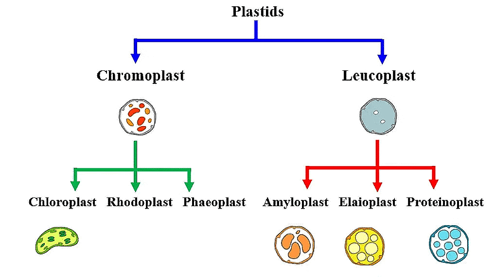 Types of Plastids
(a) Chloroplasts
Chloroplasts play a vital role in capturing light energy for the process of photosynthesis, utilizing pigments like chlorophyll and carotenoids. They are organelles enclosed by a double membrane. Notably, the inner membrane of chloroplasts has relatively low permeability.
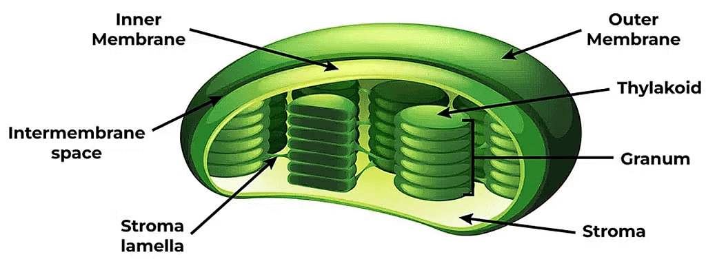 Chloroplast
- Within the chloroplast, there is an inner space delimited by the inner membrane. This space contains flattened membranous sacs known as thylakoids, which house the chlorophyll necessary for photosynthesis.
- Thylakoids are organized into stacks called grana and are interconnected by flat membranous tubules known as stroma lamellae. The lumen is the space enclosed by the thylakoid membrane.
- The stroma, another part of the chloroplast, contains enzymes essential for synthesizing carbohydrates and proteins. Additionally, it houses circular DNA molecules and ribosomes. Chlorophyll pigments are concentrated within the thylakoids, and it's worth noting that the ribosomes found in chloroplasts are smaller (70S) compared to those in the cytoplasm (80S).
(b) Chromoplasts: Chromoplasts contain fat-soluble carotenoid pigments such as carotene, xanthophylls, and others. These pigments give the plant parts a yellow, orange or red colour.
(c) Leucoplasts: Leucoplasts are colourless plastids of varied shapes and sizes that store nutrients. There are three types of leucoplasts:
(i) Amyloplasts store carbohydrates (starch), e.g., potato
(ii) Elaioplasts store oils and fats
(iii) Aleuroplasts store proteins
7. Ribosomes
Ribosomes were initially observed as dense particles under the electron microscope by George Palade in 1953. They consist of ribonucleic acid (RNA) and proteins and are not enclosed by any membrane. Ribosomes are tiny structures made up of ribonucleic acid (RNA) and proteins.
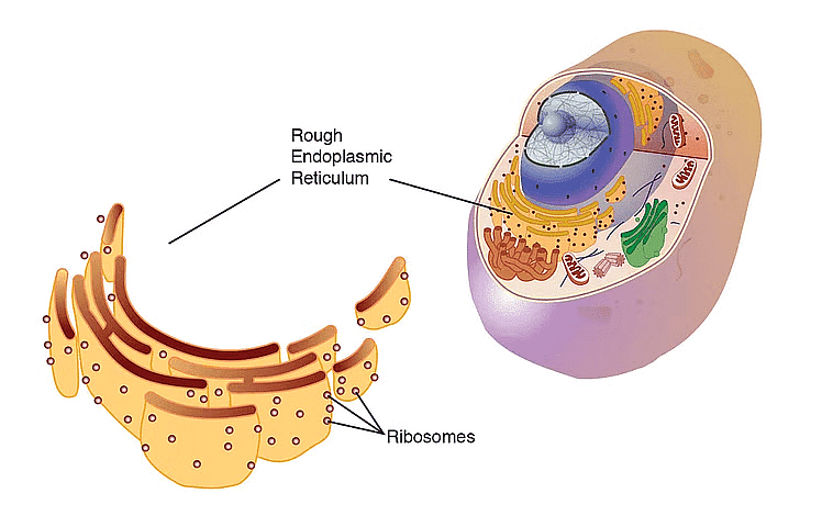 Ribosomes
Eukaryotic ribosomes have a sedimentation coefficient of 80S, while prokaryotic ribosomes have a sedimentation coefficient of 70S. Each ribosome is composed of two subunits: a larger subunit and a smaller subunit.
- In 80S ribosomes, the two subunits are 60S and 40S, while in 70S ribosomes, they are 50S and 30S. The "S" in these values represents the sedimentation coefficient, which indirectly indicates the ribosome's density and size.
- It's important to note that both 70S and 80S ribosomes consist of two subunits.
- Ribosomes are tiny structures made up of ribonucleic acid (RNA) and proteins.
8. Cytoskeleton
The cytoskeleton is a complex system of proteinaceous filamentous structures, including microtubules, microfilaments, and intermediate filaments, found within the cytoplasm.
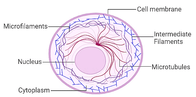 Cytoskeleton
Collectively, these components are known as the cytoskeleton, and they serve various essential roles within the cell. These functions encompass providing mechanical support, enabling cell motility, and maintaining the cell's shape.
9. Cilia and Flagella
Cilia (singular: cilium) and flagella (singular: flagellum) are slender, hair-like projections extending from the cell membrane. Cilia are relatively small structures that function like oars, generating movement in either the cell or the surrounding fluid. Flagella, on the other hand, are longer and primarily responsible for propelling the cell. It's worth noting that prokaryotic bacteria also possess flagella, but their structure differs from that of eukaryotic flagella.
 Cilia and Flagella
Under the electron microscopic study of cilia and flagella show that:
- They are covered with plasma membrane
- Their core, called the axoneme, consists of microtubules running parallel to the long axis
- The axoneme usually has nine doublets of radially arranged peripheral microtubules, and a pair of centrally located microtubules
- This arrangement of axonemal microtubules is referred to as the 9+2 array
- The central tubules are connected by bridges and enclosed by a central sheath, which is connected to one of the tubules of each peripheral doublet by a radial spoke
- There are nine radial spokes
- The peripheral doublets are also interconnected by linkers
10. Centrosome and Centrioles
The centrosome is an organelle typically composed of two cylindrical structures known as centrioles, surrounded by a less structured pericentriolar material. Within the centrosome, both centrioles are oriented perpendicular to each other and exhibit an organization resembling a cartwheel.
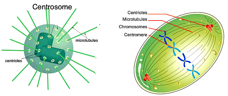 Centrosome and Centrioles
- These centrioles consist of nine evenly spaced peripheral fibrils made of tubulin protein, with each peripheral fibril being a triplet.
- These adjacent triplets are interconnected. The central portion of the proximal region of the centriole is also proteinaceous and is referred to as the hub. It is linked to the tubules of the peripheral triplets by radial spokes composed of protein.
- Centrioles play essential roles, such as serving as the basal body for cilia or flagella and forming spindle fibers that give rise to the spindle apparatus during cell division in animal cells.
11. Nucleus
The concept of the nucleus as a cellular organelle was first introduced by Robert Brown back in 1831. Later on, Flemming coined the term "chromatin" to describe the material within the nucleus that could be stained with basic dyes.
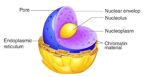 Nucleus
- During the interphase, which is the phase when a cell is not actively dividing, the nucleus displays a complex structure. It contains highly extended and intricate nucleoprotein fibers known as chromatin, a nuclear matrix, and one or more spherical bodies called nucleoli.
- Typically, the outer membrane of the nuclear envelope remains continuous with the endoplasmic reticulum and is also studded with ribosomes.
- The nuclear envelope features small pores at various locations, formed by the fusion of its two membranes. These nuclear pores serve as passages through which RNA and protein molecules can move bidirectionally between the nucleus and the cytoplasm.
Chromosomes
Chromosomes are thread-like structures found in the nucleus of eukaryotic cells, such as those in humans and many other organisms. They are made up of DNA, which contains the genetic information or genes that carry instructions for various cellular functions and traits.
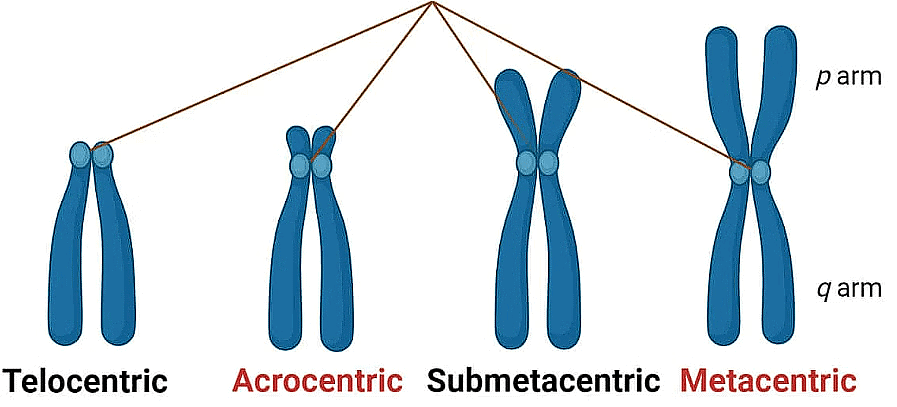 Types of Chromosome
- Chromosomes are essential for the process of cell division, as they help ensure that genetic information is accurately passed from one generation of cells to the next during processes like mitosis and meiosis.
- In humans, the normal number of chromosomes in a cell is 46 (23 pairs), and alterations in the structure or number of chromosomes can lead to genetic disorders.
12. Microbodies
Microbodies are small, membrane-bound vesicles found in both plant and animal cells. These microbodies are notable for containing a variety of enzymes within their structure. These enzymes are involved in diverse cellular processes and metabolic reactions. Microbodies are essential for compartmentalizing specific enzymatic activities, allowing cells to carry out various biochemical reactions efficiently and independently within these specialized vesicles.
Some Important Differences
1: Difference between extrinsic protein and intrinsic protein

2: Difference between primary cell wall and secondary cell wall
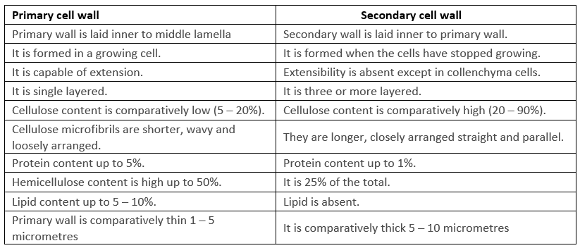
3: Difference between Prokaryotic Cells and Eukaryotic Cells
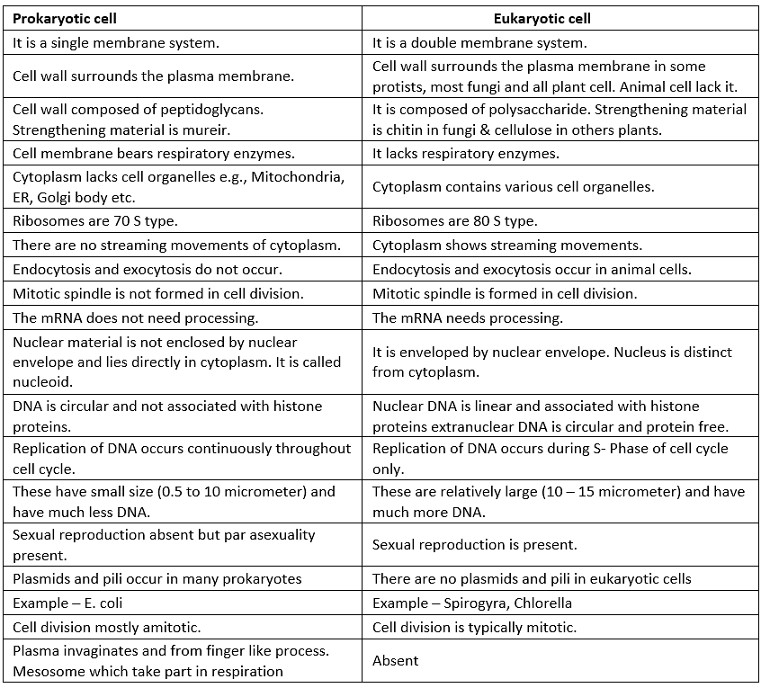
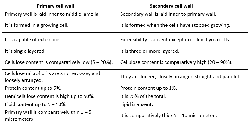
4: Difference between the primary cell wall and secondary cell wall
Additional Information
1. Plant Cell
Plant cells are fundamental to the structure and function of plants. They possess unique organelles like chloroplasts and a central vacuole that are not found in animal cells.
- These features enable them to perform photosynthesis, store water and nutrients, and provide structural support, all of which are essential for plant growth and survival.
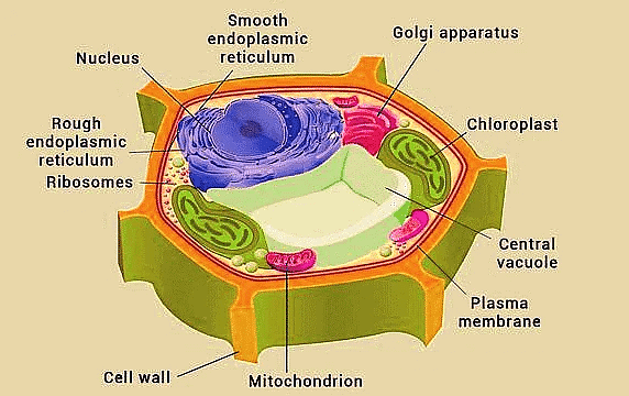 Plant Cell
2. Animal Cell
An animal cell is the fundamental structural and functional unit of animal organisms. It is a microscopic, membrane-bound structure that serves as the basic building block of animal tissues and organs.
- An animal cell is typically small and has a complex internal structure.
- It is surrounded by a semi-permeable cell membrane, also known as the plasma membrane, which encloses the cell's contents and separates it from the extracellular environment.
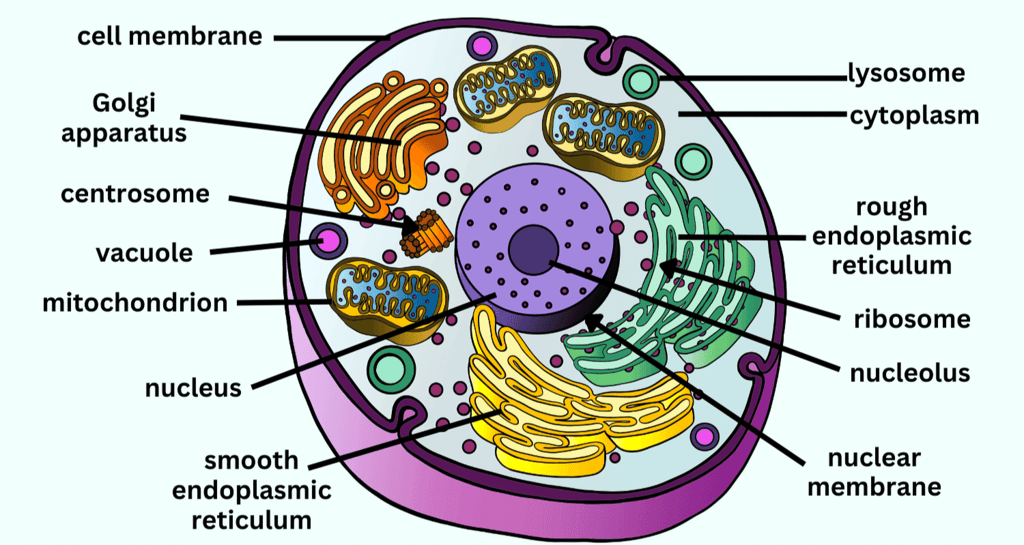 Animal Cell
|
150 videos|398 docs|136 tests
|
FAQs on Cell: The Unit of Life Chapter Notes - Biology Class 11 - NEET
| 1. What is a cell and why is it considered the basic unit of life? |  |
| 2. What are the key components of a prokaryotic cell? |  |
| 3. How do eukaryotic cells differ from prokaryotic cells? |  |
| 4. What is cell theory and what are its main tenets? |  |
| 5. Why are cells important in the study of biology? |  |


