Cell Cycle & Mitosis

Cell division is a fundamental process in all living organisms. Every large organism begins life from a single cell that grows and divides repeatedly. Growth and reproduction are characteristics of all cells and living organisms. Each cell divides into two daughter cells, which can further grow and divide, forming structures with millions of cells through coordinated cycles of growth and division.
1. Cell Cycle
The cell cycle is the sequence of events by which a cell duplicates its genome, synthesizes other cellular constituents, and divides into two daughter cells. It involves three coordinated processes: DNA replication, cell growth, and cell division. These events are under genetic control to ensure correct division and formation of progeny cells with intact genomes.
 Phases of cell cycle
1.1 Phases of Cell Cycle
The cell cycle is divided into two basic phases:
- Interphase: The phase between two successive M phases where the cell prepares for division
- M Phase (Mitosis phase): The phase when actual cell division occurs
1.1.1 Duration of Cell Cycle
- Human cells: Divide once in approximately 24 hours
- Yeast cells: Progress through cell cycle in about 90 minutes
- M Phase duration: Lasts only about 1 hour in human cells
- Interphase duration: Lasts more than 95% of the total cell cycle duration
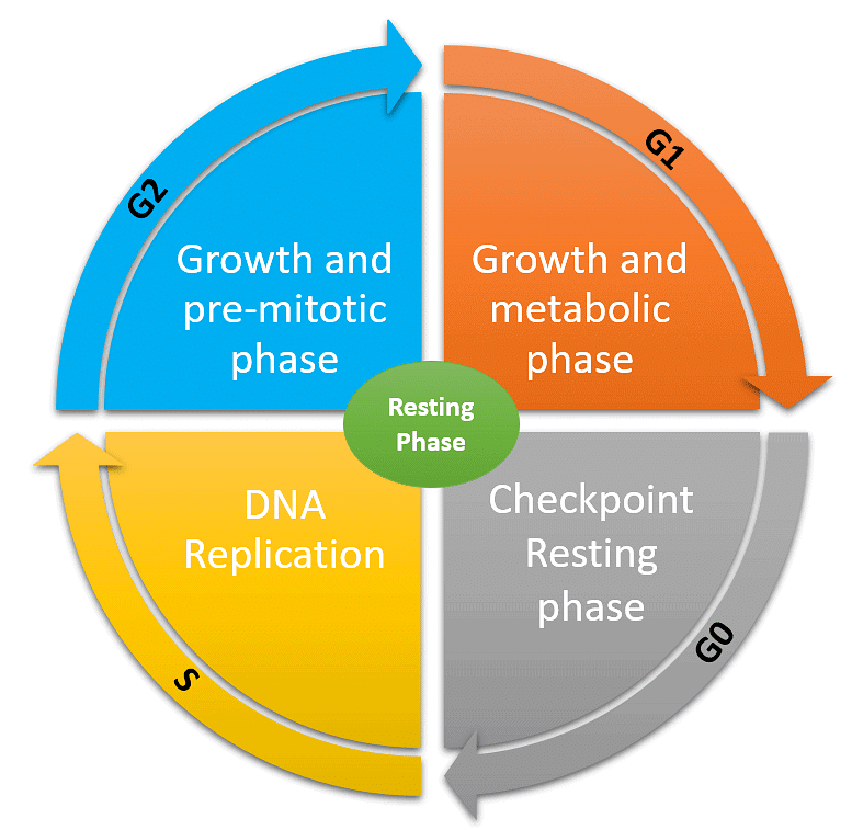
1.2 Interphase
Interphase is often called the "resting phase" but the cell is actually preparing for division through cell growth and DNA replication. It is divided into three sub-phases:
 Phases of Cell Cycle
1.2.1 G₁ Phase (Gap 1)
- Interval between mitosis and initiation of DNA replication
- Cell is metabolically active and continuously grows
- No DNA replication occurs
- DNA content remains 2C
- Chromosome number remains 2n (diploid)
1.2.2 S Phase (Synthesis)
- Period during which DNA synthesis or replication takes place
- Amount of DNA per cell doubles from 2C to 4C
- Chromosome number remains same (2n)
- In animal cells, centriole duplicates in the cytoplasm during S phase
- DNA replication begins in the nucleus
1.2.3 G₂ Phase (Gap 2)
- Proteins are synthesized in preparation for mitosis
- Cell growth continues
- DNA content is 4C
- Chromosome number remains 2n
NEET PYQ from this Topic:
Match List-I with List-II: (NEET 2024)
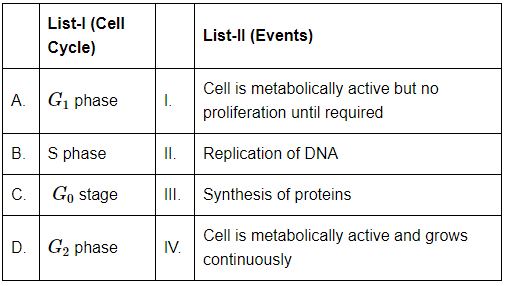 Choose the correct answer from the options given below :
(a) A-I, B-II, C-IV,D-III
(b) A-IV, B-II, C-I, D-III
(c) A-II, B-IV, C-I, D-III
(d) A-I, B-III, C-II, D-IV
1.3 G₀ Phase (Quiescent Stage)
- Some cells exit G₁ phase to enter an inactive stage called G₀
- Cells remain metabolically active but do not proliferate
- Examples: Heart cells (do not divide in adult animals)
- Many cells divide only occasionally when needed to replace injured or dead cells
- Cells can re-enter cell cycle from G₀ depending on organism's requirement
Try yourself: During which phase of the cell cycle does the DNA get synthesized?
1.4 Mitosis in Different Cell Types
- In animals: Mitotic cell division is seen only in diploid somatic cells
- Exception in animals: Male honey bees (haploid cells divide by mitosis)
- In plants: Mitosis occurs in both haploid and diploid cells
2. M Phase (Mitosis)
M Phase is the most dramatic period involving major reorganization of virtually all cell components. It is also called equational division because the chromosome number in parent and progeny cells remains the same. M Phase consists of karyokinesis (nuclear division) and cytokinesis (cytoplasmic division).
2.1 Karyokinesis
Nuclear division is divided into four stages. These stages represent a progressive process without clear-cut boundaries between them:
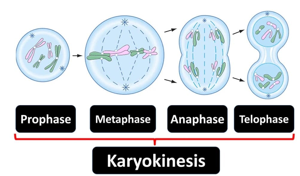
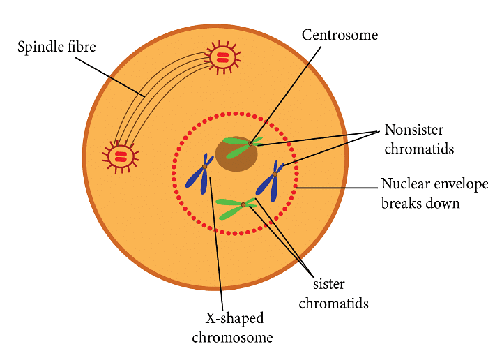
2.1.1 Prophase
First stage of karyokinesis that follows S and G₂ phases of interphase.
- Chromatin condensation: Chromosomal material initiates condensation and becomes untangled
- Chromosome structure: Compact mitotic chromosomes are composed of two chromatids attached at the centromere
- Centrosome movement: Duplicated centrosomes begin moving towards opposite poles
- Aster formation: Each centrosome radiates out microtubules called asters
- Mitotic apparatus: Two asters together with spindle fibres form the mitotic apparatus
- Organelle disappearance: Golgi complex, endoplasmic reticulum, nucleolus, and nuclear envelope are not visible
 Prophase
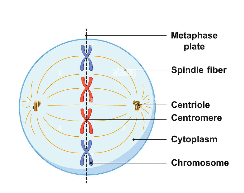
2.1.2 Metaphase
Starts with complete disintegration of nuclear envelope. Chromosomes spread through the cytoplasm.
- Chromosome condensation: Condensation is completed; chromosomes are easily studied under microscope
- Chromosome structure: Metaphase chromosome consists of two sister chromatids held together by centromere
- Kinetochore: Small disc-shaped structures at the surface of centromeres serving as attachment sites for spindle fibres
- Chromosome alignment: All chromosomes lie at the equator (metaphase plate)
- Spindle attachment: One chromatid connects to spindle fibres from one pole, sister chromatid connects to opposite pole through kinetochores
- Best stage: Morphology of chromosomes is most easily studied at this stage
 Metaphase
Spindle fibers attach to kinetochores of chromosomes during (NEET 2024)
(a) Prophase
(b) Metaphase
(c) Anaphase
(d) Telophase
2.1.3 Anaphase
Each chromosome at the metaphase plate splits simultaneously.
- Centromere splitting: Centromeres split and chromatids separate
- Daughter chromosomes: Two daughter chromatids (now called daughter chromosomes) migrate towards opposite poles
- Chromosome orientation: Centromere remains directed towards the pole (leading edge)
- Arms trail behind: Arms of chromosome trail behind during movement
2.1.4 Telophase
Final stage of karyokinesis begins when chromosomes reach their respective poles.
- Chromosome decondensation: Chromosomes decondense and lose their individuality
- Chromatin clustering: Chromatin material collects at each of the two poles
- Nuclear envelope formation: Nuclear envelope develops around chromosome clusters at each pole
- Two daughter nuclei formed: Two separate nuclei form at opposite poles
- Organelle reformation: Nucleolus, Golgi complex, and ER reform
NEET PYQ from this Topic:
Q1: Which of the following options gives the correct sequence of events during mitosis? (NEET 2017)
(a) Condensation → Nuclear membrane disassembly → Arrangement at equator → Centromere division → Segregation → Telophase
(b) Condensation → Crossing over → Nuclear membrane disassembly → Segregation → Telophase
(c) Condensation → Arrangement at equator → Centromere division → Segregation → Telophase
(d) Condensation → Nuclear membrane disassembly → Crossing over → Segregation → Telophase
2.2 Cytokinesis
Separation of cytoplasm that divides the cell into two daughter cells, completing cell division after karyokinesis.
2.2.1 Cytokinesis in Animal Cells
- Furrow formation: A furrow appears in the plasma membrane
- Deepening: Furrow gradually deepens
- Division completion: Furrow ultimately joins in the centre, dividing the cytoplasm into two
2.2.2 Cytokinesis in Plant Cells
- Different mechanism: Plant cells have relatively inextensible cell wall, so different mechanism is used
- Cell plate formation: Wall formation starts in the centre of the cell
- Outward growth: New wall grows outward to meet existing lateral walls
- Middle lamella: Cell plate is a simple precursor representing the middle lamella between walls of two adjacent cells
2.2.3 Organelle Distribution
- Organelles like mitochondria and plastids get distributed between two daughter cells during cytoplasmic division
2.2.4 Exception: Syncytium Formation
- In some organisms, karyokinesis is not followed by cytokinesis
- Results in multinucleate condition
- Leads to formation of syncytium
- Example: Liquid endosperm in coconut
NEET PYQ from this Topic:
Following are the stages of cell division : (NEET 2024)
A. Gap 2 phase
B. Cytokinesis
C. Synthesis phase
D. Karyokinesis
E. Gap 1 phase
Choose the correct sequence of stages from the options given below :
(a) C-E-D-A-B
(b) E-B-D-A-C
(c) B-D-E-A-C
(d) E-C-A-D-B
 The correct sequence will be
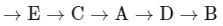
3. Significance of Mitosis
Mitosis (equational division) is usually restricted to diploid cells, but in some lower plants and social insects, haploid cells also divide by mitosis.
3.1 Key Functions of Mitosis
3.1.1 Production of Identical Daughter Cells
- Mitosis produces diploid daughter cells with identical genetic complement
- Ensures genetic stability across cell generations
3.1.2 Growth of Multicellular Organisms
- Growth of multicellular organisms is due to mitosis
- Increases cell number in tissues and organs
3.1.3 Maintenance of Nucleo-cytoplasmic Ratio
- Cell growth disturbs the ratio between nucleus and cytoplasm
- Cell division becomes essential to restore the nucleo-cytoplasmic ratio
3.1.4 Cell Repair and Replacement
- Cells of upper layer of epidermis are constantly replaced
- Cells of lining of gut are constantly replaced
- Blood cells are being constantly replaced
- This continuous replacement is achieved through mitosis
3.1.5 Continuous Growth in Plants
- Mitotic divisions in meristematic tissues (apical and lateral cambium)
- Results in continuous growth of plants throughout their life
Understanding the cell cycle and mitosis is crucial for comprehending how organisms grow, repair damaged tissues, and maintain genetic continuity. The coordinated events of interphase and M phase ensure that each daughter cell receives an exact copy of the genetic material, maintaining the integrity of biological information across generations of cells.
FAQs on Cell Cycle & Mitosis
| 1. What exactly happens during the S phase of the cell cycle and why is it so important? |  |
| 2. How do checkpoints in the cell cycle actually prevent damaged cells from dividing? | |
| 3. What's the difference between cytokinesis and mitosis, and why do students always get these confused? | |
| 4. Why do cancer cells ignore the G1 checkpoint and keep dividing uncontrollably? | |
| 5. Can I actually see chromosomes condensing during prophase under a microscope, and what exactly am I looking at? | |


