MCAT Exam > MCAT Notes > Biology for MCAT > Human Embryogenesis
Human Embryogenesis | Biology for MCAT PDF Download
| Table of contents |

|
| Introduction |

|
| The Journey Begins: From Zygote to Morula |

|
| The Formation of Blastocyst and Cell Differentiation |

|
| The Significance of Germ Layers |

|
| Neurulation: The Formation of Tubes |

|
Introduction
Embryogenesis is a remarkable process that spans the first eight weeks of development after fertilization. Within this relatively short period, a single cell evolves into a complex organism with a multi-level body plan. During embryogenesis, the circulatory, excretory, and neurologic systems begin to take shape. While this process may seem overwhelming, breaking it down into smaller, more manageable ideas can help us grasp the intricacies of human development.
The Journey Begins: From Zygote to Morula
Step 1: Fertilization occurs when an egg and a sperm cell fuse, resulting in the formation of a zygote.
Step 2: The zygote undergoes rapid cell division known as cleavage, which takes place within the first 12 to 24 hours.
During this initial stage, the zygote's primary focus is to multiply rapidly, leading to exponential growth in cell numbers. However, due to the speed of division, the individual cells do not have sufficient time to grow. Consequently, at the 32-cell stage called the morula, the size remains the same as the original zygote. The zona pellucida, a protective membrane surrounding the egg cell, also limits the growth potential during this period.
The Formation of Blastocyst and Cell Differentiation
Step 3: Blastulation occurs as the mass of cells forms a hollow ball, known as the blastocyst.
Step 4: Cell differentiation takes place, and cavities begin to form.
Around day 4, cell division continues while cells also begin to differentiate and acquire specific forms and functions. Differentiation involves cells following a specific path towards becoming a particular type of cell, such as an ear or kidney cell. The process of differentiation typically progresses in one direction. As development progresses, two distinct layers develop within the blastocyst: the outer trophoblast and the inner cell mass. The inner cell mass is pushed to one side of the trophoblast, resulting in the formation of a fluid-filled cavity called the blastocoel. This configuration resembles a snow globe. The trophoblast will eventually develop into structures facilitating embryo implantation in the uterus, while the inner cell mass, also known as the embryoblast, will further differentiate and contribute to the formation of the embryo.
 During this phase, the zona pellucida begins to disappear, allowing the blastocyst to grow and change shape. It is important to note that terminology may differ for non-mammalian animals, but for our discussion on human development, we will focus on the relevant terms.
The Significance of Germ Layers
Step 5: Gastrulation occurs, leading to the formation of three germ layers and the transformation of the cell mass into a gastrula.
Step 5a: The primitive streak, a prominent structure, forms during gastrulation.
Step 5b: The notochord, a defining feature, develops.
Week 3 of embryonic development involves gastrulation, a critical phase in which the three germ layers develop. These germ layers play a vital role in the formation of various organizational tubes in our bodies. The ectoderm, mesoderm, and endoderm represent these germ layers.
- The ectoderm, derived from the outer layer, contributes to the formation of the epidermis (outer layer of skin), hair, nails, brain, spinal cord, and peripheral nervous system. The mesoderm, originating from the middle layer, gives rise to muscle, bone, connective tissue, the notochord, kidney, gonads, and the circulatory system. Lastly, the endoderm, derived from the innermost layer, forms the epithelial lining of the digestive tract, including the stomach, colon, liver, pancreas, bladder, and lungs.
- Germ layers are established through the formation of the primitive streak, which appears around day 16. This streak serves as a midline reference, separating the left and right sides of the developing body. Deuterostomes, like humans, exhibit bilateral symmetry, allowing for a mirrored split along this midline.
- Below the primitive streak, the mesoderm, specifically the axial mesoderm, forms the notochord. The notochord plays a crucial role in determining the major axis of the body and eventually develops into intervertebral discs.

Neurulation: The Formation of Tubes
Step 6: Tubes begin to form, resulting in the formation of a neurula.
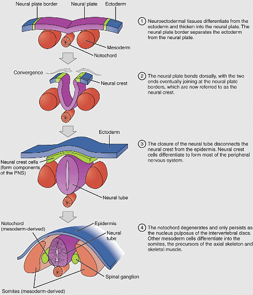
Step 6a: The notochord induces the formation of the neural plate.
Step 6b: The neural plate folds, giving rise to the neural tube and neural crest.
Step 7: The mesoderm differentiates into five distinct categories.
- Although progress has been made in the development of the germ layers, the process of tube formation is yet to occur. To initiate this critical step, the notochord stimulates the ectoderm above it to form a thick, flat plate called the neural plate. The neural plate extends along the rostral-caudal axis before folding back on itself to create the neural tube. The borders of the original neural plate develop into the neural crest, often referred to as the fourth germ layer. The neural tube ultimately gives rise to the brain and spinal cord, while the neural crest contributes to various structures, including the sympathetic and parasympathetic nervous systems, melanocytes, Schwann cells, and certain bones and connective tissue in the face.

- Simultaneously, the mesoderm undergoes further differentiation, resulting in the formation of distinct categories: axial, paraxial, intermediate, and lateral plate mesoderms. The axial mesoderm gives rise to somites, which eventually differentiate into muscle, cartilage, bone, and dermis, contributing to the segmented body plan. The intermediate mesoderm forms the urogenital system, including the kidneys, gonads, adrenal glands, and connecting ducts. The lateral plate mesoderm develops into the heart (the first organ to develop!), blood vessels, the body wall, and the muscle in our organs.
- During this period, the endoderm also undergoes tube formation, resulting in the development of the digestive tract. The digestive tract is divided into the foregut, midgut, and hindgut, each with its specific nerve and blood supply. Organs associated with the digestive tract initially develop as outpouchings from this tube. The foregut gives rise to the esophagus, stomach, part of the duodenum, and the respiratory bud, which eventually develops into the lungs. The midgut encompasses the second half of the duodenum to the transverse colon. The hindgut contributes to the remaining portions of the GI tract, including the transverse colon, descending colon, sigmoid colon, and rectum.
Conclusion
By the eighth week of human development, a meticulous sequence of events has occurred, resulting in the establishment of the body's intricate tube systems. The primitive heart has been beating for nearly five weeks, and the embryo is well on its way towards further development. Understanding the process of human embryogenesis provides insights into the complexity of life's beginnings and highlights the remarkable journey from a single cell to a highly organized and complex organism.
The document Human Embryogenesis | Biology for MCAT is a part of the MCAT Course Biology for MCAT.
All you need of MCAT at this link: MCAT
|
233 videos|16 docs
|

|
Explore Courses for MCAT exam
|

|
Signup for Free!
Signup to see your scores go up within 7 days! Learn & Practice with 1000+ FREE Notes, Videos & Tests.
Related Searches


