Cytoskelaton and Microtubules | Botany Optional for UPSC PDF Download
| Table of contents |

|
| Introduction |

|
| Microfilaments |

|
| Intermediate Filaments |

|
| Microtubules |

|
| Flagella, Cilia, and Centrosomes |

|
Introduction
Imagine a scenario where someone sneaks in during the night and steals your skeleton. Biologically speaking, this is highly unlikely, but if it were to occur, the consequences would be severe. The loss of your skeleton would result in a dramatic alteration in your body's structure. Your external appearance would change, your internal organs might shift out of place, and your ability to move, walk, and talk would be greatly compromised.
Remarkably, the same holds true for a cell. While we often perceive cells as shapeless entities, they are, in fact, intricately structured, much like our own bodies. Cells possess a network of filaments known as the cytoskeleton, aptly named the "cell skeleton." This cytoskeletal framework not only provides structural support to the cell's plasma membrane but also dictates its overall shape. Moreover, it plays a pivotal role in positioning organelles correctly, acts as tracks for vesicle transport, and facilitates cell movement in various cell types.
In eukaryotic cells, the cytoskeleton comprises three types of protein fibers: microfilaments, intermediate filaments, and microtubules. In this article, we will delve into each of these filament types and explore some specialized cellular structures associated with the cytoskeleton.
Microfilaments
- Among the trio of cytoskeletal protein fibers, microfilaments boast the slimmest dimensions, with a mere 7 nm in diameter. These filaments consist of interconnected monomers of a protein called actin, forming a structure resembling a double helix. Due to their composition of actin monomers, microfilaments are also referred to as actin filaments. Notably, actin filaments exhibit directionality, signifying the presence of two structurally distinct ends.
- Actin filaments serve several critical functions within the cell. They act as tracks for the movement of a motor protein named myosin, which can also form filaments. This association with myosin implicates actin in numerous cellular processes that require motion. For instance, during animal cell division, an actin and myosin ring constricts the cell to yield two daughter cells. Muscle cells are rich in actin and myosin, forming organized structures called sarcomeres. Contraction occurs when actin and myosin filaments within sarcomeres slide past each other in synchrony.
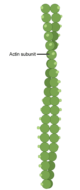
- Furthermore, actin filaments function as internal highways, facilitating the transport of cargoes, including protein-laden vesicles and even organelles. These cargoes are conveyed by individual myosin motors, which traverse along bundles of actin filaments. Remarkably, actin filaments can swiftly assemble and disassemble, a property pivotal to their role in cell motility, such as the movement of white blood cells in the immune system.
- Lastly, actin filaments play an indispensable role in cellular structure. In most animal cells, a network of actin filaments resides in the cytoplasm's peripheral region, intricately linked to the plasma membrane by specialized connector proteins. This network bestows shape and structural integrity upon the cell.
Intermediate Filaments
Intermediate filaments represent a distinct category of cytoskeletal elements, composed of multiple strands of fibrous proteins tightly coiled together. These filaments, with an average diameter of 8 to 10 nm, occupy a middle ground between microfilaments and microtubules.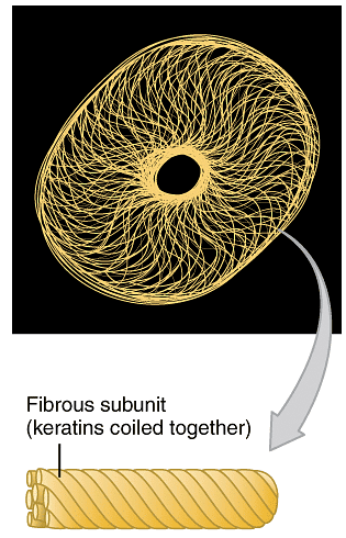 Various proteins form different types of intermediate filaments, with keratin being one prominent example. Keratin is a fibrous protein found in hair, nails, and skin. You may have encountered shampoo advertisements claiming to enhance the keratin in your hair. Unlike actin filaments, which exhibit rapid growth and disassembly, intermediate filaments serve as permanent structural elements in the cell. They are specialized to endure tension and fulfill roles such as maintaining cellular shape and anchoring the nucleus and other organelles in place.
Microtubules
Despite their name's "micro" connotation, microtubules are the most substantial among the three types of cytoskeletal fibers, boasting a diameter of approximately 25 nm. Each microtubule consists of tubulin proteins arranged to create a hollow, straw-like tube, with each tubulin protein comprising two subunits: α-tubulin and β-tubulin. Similar to actin filaments, microtubules exhibit dynamic properties, capable of rapid growth and shrinkage through the addition or removal of tubulin proteins. Microtubules also display directional polarity, characterized by structurally distinct ends. Microtubules serve a fundamental structural role within the cell, contributing to its ability to withstand compressive forces. Beyond structural support, microtubules play various specialized roles. They serve as tracks for motor proteins known as kinesins and dyneins, responsible for transporting vesicles and other cargoes throughout the cell. During cell division, microtubules assemble into a structure called the spindle, which orchestrates the separation of chromosomes.
Flagella, Cilia, and Centrosomes
Microtubules play integral roles in three specialized eukaryotic cell structures: flagella, cilia, and centrosomes. It is worth noting that prokaryotic cells also possess flagella for locomotion, although their structure differs significantly from eukaryotic flagella.
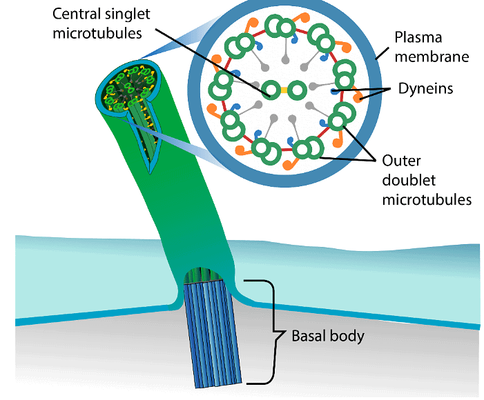
Flagella and Cilia: These structures are elongated and hair-like, extending from the cell surface and facilitating cell movement. Flagella are typically singular or present in small numbers, whereas motile cilia occur in larger quantities on the cell surface. In tissues formed by cells with motile cilia, their coordinated beating assists in moving materials across the tissue's surface. Notably, both flagella and motile cilia share a common structural pattern, featuring 9 pairs of microtubules arranged in a circle, with two additional microtubules at the center, known as a 9 + 2 array. Motor proteins called dyneins move along these microtubules, generating the force necessary for flagellum or cilium beating. Basal bodies, located at the base of these structures, play crucial roles in assembly and regulation.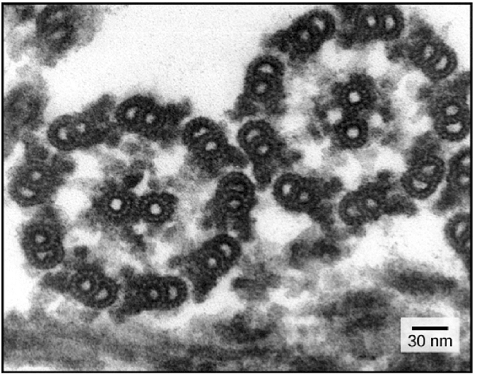
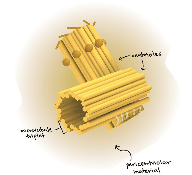
 Centrosomes: Comprising two centrioles arranged at right angles to each other, centrosomes serve as microtubule organizing centers in animal cells. Centrioles consist of nine triplets of microtubules held together by supporting proteins. Their exact function in orchestrating microtubule dynamics during cell division remains under investigation.
In conclusion, the cytoskeleton is a remarkable framework that lends structure and functionality to eukaryotic cells. Microfilaments, intermediate filaments, and microtubules, along with specialized structures like flagella, cilia, and centrosomes, collectively ensure the proper functioning of cells, contributing to their diverse and intricate activities. Understanding the cytoskeleton's roles and components offers profound insights into the biology of life at the cellular level.
|
179 videos|143 docs
|
FAQs on Cytoskelaton and Microtubules - Botany Optional for UPSC
| 1. What are microfilaments? |  |
| 2. What are intermediate filaments? |  |
| 3. What are microtubules? |  |
| 4. What are flagella, cilia, and centrosomes? |  |
| 5. How do microtubules contribute to the cytoskeleton? |  |


