Short & Long Question Answers with Solution: Body Fluids and Circulation - NEET PDF Download
Short Answer Type Questions
Q1: What are the two types of the circulatory system?
Ans: The circulatory system can be categorized into two main types:
- Open Circulatory System: This type of circulatory system is exclusive to insects. In an open circulatory system, blood circulates through various parts of the body cavity, rather than within closed vessels.
- Closed Circulatory System: This type of circulatory system is present in all vertebrates. In a closed circulatory system, blood flows through closed tubes known as blood vessels, ensuring a more controlled and efficient circulation throughout the body.
Q2: What is the significance of the AV node and AV bundle in the functioning of the heart?
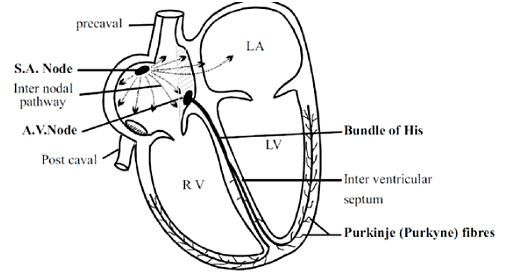
Ans: The atrioventricular node, abbreviated as the AV node, plays a crucial role in the heart's electrical conduction system. It enhances the signals that originate from the SA node. These signals need reinforcement as they tend to weaken while traveling through the heart's atria since the ventricles, where the signals need to reach, are relatively distant from the SA node. From the AV node, specialized structures known as atrioventricular bundles, or AV bundles for short, arise and serve as conduits for transmitting cardiac impulses to the muscular walls of the ventricles. This ensures coordinated and efficient contraction of the heart's ventricles, allowing for effective pumping of blood throughout the body.
Q3: What is pulse pressure?
Ans: Pulse pressure refers to the numerical difference between systolic blood pressure (the highest pressure in the arteries when the heart beats) and diastolic blood pressure (the lowest pressure in the arteries when the heart is at rest between beats). In adults, the normal value for pulse pressure is typically around 40 millimeters of mercury (mm Hg). Pulse pressure is an important parameter as it provides insights into the health and elasticity of the arteries and can be an indicator of various cardiovascular conditions when it deviates from the normal range.
Q4: Define blood and lymph.
Ans: Blood and lymph are two vital fluids within the human body. Blood is classified as a fluid connective tissue, comprising plasma, blood cells, and platelets. On the other hand, lymph is a clear fluid that circulates through the lymphatic system, which includes lymph nodes and lymphatic vessels.
Q5: Why is blood considered a connective tissue?
Ans: Blood originates from the mesodermal layer during development and consists of a fluid matrix known as plasma along with various types of cells. It circulates continuously throughout the body, playing a crucial role in the transportation of substances.
Q6: What are the causes of Anaemia?
Ans: The common symptoms of anaemia are:
- Pica.
- Insomnia.
- Jaundice.
- Pale skin.
- Leg cramps.
- Constipation.
- Abdominal pain.
- Severe joint pains.
- Shortness of breath.
- Headache and Dizziness.
- Susceptibility to infection.
- Fatigue and Loss of energy.
Q7: The Sino-atrial node is called the pacemaker of our heart. Why?
Ans: The human heart typically pulsates at a rate of 70 to 75 beats per minute. The SA node, or sinoatrial node, plays a vital role in initiating and sustaining the heart's rhythmic contractions. It serves as the pacemaker of the heart as it consists of specialized cardiac muscle fibers that conduct impulses similar to nerve fibers.
Q8: What are the symptoms of Hypertension?
Ans: The common symptoms of hypertension are:
- Dizziness.
- Heart attack.
- Visual Changes.
- Shortness of Breath.
- Narrowing of blood vessels and the formation of plaques in the blood vessels.
Q9: List out the functions of:
- Lymphatic System.
- Pulmonary vein.
- Lymphocytes.
Ans:
- Lymphatic System: This system is responsible for the transportation of white blood cells to and from the lymph nodes and into the bones.
- Pulmonary vein: It carries oxygenated blood from the lungs back to the heart.
- Lymphocytes: These cells serve as the body's defense mechanism by protecting against invading foreign substances.
Q10: Why do we call our heart myogenic?
Ans: The heart is termed myogenic because it generates heartbeats through muscle activity rather than nerve activity. The heart's functions are internally regulated, meaning they are autonomously controlled by specialized muscles.
Q11: Why are thrombocytes necessary for blood coagulation?
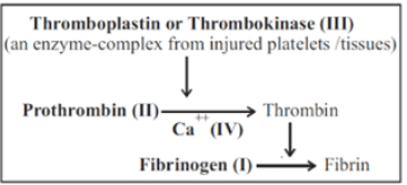
Ans: Platelets, also called thrombocytes, are blood components produced in the bone marrow and have a lifespan of approximately one week. When there is an injury that causes bleeding, platelets are released to initiate the clotting process by producing thromboplastin. The presence of thromboplastin, along with calcium ions, activates prothrombokinase. This initiates a series of reactions that result in the formation of a blood clot, which plugs the injured blood vessel, preventing further blood loss.
Q12: The walls of the ventricles are much thicker than atria. Explain.
Ans: The ventricles have thicker walls compared to the atria because they need to generate higher pressure to pump blood to various parts of the body. Specifically, the left ventricle has a thicker wall compared to the right ventricle. This is because the left ventricle is responsible for pumping oxygenated blood to supply all body tissues, and it needs to generate more force to accomplish this, whereas the right ventricle pumps deoxygenated blood to the nearby lungs, requiring less force.
Q13: What is the functional role of the lymphatic system?
Ans: The portal vein plays a crucial role in the circulation of blood from the intestines to the liver before it enters the systemic circulation.
Its significance lies in several key functions:
- Transporting nutrient-rich blood from the digestive system, containing glucose, amino acids, and various nutrients, to the liver. This allows the liver to utilize excess glucose and fats when needed, especially during periods of starvation.
- Converting toxic ammonia into urea, which can be safely eliminated from the body by the kidneys, reducing the risk of ammonia-related toxicity.
- Producing essential proteins like fibrinogen within the liver, which are then released into the bloodstream, contributing to various physiological processes in the body.
Q14: Answer the questions below:
(a) Which is the site where RBCs are formed?
(b) Name the part of the heart that initiates and maintains the rhythmic activity
(c) What is the heart of crocodiles is specific amongst reptilians?
Ans: (a) Marrow within bones
(b) SA node
(c) Reptiles typically possess a three-chambered heart, with the exception of crocodiles, which feature a four-chambered heart due to the partial division of the ventricle through a septum.
Q15: Explain the consequences of a situation in which blood does not coagulate.
Ans: Blood coagulation is the body's natural response to injury, serving to prevent excessive bleeding. Haemophilia, a genetic disorder, occurs when there is a deficiency in essential clotting factors such as prothrombin, fibrinogen, and vitamin K. This deficiency hinders the blood's ability to coagulate properly and can lead to severe and uncontrolled bleeding, potentially resulting in death.
Q16: Give a reason why the walls of ventricles are thicker than atria.
Ans: The ventricular walls are thicker because they need to pump blood to different organs.
Q17: Differentiate between the tricuspid and bicuspid valves.
Ans:
- The tricuspid valve has three muscular flaps and is responsible for regulating the opening between the right atrium and the right ventricle.
- The bicuspid valve, also known as the mitral valve, controls the opening between the left atrium and the left ventricle. It is associated with oxygenated blood.
Q18: Name the components of the formed elements in the blood and mention one major function of each.
Ans:
- The blood consists of three main types of formed elements: erythrocytes (red blood cells), leukocytes (white blood cells), and platelets.
- Erythrocytes, also known as RBCs, contain hemoglobin, which is responsible for transporting oxygen.
- Leukocytes, or WBCs, are divided into granulocytes and agranulocytes. B lymphocytes produce antibodies, neutrophils and monocytes are involved in phagocytosis, acidophils produce antitoxins against allergens, and basophils produce heparin and histamine.
- Platelets play a crucial role in blood clotting.
Q19: What is the importance of plasma proteins?
Ans:
- Plasma, which makes up about 7-8% of the blood, contains three main types of proteins: albumin, globulin, and fibrinogen.
- Albumin is the most abundant protein and plays a key role in maintaining colloidal osmotic pressure in the blood.
- Globulins are involved in antibody formation, while fibrinogen is essential for blood clotting.
Q20: Given below are the abnormal conditions related to blood circulation. Name the disorders.
- Acute chest pain due to failure of oxygen supply to heart muscles.
- Increased systolic pressure.
Ans:
- Angina. Due to the narrowing of the coronary artery, the blood supply to the heart muscles is reduced.
- High blood pressure.
Long Answer Type Questions
Q1: State the differences between the following:
- Lymph and blood
- Eosinophils and Basophils
- Bicuspid valve and tricuspid valve
Ans: Blood and Lymph:
- Blood is a connective tissue containing leukocytes (white blood cells), erythrocytes (red blood cells), and platelets within a plasma matrix. It circulates through blood vessels.
- Lymph is another connective tissue containing only white blood cells in its plasma. It flows through the lymphatic system.
Basophils and Eosinophils:
- Basophils are characterized by a three-lobed nucleus and contain coarse granules that stain basic. They make up about 0-1% of the blood's white blood cells.
- Eosinophils have a bilobed nucleus and cytoplasmic granules that stain acidic. They are present in the blood in the range of 1-6%.
Tricuspid and Bicuspid Valve:
- The tricuspid valve separates the right atrium from the right ventricle. It has three flaps and is also known as the right atrioventricular valve.
- The bicuspid valve, also called the mitral valve, separates the left atrium from the left ventricle. It has two flaps.
Q2: Describe events in the cardiac cycle.
Ans: The heart's function is to pump blood throughout the body, and this is achieved through a series of events known as the cardiac cycle, which occur with each heartbeat. The cardiac cycle is divided into two main phases: systole (contraction) and diastole (relaxation). A single heartbeat includes both atrial and ventricular systole and diastole.
Here are the key events of the cardiac cycle:
- Atrial systole: Initiated by the SA node, a contraction wave begins in the atria. During this phase, blood from the pulmonary veins and vena cava flows into the left and right ventricles because the tricuspid and bicuspid valves are open. The semilunar valves remain closed. Atrial systole enhances the blood flow into the ventricles. Atrial systole lasts for 0.1 seconds, followed by atrial diastole, which lasts for 0.7 seconds.
- Ventricular systole: In this phase, both the atria relax while the ventricles contract. As the ventricles contract, blood pressure within them rises, causing the atrioventricular valves to close rapidly, preventing the backflow of blood from the ventricles into the atria. Subsequently, an action potential is conducted by the AV node and AV bundle to the ventricles.
These events together make up the cardiac cycle, ensuring the continuous flow of blood through the heart and into the circulatory system.
Q3: Explain:
(a) Hypertension
(b) Coronary Artery Disease
Ans: (a) Hypertension, commonly known as high blood pressure, is a prevalent condition affecting the heart, blood vessels, brain, kidneys, and eyes. A normal blood pressure reading is around 120/80 mm Hg. When it exceeds 140 mm Hg for systolic pressure and 90 mm Hg for diastolic pressure, it is considered high blood pressure or hypertension.
The causes of hypertension include:
- The blockage of coronary heart vessels.
- Smoking tobacco, which accelerates heart rate, narrows blood vessels, and raises blood pressure.
(b) Coronary Artery Disease (CAD) develops when fatty substances accumulate on the arterial walls, forming atherosclerotic plaques. This condition results in the narrowing of the artery's lumen, potentially leading to a complete blockage of blood flow, which can trigger a heart attack.
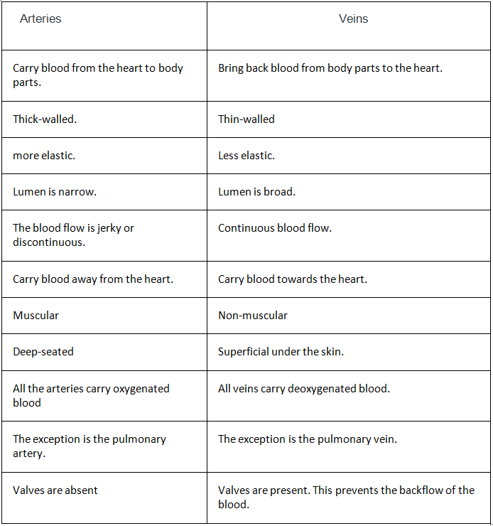
Q4: Differentiate between arteries and veins.
Ans:
Q5: Difference between P and T wave.
Ans:
- The P wave signifies the depolarization phase, while the T wave indicates repolarization.
- The P wave represents the electrical excitation of the atria, while the T wave signals the return of the ventricles to their normal state after excitation.
- The P wave corresponds to the contraction of both the atria, while the T wave marks the conclusion of systole (contraction) in the heart.
Q6: What is meant by double circulation? What is its significance?
Ans: In the context of double circulation, the blood completes two rounds through the heart:
- First round to the lungs and back: This is known as pulmonary circulation or the lesser circulation because it covers a shorter distance between the heart and the lungs.
- Second round to the body's organs and back: This is referred to as systemic circulation or the greater circulation because it encompasses a longer path between the heart and the various organs.
The significance of this dual circulation system lies in its functions:
- In pulmonary circulation, deoxygenated blood is transported to the lungs, where it receives oxygen and is carried back by the pulmonary veins into the left atrium of the heart.
- In systemic circulation, oxygen-rich blood is distributed to all parts of the body.
- It facilitates the delivery of essential nutrients and oxygen to different body tissues.
- It also aids in the removal of carbon dioxide and harmful substances from various tissues for eventual elimination from the body.
Q7: Explain heart sounds.
Ans:
- Heart sounds are produced when the heart valves close, resulting in two distinct sounds known as "Lubb" and "Dupp." These sounds repeat in a rhythmic pattern.
- The first heart sound is characterized by being longer, louder, and having a lower frequency. It is generated by the closure of the atrioventricular valves right after the start of ventricular systole. Its duration typically ranges from 0.16 to 0.90 seconds.
- Conversely, the second heart sound is sharper, with a higher frequency, and is softer and shorter in duration, approximately 0.10 seconds. This sound is produced by the closure of the semilunar valve at the conclusion of the ventricular systole.
Q8: Distinguish between open and closed circulatory systems.
Ans:
Q9: Describe the following terms and give their location.
- Bundle of His
- Purkinje fibre
Ans:
- The atrioventricular (AV) node is located near the tricuspid valve in the heart. Emerging from the AV node is a specialized bundle of muscle fibers known as the Bundle of His or Atrioventricular bundles. These bundles run through the front part of the interventricular septum before branching into smaller fibers.
- The atrioventricular bundle then gives rise to tiny fibers called Purkinje fibers. These fibers originate from myocardial tissue and are responsible for conducting electrical impulses throughout the walls of the ventricles in the heart.

Q10: Briefly describe the following:
- Anaemia
- Angina pectoris
- Atherosclerosis
- Hypertension
- Heart failure
Ans:
- Anemia refers to a reduction in the amount of hemoglobin, and there are various types of anemia:
- Nutritional anemia results from insufficient iron in the blood.
- Megaloblastic anemia is caused by deficiencies in vitamin B12 and folic acid.
- Pernicious anemia is due to a deficiency of vitamin B12, which can disrupt the formation of myelin sheaths around neurons and is a severe condition.
- Sickle cell anemia is an inherited autosomal recessive disorder characterized by the presence of sickle-shaped red blood cells (RBCs) that cannot effectively carry oxygen to tissues.
- Thalassemia, another autosomal recessive disorder, results from the inadequate synthesis of either alpha or beta hemoglobin chains.
- Angina pectoris can occur in individuals of various ages but is more common in early and middle-aged people. It occurs when the heart muscles do not receive sufficient oxygen, leading to acute chest pain triggered by exertion or exercise.
- Atherosclerosis is a condition affecting arteries and blood vessels, characterized by the deposition of calcium, cholesterol, and fibrous tissue. This process narrows the arterial lumen, causing hardening and loss of elasticity, particularly in the arteries that supply blood to the heart muscle. Atherosclerosis is also known as coronary artery disease.
- High blood pressure, known as hypertension, occurs when blood flow exerts excessive pressure on the elastic walls of arteries. Normal blood pressure typically falls within the range of 120/80 mm Hg, with 120 mm Hg representing systolic pressure (pumping pressure) and 80 mm Hg representing diastolic pressure (resting pressure). Hypertension is diagnosed when blood pressure consistently exceeds 140/90 mm Hg, and it can affect critical organs like the heart, brain, and kidneys.
- Heart failure refers to a condition in which the heart is unable to effectively pump blood to meet the body's needs, often leading to lung congestion. This condition is also referred to as congestive heart failure.
Q11: Explain the events in the cardiac cycle. Describe ‘double circulation’.
Ans: The cardiac cycle represents one complete heartbeat, encompassing the relaxation (diastole) and contraction (systole) of the cardiac muscles.
It involves a sequence of events:
- Atrial systole: The atria contract, driven by the sino-atrial node, forcing blood into the ventricles as the bicuspid and tricuspid valves remain open.
- Initiation of ventricular systole: The AV node triggers the ventricles to contract, causing the bicuspid and tricuspid valves to close, resulting in the first heart sound, "lub."
- Completion of ventricular systole: As the ventricles contract, blood is forced into the pulmonary trunk and aorta through the open semilunar valves.
- Initiation of ventricular diastole: The ventricles begin to relax, accompanied by the closure of the semilunar valves, leading to the second heart sound, "dub."
- Completion of ventricular diastole: As ventricular pressure decreases, the bicuspid and tricuspid valves open, allowing blood to flow from the atria into the ventricles. The heart's contraction prevents blood from flowing backward, as the relaxed ventricles' pressure is lower than that of the atria and veins.
In the case of double circulation in birds and mammals, two separate pathways exist. The left atrium receives oxygenated blood, while the right atrium receives deoxygenated blood. Each set of atria passes the blood on to the corresponding ventricles, which then pump it out of the heart without mixing oxygenated and deoxygenated blood.
Q12: Describe the Rh-incompatibility in humans.
Ans: Rh antigen is seen on the RBC surface of majority humans, these are called Rh-positive individuals and when the antigen is absent they are Rh-negative individuals. Both these individuals are phenotypically normal individuals. However, in these individuals, a problem emerges during pregnancy or transfusion of blood. The first blood transfusion from Rh-positive blood to the RI-T individual leads to no harm as the Rh-negative person acquires antibodies or Rh factors in their blood. During the second transfusion of blood, from Rh-positive blood to the Rh-negative individual, the antibodies already formed attack to destruct the RBC of the donor. In pregnancy, if the father’s blood is Rh-positive and the mother’s blood is Rh-negative, the blood of the fetus will be Rh-positive, which leads to serious issues. The Rh antigens of the fetus are not exposed to the Rh-positive blood of the mother during the first pregnancy, as they are separated from the placenta. But in the succeeding Rh-positive fetus, the anti-Rh factors from the mother destruct the RBCs of the fetus as the blood mixes which causes hemolytic disease in the newborn(HDN) known as erythroblastosis fetalis. This can be prevented through the administration of anti-Rh antibodies to the mother after the delivery of the first child.
Q13: State the functions of the following in blood.
- Fibrinogen
- Globulin
- Neutrophils
- Lymphocytes
Ans:
- Fibrinogen makes up approximately 0.3% of the plasma and plays a crucial role in the formation of blood clots at the site of injury.
- Globulins are categorized into three types: α, β, and γ, constituting 2 to 3% of plasma proteins. Gamma globulins are involved in antibody formation, and globulins are primarily associated with immunity, serving as a vital component of the body's defense system against infections.
- Neutrophils are phagocytic cells responsible for eliminating foreign organisms that enter the body.
- Lymphocytes come in two varieties: T cells and B cells. They are responsible for producing antibodies and play a central role in providing immunity to the body.
Q14: Thrombocytes are essential for the coagulation of blood. Explain.
Ans: Thrombocytes play a crucial role in blood clotting, and this process involves several steps:
- Platelets release thromboplastin, a vital component in clot formation.
- Blood clotting primarily depends on factors such as fibrinogen, prothrombin, thromboplastin, and calcium ions.

- Thromboplastin aids in the formation of prothrombinase, an enzyme complex that neutralizes heparin. This process leads to the conversion of prothrombin into its active form, thrombin.
- Thrombin is a proteolytic enzyme responsible for converting fibrinogen into its insoluble form, fibrin. These fibrin monomers combine to form long, adhesive fibers.
- The fibrin threads create a network that traps damaged or dead blood components.
- Clot formation occurs over the injured area, resulting in a reddish-brown appearance.
- Serum, a straw-colored fluid, separates from the clot.
- Serum lacks fibrinogen and cannot participate in clot formation.
In medical terms, an increase in platelet count is referred to as thrombocytosis, while a decrease is known as thrombocytopenic purpura.
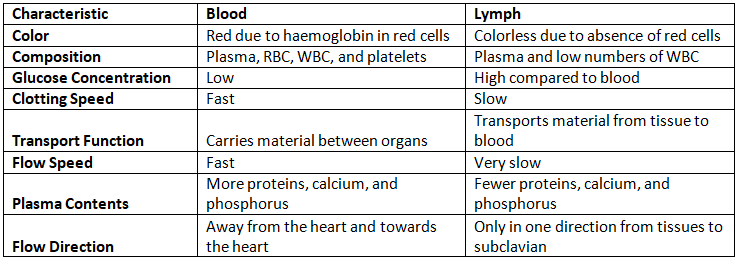
Q15: Write the differences between blood and lymph.
Ans:


