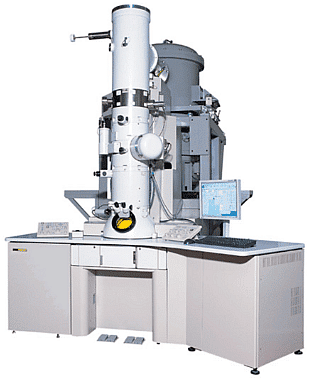
Electron microscopy (TEM, SEM) | Zoology Optional Notes for UPSC PDF Download
Transmission Electron Microscope (TEM): It is an advanced microscopy technique that uses electron beams instead of light to produce high-resolution images of specimens. 
It operates based on the principles of the light microscope but uses electrons to achieve a much higher resolution.
Historical Development: TEM
TEM was developed in 1931 by Max Knoll and Ernst Ruska in Germany. The first practical electron microscope was constructed in 1938 at the University of Toronto by Eli Franklin Burton, Cecil Hall, James Hillier, and Albert Prebus. It was further commercialized by Siemens in 1939.
Working Principles
Electron Beam Generation: TEM uses a source at the top of the microscope to emit a stream of monochromatic electrons. These electrons are produced by heating a tungsten filament at voltages ranging from 6,000 to 10,000 Volts.
Electromagnetic Lenses: Instead of glass lenses, TEM utilizes electromagnetic lenses to focus the electron beam into a thin beam. The first lens determines the spot size, while the second lens, controlled by a brightness knob, adjusts the beam size.
Specimen Interaction: The electron beam is directed through the specimen. Some electrons are scattered due to the specimen's density, while unscattered electrons hit a fluorescent screen, creating an image with varying darkness based on density.
Image Formation: The transmitted portion of the beam is focused by the objective lens to create an image. Additional components, such as objective and selected area metal apertures, can restrict the beam and enhance contrast by blocking high-angle diffracted electrons.
Image Display: The final image is enlarged through intermediate and projector lenses and is displayed on a phosphorescent image screen. Darker areas represent denser parts of the sample, while lighter areas represent less dense regions.
Sample Preparation
Sample preparation is a crucial step in TEM:
Fixation: Tissues are preserved using fixatives with matching pH and osmolarity to the living tissue. Glutaraldehyde is a common primary fixative, while Osmium Tetroxide is used as a secondary fixative.
Dehydration: Water is removed from the sample using a graded ethanol series.
Infiltration and Embedding: The sample is infiltrated with resin and placed in an embedding mold, which is then polymerized in an oven.
Sections of Embedded Material: The sample is cut into thin sections (50-100 nm thick) using an ultra microtome, placed on a copper grid, and stained with heavy metals.
Negative Staining of Isolated Material: Isolated material, like a solution with bacteria, is spread onto a support grid coated with plastic and stained with heavy metal salts, creating a "shadow" around the specimen.
Advantages over Light Microscope
- TEM uses electrons, providing a much higher resolution (up to 2 million times magnification) compared to light microscopes (limited to 2000 times).
- Unlike Scanning Electron Microscope (SEM) that bounces electrons off the surface, TEM shoots electrons through the sample, allowing for detailed internal imaging.
Scanning Electron Microscope (SEM)
A Scanning Electron Microscope (SEM) is an advanced microscopy technique that employs a focused beam of high-energy electrons to generate various signals at the surface of solid specimens, allowing for detailed imaging and elemental analysis. It offers several advantages over traditional optical microscopes.
 Historical Development
- The first SEM image was obtained by Max Knoll in 1935, revealing electron channelling contrast in silicon steel.
- In 1938, M. vonArdenne constructed a scanning transmission electron microscope by adding scan coils to a transmission electron microscope.
- The SEM was further developed by Professor Sir Charles Oatley and Gary Stewart in 1965.
- The first SEM used to examine the surface of a solid specimen was described by Zworykin et al. (1942) at RCA Laboratories in the United States.
Principles of Scanning Electron Microscopy
- SEM employs accelerated electrons with high kinetic energy that interact with the sample, resulting in signals like secondary electrons, backscattered electrons (BSE), diffracted backscattered electrons (EBSD), X-rays, visible light (cathodoluminescence–CL), and heat.
- Secondary electrons are useful for morphology and topography, while BSE illustrates composition contrasts in multiphase samples.
- X-ray generation provides elemental analysis.
- SEM can achieve high-resolution images (up to 300,000 times magnification), revealing details as small as 1 to 5 nm.
- SEM micrographs offer a large depth of field, giving a three-dimensional appearance to the images.
Components of a Scanning Electron Microscope
- Electron Gun: It generates a continuous stream of electrons for SEM operation. Electron guns are typically of two types: thermionic guns and field emission guns.
- Lenses: Instead of glass lenses, SEMs use magnetic lenses to control the electron beam's path, allowing for precise focus and magnification.
- Sample Chamber: This is where the specimen is placed in a vacuum to ensure stability and prevent interference from air particles.
- Detectors: Various detectors collect signals produced by the interaction of the electron beam with the specimen, such as secondary electron detectors and X-ray detectors.
- Vacuum Chamber: SEMs operate in a vacuum, ensuring the electron beam's stability and eliminating interference from air particles.
- Scanning Coils: These coils create a magnetic field to manipulate the electron beam's position, allowing for precise scanning.
Sample Preparation
Sample preparation for SEM analysis can vary from minimal to elaborate, depending on the sample and data requirements. Steps may include:
- Coating: Most insulating samples are coated with a thin layer of conductive material, like carbon, gold, or metal alloys, to prevent charge build-up and improve imaging.
- Vacuum Requirement: The specimen must be placed in a vacuum for stable electron beam operation. High instability results if the sample is in a gas-filled environment.
- Sputter Coating: A method used to coat specimens with a thin layer of metal, such as gold, by using argon gas and an electric field.
Scanning Process
- The electron beam, with energy ranging from a few hundred eV to 40 keV, is focused by one or two condenser lenses.
- The beam is scanned over a rectangular area of the specimen's surface in a raster fashion, synchronizing with the CRT display.
- Different signals are emitted from the specimen, including secondary electrons, which are collected by detectors and used to create the image displayed on a computer monitor.
- Image magnification in SEM depends on the ratio of the raster dimensions on the specimen and the display device, controlled by the current supplied to the scanning coils.
Scanning Electron Microscopy provides high-resolution imaging, making it valuable for various scientific and industrial applications.
|
181 videos|346 docs
|

|
Explore Courses for UPSC exam
|

|


