Molluscs: Lamellidens | Zoology Optional Notes for UPSC PDF Download
Description of Lamellidens
- Classification: Lamellidens belongs to the class Pelecypoda within the phylum Mollusca.
- Habitat: These freshwater mussels are found in ponds, lakes, and slow-moving streams worldwide, with distribution throughout the Indian subcontinent.
- Pearls: Small-sized pearls have been reported from Lamellidens in West Bengal (India) and Bangladesh.
- Habitat Behavior: Lamellidens mussels are partially embedded in mud or sand at the bottom of water bodies. They retract their foot and siphons inside the shell and tightly close the shell valves upon slight disturbance.
External Features of Lamellidens
- Shell: The hard, calcareous, horny-blackish bivalve shell is about 10 cm in length.
- Body: The body is light cream in color, soft, elongate oval with a somewhat broad and a narrow posterior end.
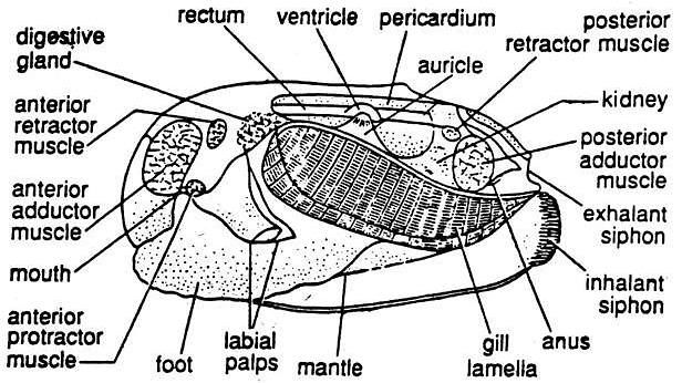
- Mantle: It consists of two equal halves, the mantle lobes, lining the inner surface of the shell and enclosing a mantle or pallial cavity.
- Siphons: Lamellidens has two siphons—the dorsal exhalant siphon with a smooth margin and the ventral inhalant siphon with a fimbriated margin—protruding beyond the shell at the posterior end.
- Foot: In its natural state, a narrow gape exists between the valves of the shell, allowing the muscular foot to project out from the anteroventral end of the body.
- Organs: The visceral mass, a pair of gills or ctenidia, two renal pores, and the anus are located in the mantle cavity.
Shell Structure of Lamellidens
- Shell Composition: The shell consists of conchiolin and calcic substances and grows along the margins with mussel growth.
- Hinge Line: The two valves are hinged along a straight dorsal hinge line with a tough, elastic hinge ligament.
- Hinge Teeth: Each valve bears hinge teeth along the hinge line, fitting with sockets on the other valve.
- Growth Rings: Concentric rings, the lines of growth, are present on the outer surface of the valve.
- Umbo: A dorsomedian elevated region, the umbo, is the thickest and oldest part of the shell. Lines of growth originate from the umbo.
- Inner Surface: The inner surface of the shell is smooth, pearly in color with a faint bluish tinge.
- Pallial Line: A narrow streak, the pallial line, marks the outer border of the mantle lobe on the inner surface.
- Muscle Scars: Scars on the inner surface indicate the attachment of various muscles:
Anterodorsal Scars:
- Anterior adductor muscle (large and nearly round)
- Anterior retractor muscle (small and posterodorsal to the adductor)
- Protractor muscle (small and posterior to the adductor)
Posterodorsal Scars:
- Posterior adductor muscle (large and oval)
- Posterior retractor muscle (small and close to the posterior adductor)

Microscopic Structure of Lamellidens Shell
Three Layers: The shell consists of three distinct layers:
- Periostracum: The thin outermost layer made of conchiolin.
- Ostracum or Prismatic Layer: The thick middle layer composed of alternate layers of conchiolin and calcic substances.
- Nacreous Layer or Mother of Pearl: The innermost layer composed of alternate layers of conchiolin and calcic substances. It is smooth and lustrous and is the layer where pearls are secreted.
Muscles of Lamellidens
- Muscle Types: The muscles are un-striated, with muscle fibers running from the body to the shell. Foot muscles are not attached to the shell.
- Adductor Muscles: There are two adductor muscles, anterior and posterior, which are large transverse bands controlling the movements of the shell valves. Contraction of these muscles closes the shells.
- Retractor Muscles: There are two retractor muscles, anterior and posterior, which run from the foot to the shell. Contraction of these muscles causes retraction of the foot.
- Protractor Muscles: Two protractor muscles, anterior and posterior, compress the visceral mass when they contract.
- Pedal Muscle: The foot consists of a complex mass of intrinsic muscles.
Locomotion in Lamellidens
- Foot Function: The foot serves as the locomotor organ.
- Foot Structure: The foot is a muscular projection that is protrusible through the gap between the shell valves, having a flattened shape from side to side and an elongated keel at the anterior end that resembles a ploughshare.
- Pedal Sinus: The foot has a large pedal sinus that receives haemolymph from the pedal artery. Contraction of foot muscles, aided by the pressure of haemolymph, forces the foot to extend forward.
- Foot Movement: The foot is used to move forward through a ploughing motion. It is repeated, allowing the mussel to progress by ploughing its foot through the substratum.

Body Cavity of Lamellidens
- In the adult, the coelom is greatly reduced and is restricted to the pericardium, gonad, and the lumen of the kidney. The haemocoel serves as channels for haemolymph circulation.
Digestive System of Lamellidens
- The digestive system includes a coiled alimentary tract, two pairs of labial palps, and a pair of digestive glands.
Alimentary Canal:
- Mouth: The mouth is a transverse slit located below the anterior adductor muscles. It is surrounded by two pairs of ciliated triangular flaps—external and internal labial palps.
- Oesophagus: A short tube runs dorsally from the mouth to open into the stomach.
- Stomach: A large, oval sac is embedded in the digestive gland. It has folds in the epithelium and receives secretions from the digestive glands. Occasionally, a mucous rod called the crystalline style, containing diastatic enzymes, is found in the stomach.
- Intestine: The intestine is a narrow tube that descends from the ventral surface of the stomach, forms two incomplete loops, ascends to the level of the stomach, turns sharply backward, and proceeds straight as the rectum.
- Rectum: The rectum pierces the pericardium and the ventricle of the heart, runs parallel to the dorsal surface of the mussel, and opens into the exhalant siphon via the anus. It has a ridge called the typhlosole.
- Digestive Glands: These are a pair of irregular light gray structures situated around the stomach, which secrete digestive enzymes and their secretions are discharged into the stomach.

Feeding:
- Lamellidens is a ciliary feeder and collects food with the help of cilia. Its diet consists of detritus and microorganisms suspended in water, with detritus making up the majority of its food.
- The cilia create a respiratory water current that carries food particles.
- Heavy particles are dropped in the mantle cavity and expelled through the exhalant siphon by cilia.
- The gills collect food particles as they are covered with a thin sheet of mucus.
Respiratory System of Lamellidens
- The respiratory organs consist of a pair of gills.
Gills Structure:
- Each gill consists of two laminae, the outer and the inner, with chambers or water tubes formed by vertical interlamellar junctions.
- The cells in the gill filaments have cilia, creating an inhalant current to bring water into the mantle cavity.
- Gaseous exchange occurs in the highly vascular gill filaments.
- Cleansing cilia sweep non-food particles off the gill filaments and are expelled through the exhalant siphon.
- The mantle is also richly supplied with haemolymph, facilitating gaseous exchange.

Circulatory System of Lamellidens
The circulatory system of Lamellidens comprises a heart, arteries, sinuses, and veins, forming an open circulatory system. The haemolymph, which contains few leucocytes, circulates through this system.
Heart and Pericardium
Heart: The heart consists of a single ventricle and two auricles. It is enclosed within a thin-walled pericardium.
- Ventricle: A muscular chamber surrounding the rectum.
- Auricles: Two thin-walled chambers connected to the ventricle via auriculoventricular apertures guarded by valves, allowing one-way flow into the ventricle.
Aortae: The aorta arises from each end of the ventricle and is named the anterior and posterior aorta:
- Anterior Aorta: Originates from the anterior end of the ventricle, running anteriorly above the rectum. It bifurcates into the right and left terminal arteries, with each branching to various organs before terminating as the pallial artery.
- Posterior Aorta: Arises from the posterior end of the ventricle, running posteriorly and ventral to the rectum, supplying organs in the posterior part of the body.
Sinuses, Veins, and Circuits
- Sinuses: Haemolymph from different body parts returns through sinuses, smaller veins, and is collected in a large longitudinal vena cava situated between the kidneys.
- Veins: Haemolymph is returned to the heart through three circuits:
- Mantle Circuit: Small veins from the mantle lobe merge into a large vein that directly returns oxygenated haemolymph to the auricle.
- Vena Cava Circuit: A small vein from the vena cava carries some deoxygenated haemolymph directly to the auricle before it branches into renal veins for the kidney.
- Ctenidial Circuit: The major portion of haemolymph is returned to the auricle through this circuit.
- Renal Veins: These carry deoxygenated haemolymph from the vena cava to the kidney.
- Afferent Branchial Veins: Carry haemolymph from the kidney to the gills.
- Efferent Branchial Veins: Transport oxygenated haemolymph from the gills to the auricles.
Excretory System of Lamellidens
The excretory system includes a pair of kidneys (also known as organs of Bojanus) and a pericardial gland (Keber's organ).
Kidney
- The kidneys consist of U-shaped tubes, with one located on each side of the visceral mass, ventral to the pericardium.
- They have two parts: the brownish, spongy, and glandular ventral arm (kidney proper), and the thin-walled, non-glandular, ciliated dorsal arm (urinary bladder). Cilia generate an outward current.
- The kidney proper opens into the pericardium through a Reno pericardial aperture, while the bladder communicates with its counterpart on the other side through a large, oval aperture.
- Each bladder opens into the mantle cavity through a small renal aperture (nephridiopore) between the visceral mass and the inner gill lamina.
Pericardial Gland of Lamellidens
- This reddish-brown glandular mass is situated just in front of the pericardium.
- It discharges excretory waste into the pericardial cavity.
Nervous System of Lamellidens
Lamellidens has a nervous system consisting of paired cerebral, pedal, and visceral ganglia, connected by commissures and connectives, and associated nerves:
Cerebral Ganglia
- Two triangular, yellowish ganglia located at the base of the labial palps, beneath the skin.
- Connected by the cerebral commissure, a slender nerve running around the anterior part of the esophagus.
Pedal Ganglia
- Two oval, pale-yellow ganglia found within the muscles of the foot, approximately 1 cm above the ventral border.
- The two ganglia fuse medially to form a bilobed mass.
Cerebropedal and Cerebrovisceral Connectives
- Cerebropedal connectives originate from the posterior border of the cerebral ganglia and extend downward and backward through the visceral mass and foot muscles, connecting with the pedal ganglia.
- Cerebrovisceral connectives arise from the posterolateral border of the cerebral ganglia and run backward, ventral to the gill lamina attachment with the visceral mass. They pass through the kidney and ultimately connect to the visceral ganglia.
Innervation
- Nerves arising from the cerebral ganglia innervate various structures, including the labial palps, the anterior part of the mantle, and adjacent areas.
- The pedal ganglia send nerves to the foot and the anterior retractor muscle.
- The visceral ganglia send nerves to different parts, including dorsal and posterior pallial nerves to the mantle lobes, posterior renal nerves to the kidneys, branchial nerves to the gills, and nerves to the alimentary canal.
- Fine nerves from cerebropedal and cerebrovisceral connectives innervate the statocysts and structures within the visceral mass, respectively.
Sensory Structures in Lamellidens
In Lamellidens, the presence of receptors and sense organs plays a crucial role in their survival and interaction with the environment. While these sense organs may not be highly developed, they serve specific functions:
1. Osphradium:
- Osphradium is a chemoreceptor.
- It consists of brown-pigmented sensory epithelium associated with each visceral ganglion.
- Its primary function is to assess the purity of the water entering the mantle cavity.
2. Statocyst:
- The statocyst serves as a balance organ.
- It is located near the pedal ganglion and takes the form of a round sac.
- Inside the statocyst, a granule called the statolith is present.
- The statocyst is innervated by the statocyst nerve, which originates from the cerebropedal connective.
- It encloses a space lined with sensory cells.
3. Scattered Sensory Cells:
- Sensory cells are distributed on the outer epithelium of the body and the inner border of the mantle.
- These cells function as tactile sense organs, enabling Lamellidens to sense their surroundings.
Reproductive System in Lamellidens
Lamellidens exhibits separate sexes, and the reproductive system is characterized by distinct features:
Male Reproductive System:
- The testes are white in color.
- Each testis is connected to a vas deferens arising from its posterolateral border.
Female Reproductive System:
- The ovaries have a reddish color.
- Each ovary is associated with an oviduct that arises from its posterolateral border.
Fertilization and Development in Lamellidens
The process of fertilization and development in Lamellidens is complex and takes place internally. Here is a step-by-step breakdown of this process:
1. Fertilization Process:
- Eggs are released from the ovaries and enter the mantle cavity through female genital pores.
- Sperms are discharged from the testes into the mantle cavity through male gonopores, and they reach the supra-branchial chamber of the female.
- Fertilization occurs in the supra-branchial chamber, resulting in fertilized eggs.
2. Egg Capsule Formation:
- Fertilized eggs are transferred to the water tubes.
- In the water tubes, they are enclosed within thick, soft, somewhat triangular, whitish capsules known as egg capsules.
3. Glochidium Larvae:
- Development continues within the egg capsules, resulting in the formation of glochidium larvae.
- Glochidium larvae possess several distinctive features, including a bivalve shell, dorsal valve unity, ventral flatness, and spines on the ventral ends of the valves.
- The larvae have a brush-like hair-bearing mantle lining, an adductor muscle connecting the valves, and the presence of a byssus gland with a long byssus thread.
Metamorphosis:
- Glochidium larvae attach themselves to the gills of freshwater fish using the byssus thread.
- They penetrate the host tissue, become ectoparasites, and undergo metamorphosis.
- During metamorphosis, the byssus thread and sense organs disappear.
- A stomodaeum is formed through ectodermal invagination behind the mouth, creating the mouth opening.
- The proctodaeum is not formed, and the anus is established through a simple process of rupture.
- The foot emerges as a median, ventral elevation behind the mouth.
- Gill rudiments appear as two papillae on each side of the mouth.
- Eventually, the larvae, now suitable for a free life, detach from the host, sink to the bottom, and gradually transform into their adult form.
|
181 videos|346 docs
|
FAQs on Molluscs: Lamellidens - Zoology Optional Notes for UPSC
| 1. What are the external features of Lamellidens? |  |
| 2. How is the shell of Lamellidens structured at a microscopic level? |  |
| 3. What are the muscles involved in the movement of Lamellidens? |  |
| 4. How does Lamellidens locomote? |  |
| 5. What is the digestive system of Lamellidens like? |  |

|
Explore Courses for UPSC exam
|

|


