UPSC Exam > UPSC Notes > Zoology Optional Notes for UPSC > Protochordata: General features Life history of Branchiostoma
Protochordata: General features Life history of Branchiostoma | Zoology Optional Notes for UPSC PDF Download
Historical Classification of Branchiostoma
- In 1974, P. S. Pallas, a German zoologist, initiated the classification of Branchiostoma, naming it Limax lanceolatus and mistakenly associating it with slugs.
- In 1836, W. Yarrell recognized the distinct nature of these creatures and named them Amphioxus lanceolatus.
- However, it was later discovered that O. G. Costa had named them Branchiostoma in 1834. Following taxonomic priority rules, Branchiostoma is accepted as the generic name, with Amphioxus retained as the common name.
Geographical Reach of Branchiostoma
- Branchiostoma boasts a nearly cosmopolitan distribution, inhabiting sandy shores across tidal zones to considerable depths.
- It thrives in both marine and estuarine environments, with a presence in tropical and temperate seas.
- The common lancelet, Branchiostoma lanceolatus, has been identified on European coasts, East African coasts, and the western and south-eastern Indian coasts.
Habits and Habitat
- Branchiostoma is a versatile dweller, residing in both marine and estuarine locales, commonly found in sandy shores.
- It thrives in sea water with salinities ranging from 15.4% to 33.1%.
- While primarily sedentary, it exhibits active swimming, moving vertically in water. Its fins play no role in swimming.
- Branchiostoma burrows in the sand, maintaining a shallow depth with the anterior end projecting for a constant water current, facilitating feeding.
- Feeding involves the ciliary mode, with Branchiostoma consuming microorganisms carried into the pharyngeal cavity by the respiratory water current.

External Anatomy
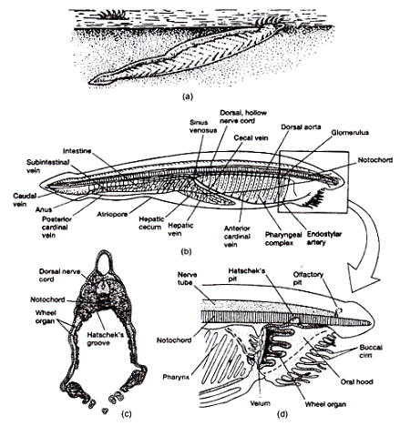
- Branchiostoma is a small, lancet-shaped creature with pointed ends, ranging from 5 to 8 cm in length.
- The body is elongated and flattened, distinguishing into the body proper and a postanal tail.
- The pointed anterior end forms a snout, below which lies the oral hood with over twenty oral cirri or tentacles.
- The mouth is concealed within the oral hood, and the anus is on the left side near the ventral fin.
- The atrium opens through an atriopore close to the anterior end of the ventral fin.
- Gill slits, not usually visible externally, line the body's sides, covered partially by lateral folds.
Body Wall Structure
- The epidermis is a single layer of columnar cells with sensory hair, devoid of gland and pigment cells.
- Beneath the epidermis are the cutis and sub-cutis layers, with canals traversing both.
- The muscle layer consists of myotomes and myocommas, aiding lateral undulation during locomotion.
- The notochord extends along the mid-dorsal line, preventing shortening during muscle contraction.
Supporting Structures
- Supporting structures include gill rods, skeletal oral ring, and fin-ray boxes for gill-bars, oral hood, and fins, respectively.
- The notochord, gill-rods, skeletal oral ring, and fin-ray boxes provide structural integrity.
Locomotion Mechanics
- Branchiostoma occasionally swims freely when disturbed, contracting longitudinal muscle fibres for forward motion.
- The notochord prevents shortening during muscle contraction, serving as a lever for efficient movement.
- Lateral undulation is facilitated by the arrangement of myotomes on either side, aiding rapid sidewise twisting.
Internal Organization
- The body is divided into pharyngeal and intestinal regions.
- Transverse sections reveal distinct structures, including the epidermis, myotomes, dorsal fin, notochord, and nerve cord.
- Specific differences exist between pharyngeal and intestinal sections.
Respiratory Mechanism
- The pharynx's vascular wall aids in respiration, with water current bringing fresh oxygen.
- Gill-bars with blood vessels efficiently absorb oxygen close to the surface.
- Doubts persist about the pharynx's respiratory role, with some emphasizing its function in food concentration.
Circulatory Symphony
- Branchiostoma's circulatory system lacks a respiratory pigment, relying on dissolved oxygen for vital functions.
- The sinus venosus collects blood from various body parts, pumping it through the ventral aorta, gill-bars, dorsal aortae, and dorsal aorta.
- Blood circulation occurs in a continuous loop from posterior to anterior and vice versa.
- The caudal vein, sub-intestinal vein, and cardinal veins contribute to this intricate circulatory dance.
In the grand saga of Branchiostoma, its evolutionary intricacies, ecological ballet, and physiological poetry unfold, marking its significance in the aquatic tapestry.
Excretory System of Branchiostoma
Nephridia:
- Branchiostoma possesses approximately 90 to 100 paired segmental nephridia, known as protonephridia.
- These are located on the dorsolateral wall of the pharynx, each corresponding to a primary gill-bar.
- Each nephridium comprises a vesicular sac with a horizontal and vertical limb, opening into the atrium.
- Elongated tubular flame cells or solenocytes within the vesicle aid in waste elimination.
Nephridium of Hatschek:
- This tube, originating from the mouth and extending to the right side of the notochord, is considered an excretory organ.
- It is an ectodermal derivative and is vascularized by the dorsal aorta.
Miscellaneous Excretory Organs:
- Brown Funnels: Paired structures located at the anterior end of the atrium, possibly acting as excretory or receptor organs.
- Atrial Wall: Groups of cells in the atrial wall also contribute to excretion.
- Gonads: Yellow masses containing uric acid in the testes contribute to excretion during gamete expulsion.
Neural System of Branchiostoma
- The nervous system comprises a hollow dorsal nerve cord above the notochord.
- The anterior part forms the brain, while the posterior part remains the spinal cord.
- Paired dorsal and ventral nerve roots extend into each body segment, with non-myelinated nerves.
- The ventricle at the anterior end gives rise to dorsal nerves conveying impulses from the oral hood and buccal cirri receptors.
- The infundibular organ, situated on the ventral wall of the ventricle, contains tall, ciliated cells, generating Reissner's fibre.
- Photoreceptive cells (eye-spots) are scattered in the spinal cord.
Receptor Organs in Branchiostoma
- Pigment Spot: An unpaired spot on the brain's anterior wall, lacking lens and not photosensitive.
- Eye-Spots: Photosensitive cells enclosed by pigment granules, distributed on the spinal cord.
- Kolliker’s Pit: A ciliated depression at the anterior brain, possibly a chemoreceptor.
- Sensory Papillae: Modified sensory papillae on oral cirri and velar tentacles act as chemoreceptors and tactile receptors.
- Infundibular Organs: Located at the ventricle floor, may act as photoreceptors.
- Epidermal Sensory Cells: Sensory cells on the dorsal body surface.
Reproductive System of Branchiostoma
- Branchiostoma exhibits separate sexes with gonads as simple pouch-like segmental organs.
- Gonoducts are absent, and gametes are released into the atrium, escaping through the atriopore.
- Fertilization and development occur in seawater, with small, isolecithal eggs.
Development of Branchiostoma
Primitive, Degenerated, and Specialized Features:
- Primitive Characters:
- Presence of a persistent notochord, single-layered epidermis, and myotomic segmentation.
- Simple alimentary canal, ciliary feeding, and a basic circulatory system.
- Degenerated Characters:
- Sedentary behavior and reduced brain and sense organs.
- Specialized Characters:
- Spacious pharynx, numerous gill-slits, and specialized feeding apparatus.
Affinities and Systematic Position of Branchiostoma
Relationships with Other Phyla:
- Annelida: Similarities include bilateral symmetry, segmented body, and segmental nephridia. Differences involve the notochord and gill-slits absent in annelids.
- Mollusca: Resemblances in feeding and respiratory mechanisms, but significant anatomical differences.
Relationship with Chordate Groups:
- Hemichordata: Structural similarities exist, but Branchiostoma is considered more advanced.
- Urochordata: Strong evidence supports a close phylogenetic relationship, especially in larval features.
- Cyclostomata: Resemblances with lamprey larvae, but differences in cranium and vertebral column structure.
Conclusion:
- Phylum Chordata: Branchiostoma is universally accepted as a chordate, possessing notochord, dorsal tubular nerve cord, and gill-slits.
- Relative Status: While its precise position within Chordata is uncertain, it shares common features with various chordate groups, showcasing evolutionary connections.
The document Protochordata: General features Life history of Branchiostoma | Zoology Optional Notes for UPSC is a part of the UPSC Course Zoology Optional Notes for UPSC.
All you need of UPSC at this link: UPSC
|
181 videos|346 docs
|

|
Explore Courses for UPSC exam
|

|
Signup for Free!
Signup to see your scores go up within 7 days! Learn & Practice with 1000+ FREE Notes, Videos & Tests.
Related Searches


