IGCSE Class 10 > Class 10 Notes > Biology for GCSE/ > The Eye
The Eye

Structure & Function of the Eye
- The eye, a sensory organ, holds receptor cells that detect light (rod cells) and color (cone cells).
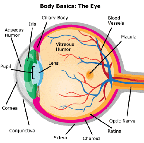
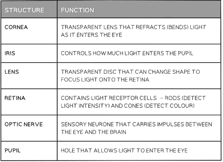
Function of the parts of the eye:
The Blind Spot
- At the point where the optic nerve joins the retina, there are no light-sensitive rod and cone cells on that part of the retina
- Light falling onto that part of the retina will not result in an image being detected. The brain 'fills in' from surrounding light so we don't see a black hole where no light has fallen.
- The brain processes visual information by 'filling in' from surrounding light to prevent the perception of black holes where no light has fallen.
- As a result of this process, a blind spot is created in our peripheral vision, rendering us unable to detect objects even if they are present.
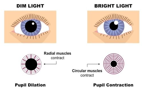
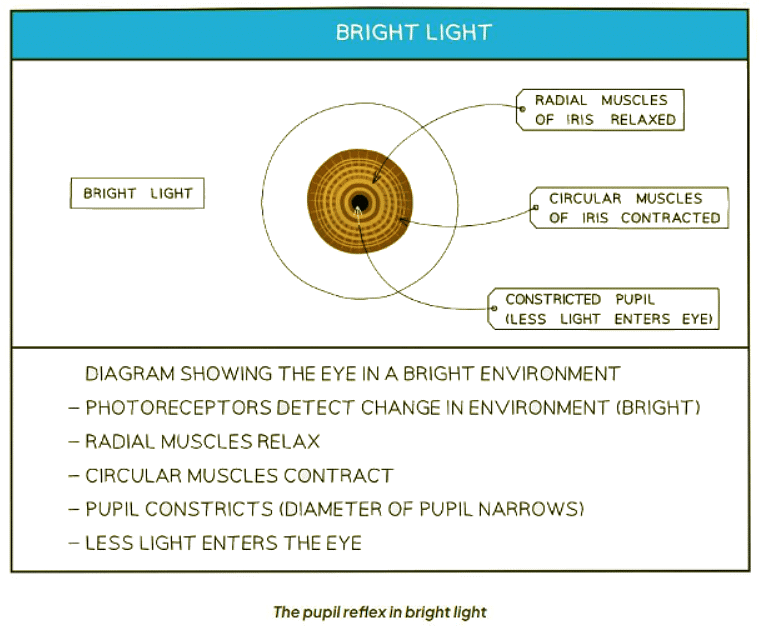
The Pupil Reflex
- This is a reflex action carried out to protect the retina from damage in bright light and protect us from not seeing objects in dim light
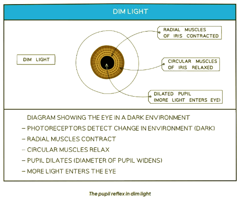
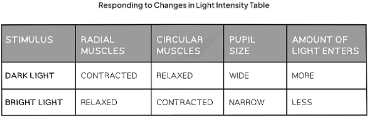
- In dim light the pupil dilates (widens) in order to allow as much light into the eye as possible
- In bright light the pupil constricts (narrows) in order to prevent too much light entering the eye and damaging the retina
- In dim lighting conditions, the pupil dilates, enlarging to permit more light to enter the eye, thus enhancing vision.
- Conversely, in bright environments, the pupil constricts, shrinking to reduce the amount of light entering the eye and safeguarding the retina from potential damage.
The Pupil Reflex - Antagonistic Muscle Action
- The pupil reflex involves two sets of muscles, the radial muscles, and the circular muscles, which work in opposition to each other to control the amount of light entering the eye.
- Radial muscles dilate the pupil, allowing more light to enter, while circular muscles constrict the pupil, reducing the amount of light entering the eye.
- When radial muscles contract, circular muscles relax, and vice versa, demonstrating antagonistic action.
Accommodation
Accommodation: The function of the eye in focusing on near and distant objects
- The process by which the lens adjusts to bring objects into focus is known as accommodation.
- The lens is flexible and can change its shape based on the tightness or looseness of the suspensory ligaments attached to it.
- Key Terms: flexible, suspensory ligaments, tight, loose
- Changes in the lens shape are controlled by the contraction or relaxation of the ciliary muscles.
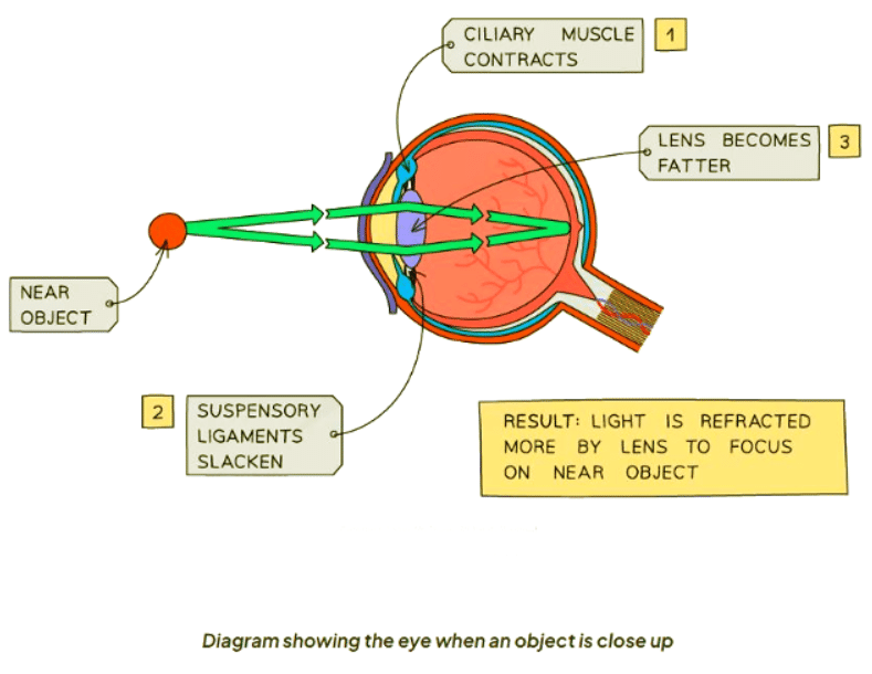
- When an object is near, the ciliary muscles contract, reducing the diameter of the muscle ring.
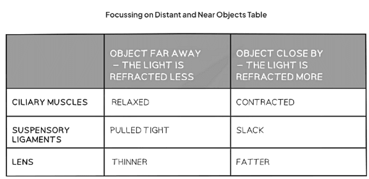
- When an object is close up:
- The ciliary muscles contract, causing the suspensory ligaments to loosen.
- This relaxation prevents the suspensory ligaments from tugging on the lens, allowing it to thicken.
- As a result, light gets refracted more effectively.
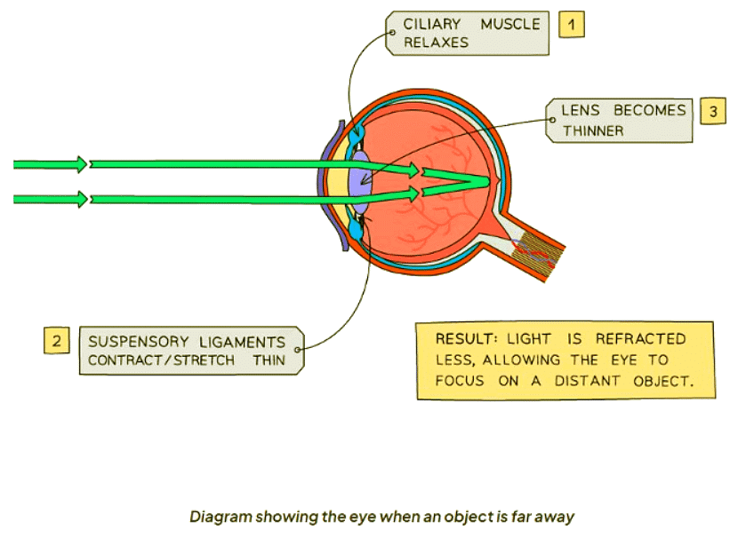
When an object is far away:
- The ciliary muscles relax, causing the suspensory ligaments to tighten.
- This tension makes the suspensory ligaments pull on the lens, resulting in its thinning.
- Consequently, light is refracted less compared to when the object is closer.
Rod & Cone Cells
- There are two main types of cells in the retina responsible for detecting light:
- Rods: These cells are highly sensitive to low light conditions.
- Cones: These cells are specialized in distinguishing between different colors, especially in bright light.
- Rods and cones have distinct functions:
- Rods are primarily activated in dim light situations.
- Cones come into play in bright light, allowing us to perceive a wide range of colors.
- Cones further specialize into three subtypes, each sensitive to a particular color of light:
- Red-sensitive cones
- Blue-sensitive cones
- Green-sensitive cones
- The fovea, a central area on the retina, contains a high density of cone cells, facilitating detailed color vision.
- Rod cells are spread throughout the retina except for the optic nerve's point of attachment, creating a region known as the blind spot:
- The blind spot lacks light-sensitive cells, making it unable to detect light or color.
- In low light conditions, such as during night vision, only rod cells are stimulated, resulting in black and white vision.
MULTIPLE CHOICE QUESTIONTry yourself: What is the function of the blind spot in the eye?
The document The Eye is a part of the Class 10 Course Biology for GCSE/IGCSE.
All you need of Class 10 at this link: Class 10
About this Document

4.73/5 Rating

Apr 29, 2026 Last updated
Related Exams
Document Description: The Eye for Class 10 2026 is part of Biology for GCSE/IGCSE preparation. The notes and questions for The Eye have been prepared according to the Class 10 exam syllabus. Information about The Eye covers topics like and The Eye Example, for Class 10 2026 Exam. Find important definitions, questions, notes, meanings, examples, exercises and tests below for The Eye.
Introduction of The Eye in English is available as part of our Biology for GCSE/IGCSE for Class 10 & The Eye in Hindi for Biology for GCSE/IGCSE course. Download more important topics related with notes, lectures and mock test series for Class 10 Exam by signing up for free. Class 10: The Eye
Description
The Eye of Biology covers all the important topics, helping you prepare for the Class 10 exam on EduRev. Start for free!
Information about The Eye
In this doc you can find the meaning of The Eye defined & explained in the simplest way possible. Besides explaining types of The Eye theory, EduRev gives you an ample number of questions to practice The Eye tests, examples and also practice Class 10 tests


Related Searches
Extra Questions, video lectures, Objective type Questions, shortcuts and tricks, The Eye, practice quizzes, past year papers, pdf , Sample Paper, Free, mock tests for examination, study material, Important questions, The Eye, ppt, Summary, Previous Year Questions with Solutions, Semester Notes, The Eye, Viva Questions, MCQs, Exam;