Revision Notes: The Nervous System | Biology Class 10 ICSE PDF Download
| Table of contents |
|
| Nervous System |
|
| Functions of the Nervous System |
|
| Neuron: The Unit of the Nervous System |
|
| Synapse |
|
| Types of Neurons |
|
| Types of Nerves |
|
| Division of the Nervous System |
|
| Reflexes |
|

Nervous System
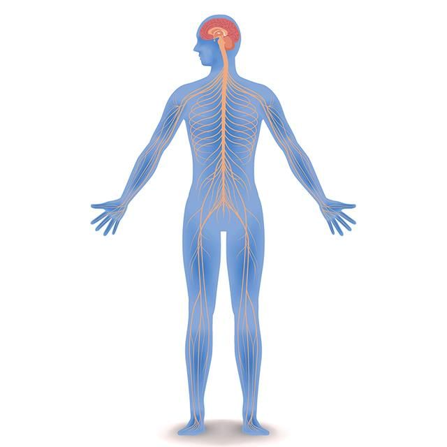
The nervous system comprises the brain, spinal cord, sense receptors, and nerves.
Functions of the Nervous System
- Keeps us informed about the outside world through sensory organs.
- Controls and harmonizes all voluntary muscular activities, such as running and writing.
- Enables us to remember, think, and reason.
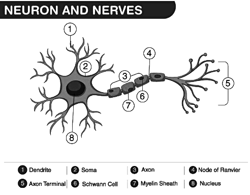
Neuron: The Unit of the Nervous System
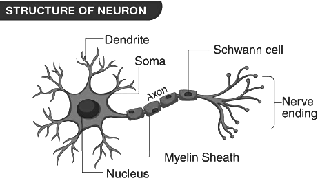
The three main parts of the neuron are as follows:
- Cell Body: It contains a well-defined nucleus and granular cytoplasm.
- Dendrites: These are branched cytoplasmic projections of the cell body.
- Axon:
- It is a long process extending from the cell body.
- The axon is covered by a myelin sheath, which has gaps known as Nodes of Ranvier.
Some Basic Terms
- Stimulus: An agent or sudden change in the external or internal environment that causes a change in an organism or any of its body parts.
- Response: The change in an organism resulting from a stimulus.
- Impulse: A wave of irritability, or an electrical disturbance, that sweeps over a nerve cell.
- Receptors: Nerve cells that generate waves of impulses towards the central nervous system upon receiving a stimulus.
- Effectors: Muscles or glands that contract or secrete substances upon receiving an impulse from the brain or spinal cord.
Synapse
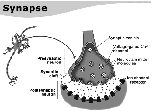
- A synapse is the junction where the terminal branches of an axon from one neuron connect with the dendrites of another neuron.
- When a nerve impulse reaches the axon terminal of a neuron, a neurotransmitter called acetylcholine is released from the bulbs present in the axon.
- To prepare the synapse for the next nerve impulse, acetylcholine is subsequently broken down by an enzyme.
Transmission of Nerve Impulse
- When a nerve fibre is at rest, its outer side carries a positive charge because there are more Na+ ions outside the axon membrane. This state is known as the polarised state or polarisation of the nerve fibre.
- Upon stimulation, the axon membrane at the site of stimulation becomes more permeable to Na+ ions. As a result, Na+ ions move inward, causing a loss of polarisation. This is referred to as the depolarised state or depolarisation of the nerve fibre. The region undergoing depolarisation is called the excited region.
- The point of depolarisation acts as a stimulus for the adjacent region of the axon membrane, prompting it to depolarise as well. Meanwhile, the previous region of the membrane undergoes repolarisation due to the active transport of Na+ ions back to the outside of the membrane.
Types of Neurons
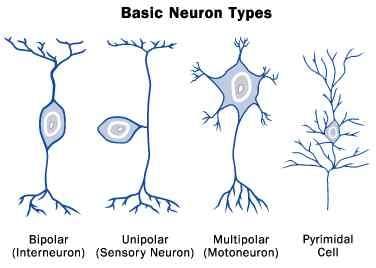
- Sensory Neurons: These neurons are responsible for transmitting impulses from sensory receptors (such as those in the skin, eyes, ears, nose, and tongue) to the central nervous system, which includes the brain and spinal cord.
- Motor Neurons: Motor neurons carry impulses from the central nervous system to effectors, which are typically muscles or glands. This process initiates a response, such as muscle contraction or gland secretion.
- Associated Neurons: Also known as interneurons or relay neurons, these neurons connect sensory and motor neurons within the central nervous system. They play a crucial role in processing information and coordinating responses.
Types of Nerves
A nerve consists of a bundle of nerve fibers (axons) from different neurons, all enclosed in a tubular sheath. Ganglia are clusters of nerve cell bodies, from which nerve fibers may emerge or enter.
Types of Nerves:
- Sensory Nerves: These nerves contain sensory fibers that transmit sensory information from the body to the central nervous system.
- Motor Nerves: Motor nerves contain motor fibers that carry commands from the central nervous system to the muscles, triggering movement.
- Mixed Nerves: These nerves carry both sensory and motor fibers, allowing for the transmission of sensory information to the central nervous system and the relay of motor commands to the muscles.
Division of the Nervous System
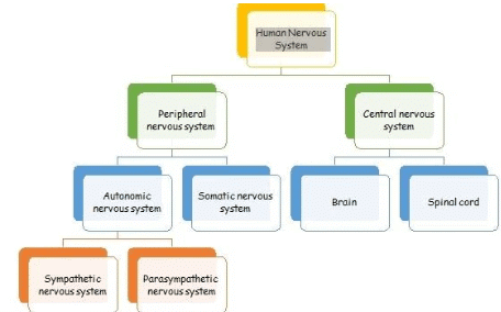
The nervous system is divided into two main parts: (i) Central Nervous System (CNS) (ii) Peripheral Nervous System (PNS)
The Central Nervous System
The central nervous system comprises the brain and the spinal cord.
The Brain
- The brain is protected within the cranium, also known as the skull. In adults, the brain weighs approximately 1.35 kg. It is safeguarded by three protective membranes called the meninges : dura mater, arachnoid, and pia mater.
- The space between these membranes, along with the central spaces of the brain and the central canal of the spinal cord, is filled with cerebrospinal fluid. This fluid acts as a cushion, protecting the brain from shocks.
Three Primary Regions of the Brain
1. Forebrain:
- The forebrain is responsible for higher cognitive functions such as intelligence, memory, consciousness, willpower, and voluntary actions.
- The thalamus is a part of the forebrain that relays sensory information, including pain and pressure signals, to the cerebrum.
- The hypothalamus regulates essential bodily functions, including body temperature and the activity of the pituitary gland.
2. Midbrain:
- The midbrain is a small, tube-like structure responsible for reflexes associated with the eyes and ears.
3. Hindbrain:
- The cerebellum in the hindbrain coordinates muscular activity and maintains body balance.
- The pons serves as a communication pathway, carrying impulses between the two hemispheres of the brain and coordinating muscular movements on both sides of the body.
- The medulla oblongata regulates vital functions of internal organs, including heartbeat, breathing, and other autonomic activities.
Parts of the Brain
1. Cerebrum
- The cerebrum is the largest part of the brain and is divided into two hemispheres.
- These hemispheres are connected by a bundle of nerve fibers called the corpus callosum.
- The outer layer of the cerebrum, known as the cortex, is made up of neuron cell bodies and appears greyish in color, which is why it is called grey matter.
- The grey matter has many folds, called gyri, and grooves, known as sulci, which increase its surface area.
- Beneath the cortex, the medulla contains the axons of nerve fibers and is called white matter due to its lighter color.
2. Cerebellum
- The cerebellum is located at the base of the cerebrum.
- In a cross-section, its white matter resembles a branching tree, which is a characteristic feature.
3. Medulla Oblongata
- The medulla oblongata is situated at the base of the skull and has a roughly triangular shape.
- It is an extension of the brain and continues downwards as the spinal cord.
- The medulla oblongata is vital for survival, and injury to this part of the brain can lead to death.
The Spinal Cord
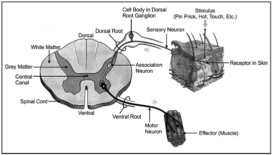
- The spinal cord is housed within the neural canal of the vertebrae.
- Similar to the brain, the spinal cord is protected by three layers of meninges : the dura mater, arachnoid, and pia mater.
- In the spinal cord, the grey matter is located on the inner side, while the white matter is on the outer side.
- The grey matter contains the cell bodies of neurons, and the white matter consists of myelinated axons.
Functions of the Spinal Cord:
- The spinal cord is responsible for reflex actions that occur below the neck.
- It conducts sensory impulses from the skin and muscles to the brain.
- The spinal cord also transmits motor signals from the brain to the muscles of the trunk and limbs.
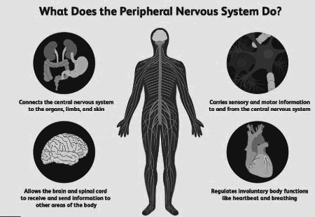
Peripheral Nervous System
The peripheral nervous system (PNS) is made up of nerves that carry impulses to and from the central nervous system (CNS), which includes the brain and spinal cord.
1. Somatic Nervous System: This part of the PNS controls voluntary movements and transmits sensory information from the body to the CNS. It includes:
- Cranial Nerves: Nerves that emerge directly from the brain and brainstem, responsible for various functions such as smell, vision, and facial movements.
- Spinal Nerves: Nerves that emerge from the spinal cord, carrying signals to and from the rest of the body.
2. Autonomic Nervous System: This part of the PNS regulates involuntary bodily functions, such as heart rate and digestion. It is further divided into:
- Sympathetic System: Prepares the body for "fight or flight" responses during stressful situations.
- Parasympathetic System: Promotes "rest and digest" activities, helping the body to relax and conserve energy.
Somatic Nervous System
Cranial Nerves: There are 12 pairs of cranial nerves that emerge from the brain.
Spinal Nerves: There are 31 pairs of spinal nerves, which are distributed as follows:
- 8 pairs in the neck region
- 12 pairs in the thorax
- 5 pairs in the lumbar region
- 5 pairs in the sacral region
- 1 pair in the coccygeal region
Autonomic Nervous System
The autonomic nervous system is responsible for controlling the involuntary actions of internal organs.
- Sympathetic Nervous System: Nerves of the sympathetic nervous system arise from the spinal cord between the neck and waist regions.
- Parasympathetic Nervous System Nerves of the parasympathetic nervous system are located anteriorly in the head and neck and posteriorly in the sacral region.
Opposite Effects of the Two Systems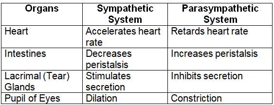
Reflexes
Reflex action is a quick, automatic, and involuntary response in the body triggered by a stimulus.
Difference between Reflexes/Involuntary Actions and Voluntary Actions
Reflexes (Involuntary Actions)
- Initiation: Reflexes are triggered by stimuli such as touch, pain, pressure, heat, or light.
- Origin of Commands: Commands for reflexes originate in the spinal cord, autonomic nervous system, and sometimes in the brain.
- Examples: Shivering in cold, sweating in heat, heartbeat, withdrawing hand from hot surface, pupil dilation in darkness.
Voluntary Actions
- Initiation: Voluntary actions are initiated by a conscious thought.
- Origin of Commands: Commands for voluntary actions originate in the brain.
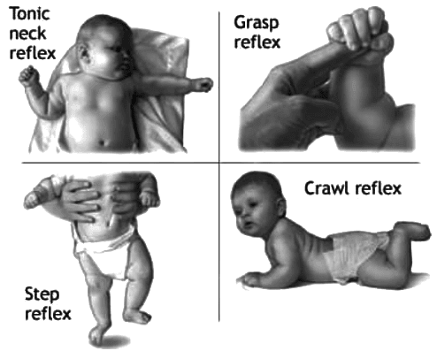
Types of Reflexes
Natural (Inborn) Reflex
- Definition: Reflexes that are present from birth and do not require previous experience or learning.
- Characteristics: These reflexes are similar in all humans and are not influenced by individual experiences.
- Examples: Salivation, peristalsis (movement of food through the digestive tract), swallowing.
Conditioned (Acquired) Reflex
- Definition: Reflexes that develop during an individual’s lifetime based on experiences and learning.
- Characteristics: These reflexes vary from person to person, as they depend on individual experiences and learning.
- Examples: Salivating at the smell of food (conditioned response).
Pavlov’s Experiment
Nervous Pathways in Reflexes
Reflex actions require a quick response, so the pathway for receiving and sending information is short. A reflex arc consists of the following components:
- Stimulus: A change in the environment is detected by sensory receptors in the sense organs.
- Afferent (Sensory) Nerve Fibre: The sensory information is transmitted to the central nervous system (CNS) through afferent nerve fibres.
- Central Nervous System (CNS): The sensory information is processed in the CNS, which includes the spinal cord and brain.
- Efferent (Motor) Nerve Fibre: The CNS sends a response signal through efferent nerve fibres to the appropriate muscle or gland.
- Muscle/Gland: The muscle or gland carries out the response, such as contracting or secreting substances.
- Response: The action is executed, completing the reflex arc.
The Sense Organs
Sense organs allow us to perceive and be aware of environmental changes. A receptor is a specialized cell or tissue that responds to specific stimuli.
- Mechanoreceptors: Receptors that detect touch or pressure on the skin caused by mechanical changes.
- Chemoreceptors: Receptors that are responsible for taste (on the tongue) and smell (in the nose) due to chemical interactions.
- Photoreceptors: Receptors that respond to light, found in the rods and cones of the retina.
- Thermoreceptors: Receptors that detect changes in temperature (heat and cold) in the skin.
The Eyes
- The eyes are positioned in deep cavities called orbits.
- The upper and lower movable eyelids protect the front part of the eyes.
There are 6-12 tear glands that serve the following functions:
Lubricate the surface of the eye.
Remove dust particles.
The conjunctiva is a thin membrane that covers the front surface of the eye.
Conjunctivitis, an eye disease, is caused by viral infection of the conjunctiva.
Structure of the Eyeball
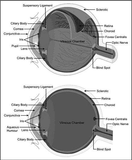 The eyeball consists of three concentric layers:
Sclera (Outer Layer)
The white part of the eye is the sclera.
The sclera covers the colored part of the eye, known as the cornea.
Choroid (Middle Layer)
This layer is rich in blood vessels that provide nourishment.
The choroid expands at the front to form the ciliary body.
The iris, also part of the choroid, partially covers the lens and leaves a central opening called the pupil.
Iris muscles control the size of the pupil, regulating the amount of light entering the eye.
Retina (Inner Layer)
The retina contains two types of sensory cells: rods and cones.
Rod cells are sensitive to dim light but don't perceive color.
Cone cells are sensitive to bright light and are responsible for color vision.
Comparison between Rods and Cones
Rods:
More numerous.
Located at the retina's periphery.
Produce light-sensitive pigment rhodopsin quickly.
Cones:
Fewer in number.
Found in the retina's center.
Produce light-sensitive pigment iodopsin more slowly.
Yellow Spot and Blind Spot
Yellow Spot
Contains the highest concentration of sensory cells, particularly cones.
The area responsible for color vision and sharpest sight.
Blind Spot
Lacks sensory cells.
No vision occurs in this region.
Lens
- The lens is transparent, biconvex, and crystalline.
- It is suspended by a ligament that connects it to the ciliary body.
Aqueous and Vitreous Chambers
The lens divides the inner eye into two chambers:
Aqueous Chamber:
- Located between the lens and the cornea.
- Contains a watery liquid called aqueous humor.
Vitreous Chamber:
- Situated behind the lens.
- Filled with a jelly-like fluid called vitreous humor.
- It helps refract light.
Four Major Steps in Seeing an Object
- Light rays from an object enter the eye through transparent structures.
- The cornea bends the rays slightly, and the lens focuses them further.
- The resulting image on the retina is inverted and real.
- The rods and cones convert light into chemical changes, generating nerve impulses. These impulses travel to the cerebrum via the optic nerve, where the brain interprets the image as upright.
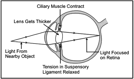
Accommodation Vision
Accommodation refers to the process of focusing on objects at different distances, which is achieved by changes in the lens curvature:
For distant vision, the lens flattens, held by suspensory ligaments.
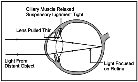
For near vision, the lens becomes round and convex, as the ciliary muscles contract, reducing ligament tension.
Light and Dark Adaptation
Dark Adaptation:
Difficulty seeing in a dark environment after leaving a brightly lit area.
Light Adaptation:
Temporary dazzle when moving from a dark area to a brightly lit one.
Common Eye Defects
Myopia (Shortsightedness):
- Clear vision for near objects, but distant ones are blurry.
- Caused by an overly curved lens.
- Corrected with concave lenses.
Hyperopia (Longsightedness):
- Difficulty seeing close objects.
- Caused by a flatter lens.
Astigmatism:
Some parts of an object are in focus, while others appear blurry.
Presbyopia:
Occurs in older individuals, with difficulty seeing nearby objects.
Cataract:
Clouding of the lens reduces vision.
Color Blindness:
Inability to distinguish certain colors, like red and green.
Night Blindness:
Difficulty seeing in dim light, often due to insufficient rhodopsin in rods.
Squint:
Misalignment of the eyes, leading to "crossed eyes."
Stereoscopic Vision
Humans, monkeys, and apes can perceive depth and distance, thanks to the simultaneous use of both eyes, producing overlapping images that create a 3D effect.
After-images
After staring at a brightly colored object, the same color will persist briefly when looking at a dark surface. This is known as persistence or after-image.
The Ear
The human ear is divided into three main sections: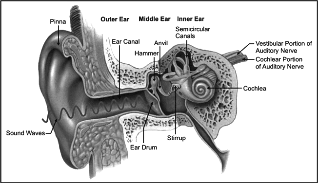
Outer Ear:
Includes the pinna (auricle) and auditory canal.
Middle Ear:
Contains the ear ossicles: malleus (hammer), incus (anvil), and stapes (stirrup).
The eustachian tube connects the middle ear to the throat.
Inner Ear:
Known as the membranous labyrinth, containing the cochlea and semicircular canals.
The cochlea has three parallel canals, with the middle canal containing the organ of Corti, responsible for hearing.
Functions of the Ear
The ear serves two primary functions: hearing and balance.
Hearing:
Sound waves are collected by the pinna and channeled through the auditory canal to the eardrum, causing it to vibrate.
The vibration travels through the ear ossicles, reaching the oval window membrane and setting cochlear fluid in motion.
Cochlear sensory cells send impulses via the auditory nerve.
Balance:
Sensory cells in the semicircular canals detect motion (dynamic equilibrium).
Sensory cells in the utriculus and sacculus detect position (static equilibrium).
Hearing Impairment
Impairments can include issues with detecting or processing sound, leading to partial or total hearing loss.
The Sense of Taste
Taste is perceived through the taste buds located on the tongue, which contain sensory cells. Substances enter the taste bud pore, stimulating sensory hairs that send signals to the brain.
The Sense of Smell
Smell is detected by sensory cells in the nasal chamber, which have hair-like projections that react to particles dissolved in the mucous. The signals are transmitted to the brain via the olfactory nerve.
|
55 videos|110 docs|21 tests
|
FAQs on Revision Notes: The Nervous System - Biology Class 10 ICSE
| 1. What are the main functions of the nervous system? |  |
| 2. What is a neuron and why is it important in the nervous system? | |
| 3. What is a synapse and how does it function? | |
| 4. What are the different types of neurons? | |
| 5. What is the difference between the central nervous system and the peripheral nervous system? | |


Previous Year Questions with Solutions
,Objective type Questions
,Important questions
,Revision Notes: The Nervous System | Biology Class 10 ICSE
,Viva Questions
,Free
,Sample Paper
,Exam
,study material
,ppt
,past year papers
,MCQs
,shortcuts and tricks
,Revision Notes: The Nervous System | Biology Class 10 ICSE
,Semester Notes
,Summary
,Extra Questions
,video lectures
,Revision Notes: The Nervous System | Biology Class 10 ICSE
,mock tests for examination
,practice quizzes
;


Revision Notes: The Nervous System Free PDF Download
Importance of Revision Notes: The Nervous System
Revision Notes: The Nervous System
Revision Notes: The Nervous System Class 10 Questions
Study Revision Notes: The Nervous System on the App
|
© EduRev
|
Education Revolution
|


|


within 7 days!