NEET PG Exam > NEET PG Notes > Dermatology and Venereology > Chapter Notes: Hansen’s Disease
Hansen’s Disease Chapter Notes | Dermatology and Venereology - NEET PG PDF Download
| Table of contents |
|
| Organism |
|
| Types of Leprosy |
|
| Diagnosis |
|
| Treatment |
|

Organism
- Mycobacterium leprae grows best at 30°C, below human core temperature, leading to lesions in cooler superficial areas like ear lobes, nose, testes, liver, and superficial nerves (e.g., ulnar, facial).
- Prefers cooler areas, avoiding deeper, warmer nerves.
- Best cultivated in armadillos, but also in mouse footpads, nude mice, and thymectomized mice.
- Phenolic glycolipid-1 (PGL-1) is a unique glycolipid on the bacillus surface, contributing to pathogenesis.
- Bacilli doubling time is 11-13 days (average 12 days).
Classification
Ridley-Jopling Classification
- Based on four criteria: Clinical, Bacteriological (slit skin smear - SSS), Histological (skin biopsy), and Immunological (lepromin testing).
- Classifies leprosy into five types: Tuberculoid (TT), Borderline Tuberculoid (BT), Borderline-Borderline (BB), Borderline Lepromatous (BL), Lepromatous-Polar (LLp).
- Includes a subpolar form (LLs) between IL-polar and BL.
- Borderline leprosies include BT, BB, and BL.
- Does not include indeterminate or pure neural types of Hansen's disease.
Pathogenesis
- BB is the most unstable form of leprosy.
- Stable leprosy (TT, LLp) does not change or shift immunologically.
- Unstable leprosies (BT, BB, BL) can shift between each other.
Disease Behavior
- TT has strong T-cell immunity (Th-1 response), forming tuberculoid granulomas, killing many bacilli, making it paucibacillary (PB).
- LL has absent T-cell immunity and high B-cell immunity (Th-2 response), leading to numerous bacilli in macrophages (foam cells), making it multibacillary (MB).
- Without treatment, immunity slides toward LL (downgrading), reducing tuberculoid granulomas and increasing foam cells.
- TT is stable and does not downgrade; BT, BB, BL can downgrade to LL-subpolar (LLs), but LLs does not downgrade to LL-polar (LLp).
- Treatment improves immunity (upgrading): T-cell improvement causes Type 1 lepra reaction; B-cell improvement causes Type 2 lepra reaction.
- BT and BB show Type 1 lepra reaction post-treatment due to increased T-cell immunity.
- LLs shows Type 2 lepra reaction post-treatment due to increased B-cell immunity.
- BL may show either Type 1 or Type 2 lepra reactions post-treatment.
- TT and LLp are stable and typically do not show reactions.
Types of Leprosy
1. Indeterminate Hansen's Disease
- Early form where the immune system has not yet determined its response to the bacillus.
- Many patients self-heal; some progress to TT, BT, BL, or LL.
- Presents as a solitary, ill-defined, hypopigmented macule or patch on cheeks, arms, thighs, or buttocks.
- Normal sensations and sweating; peripheral nerves not enlarged.
- Histology shows periadnexal and perineural lymphocytic infiltrate without granulomas; few or no bacilli seen on biopsy.
2. Tuberculoid Leprosy (TT)
- Characterized by tuberculoid granulomas forming in the dermis and cutaneous nerves, driven by a Th-1 immune response.
- Exhibits a robust cell-mediated immune response.
- Nerve granulomas cause compression, leading to symptoms such as sensory loss, loss of sweating, and hypopigmentation (occasionally erythematous) in the affected skin area.
- Typically involves a single thickened nerve and a single skin lesion, though up to three lesions may occur.
- The characteristic lesion is a plaque with a well-defined, hypopigmented (sometimes erythematous) elevated border that slopes to a flattened center, resembling a "saucer right side up."
- Well-defined lesion borders reflect strong immunity, in contrast to the ill-defined borders seen in lepromatous leprosy (LL).
3. Borderline Tuberculoid (BT)
- Most common type in India.
- Similar to TT but with more lesions (3-10) and few thickened nerves.
- Features 'satellite lesions' around the main lesion.
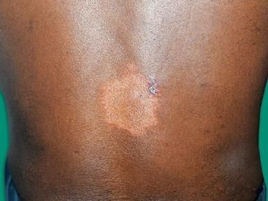 Satellite lesion in BT
4. Mid-Borderline, Borderline-Borderline (BB)
- Many countable lesions (10-30) and many thickened nerves.
- Lesions increasingly cross the midline.
- Shares features of TT (well-defined inner border) and LL (ill-defined outer border).
- Lesions described as inverted saucer, Swiss-cheese, punched out, dimorphic, or annular, with normal skin at the center.
- Sensations and sweating improve as granuloma size reduces and foam cells increase, reducing nerve compression.
- Most unstable form of leprosy.
5. Borderline Lepromatous (BL)
- Numerous small, uncountable lesions and numerous thickened nerves.
- Lesions cross the midline, becoming almost completely symmetrical.
- Sensations and sweating return to near normal due to minimal nerve compression from fewer granulomas and more foam cells.
- Features inverted saucer, Swiss-cheese, punched out, or dimorphic lesions.
6. Lepromatous Leprosy (LL)
- Dominated by foam cells (lepra cells) on biopsy due to Th-2 response with IL-4 and IL-10 prominence.
- Diffuse skin infiltration with ill-defined borders due to lack of immune response, allowing bacilli dissemination.
- Nodules and infiltration thicken skin, causing prominent skin folds.
- Perfectly symmetrical lesions; no granulomas, so no initial nerve compression, maintaining normal sensations and sweating early on.
- Gradual bacilli invasion of nerves leads to peripheral neuropathy and glove-and-stocking anesthesia.
- Earliest signs include nasal stuffiness and epistaxis; nasal discharge is a primary infection source.
- Other features: ear lobe infiltration (buddha ears), madarosis, gynecomastia (testicular involvement), saddle nose, nasal septal perforation.
- Histopathology shows foam cells in the dermis with an uninvolved papillary dermis (grenz zone, best seen in LL).
Histoid Leprosy
- A type of LL, often with dapsone resistance, more common with past dapsone monotherapy.
- Presents with shiny papules and nodules with intervening normal skin.
- Skin biopsy shows spindle-shaped foam cells.
Lucio Leprosy
- Non-nodular LL, also called 'lepra bonita' (pretty leprosy).
- Common in Mexico and Central America.
- Features diffuse skin infiltration, no wrinkles, and a shiny, youthful face.
Nerve Involvement in Leprosy
- 100% of patients have nerve involvement.
- Toward TT: rapid nerve compression, non-symmetrical involvement, no glove-and-stocking anesthesia.
- Toward LL: slower peripheral nerve invasion, leading to symmetrical peripheral neuropathy and glove-and-stocking anesthesia.
- Trophic ulcers occur due to loss of peripheral sensations.
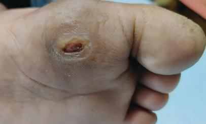 Trophic ulcer
 - Pure neural Hansen’s occurs when only nerves are involved without clinical skin involvement.
- Order of sensory loss: hot and cold differentiation > cold > hot > light touch > pain > deep touch.
Eye Involvement in Leprosy
- 7th nerve involvement causes corneal erosions, exposure keratitis, iridocyclitis, and lagophthalmos.
- In BL and LL, the iris may show micro-granuloma deposits ('iris pearls').
Systemic Involvement in Leprosy
- Secondary amyloidosis with renal impairment is common.
- Renal involvement: secondary amyloidosis is the most common lesion; among glomerulonephritis, mesangioproliferative glomerulonephritis > MPGN > MPN.
- CNS, female reproductive system, lungs, prostate, and breasts are not involved.
- Between ovary and uterus, the uterus is less likely to be involved.
Histopathology in Leprosy
- Uses routine H&E stains and Fite-Faraco stain for acid-fast bacilli (AFB).
- From TT to LL: tuberculoid granulomas decrease, while foam cells, grenz zone formation, and bacilli numbers increase.
- Foam cells are dermal histiocytes filled with lipids, appearing foamy on histology.
Reactions in Leprosy
Type 1 Lepra Reaction (Reversal/Upgrading)
- Occurs during or within 3 years post-treatment, primarily cell-mediated (Type 4 hypersensitivity).
- Mainly in borderline spectrum (BT, BB, BL), most severe in BL.
- Involves inflammation of existing lesions (red, painful, edematous) and/or neuritis (red, painful, inflamed nerves).
- Sudden neuritis can cause permanent nerve damage; nerve abscesses (most in BT) may form.
- Severe cases may cause ulceration (Lazarine leprosy).
- No systemic inflammation (no fever).
- Treatment for skin reaction: NSAIDs, oral steroids (DOC, 1 mg/kg prednisolone tapered over 12 weeks), azathioprine, chloroquine, methotrexate, cyclosporine.
- Treatment for neuritis: splinting of limb, oral steroids (DOC), incision and drainage for nerve abscesses (especially ulnar nerve) to restore function.
Type 2 Lepra Reaction (Erythema Nodosum Leprosum - ENL)
- Primarily humoral (Type 3 hypersensitivity) with immune complex deposits in tissues and vessels (leukocytoclastic vasculitis).
- Cytokine involved: TNF-α.
- Presents with new, tender, red papules or nodules on extremities and face; existing lesions unchanged.
- Systemic inflammation: fever, arthralgia, conjunctivitis, keratitis, iritis, synovitis, nephritis, hepatosplenomegaly, orchitis, generalized lymphadenopathy.
- Treatment for first episode: prednisolone (1 mg/kg, tapered over 12 weeks, DOC).
- Treatment for recurrent/chronic ENL: prednisolone + thalidomide or prednisolone + clofazimine.
- Thalidomide is the fastest-acting drug (suppresses ENL in 48-72 hours) but not supported by WHO due to risks of phocomelia and peripheral neuropathy.
Lucio Phenomenon
- Seen only in Lucio leprosy.
- Caused by severe ischemia from thrombosis of dermal vessels, leading to hemorrhagic infarcts with serrated margins and ulceration (ulceronecrotic lesions).
- Accompanied by systemic inflammation: fever, arthralgia, conjunctivitis, keratitis, iritis, synovitis, nephritis, hepatosplenomegaly, orchitis, generalized lymphadenopathy.
- Treatment: NSAIDs (for mild cases), oral steroids (DOC), thalidomide, clofazimine, azathioprine, chloroquine, colchicine.
Diagnosis
WHO Definition of Leprosy
- In endemic countries, leprosy is diagnosed if one or more cardinal signs are present: hypopigmented or reddish skin lesions with definite sensory loss, thickened nerves, positive skin smears.
- Mnemonic: LEProsy - L = Loss of sensation, E = Enlarged nerves, P = Positive AFB on smear.
- Skin biopsy confirms diagnosis; nerve biopsy used for pure neural Hansen’s.
Slit Skin Smear (SSS)
- Slits made on skin with a blade, stained for AFB; WHO recommends 4 sites (minimum 3: one ear lobe, two active lesions).
- AFB staining patterns: Solid bacilli (S) = viable, Fragmented (F) = dead, Granular (G) = dead.
- Bacterial Index (BI): Counts S + F + G (dead + living) on a 0-6 scale, indicating total bacillary load; remains positive post-therapy.
- Morphological Index (MI): Counts only solid (living) bacilli, reported as a percentage (e.g., 50% means 50% bacilli are alive); better for treatment efficacy and drug resistance, becomes negative post-therapy.
- BI reduces by 1+ annually on treatment; relapse diagnosed if BI increases by 2+ from baseline.
- SSS negative = paucibacillary (PB); SSS positive = multibacillary (MB).
- SSS may yield false negatives due to superficial sampling; histological biopsy is the gold standard.
- Best site for SSS: untreated cases - ear lobes; treated cases - dorsa of fingers.
- SSS detects bacilli only if >10,000/gm of tissue, so negative in TT, indeterminate, and pure neural Hansen’s.
- Lepromin Test
- Intradermal antigen injection to assess cell-mediated immunity (CMI).
- Prognostic test, useful for classifying leprosy (negative = lepromatous side, positive = tuberculoid side).
- Not diagnostic, as it is positive in healthy individuals.
- Readings: Fernandez response (within 48 hours), Mitsuda response (at 4 weeks, better CMI indicator).
- FNAC of Nerves
- Used for pure neural leprosy; aspirate stained for cells and bacilli.
Treatment
Most patients have established nerve damage, so hypoesthesia and crib change may persist post-treatment; patients need counseling on this.
Drugs in Leprosy
- Dapsone: Weakly bactericidal, inhibits dihydrofolic acid. Side effects: anemia, dapsone hypersensitivity syndrome (erythroderma, fever, lymphadenopathy, hepatitis after 5 weeks).
- Rifampicin: Powerful bactericidal, cornerstone of leprosy management. Monthly dosing suffices due to M. leprae’s slow 12-day doubling time. Resistance is a major concern. WHO guideline for rifampicin-resistant patients: Intensive phase (6 months, daily supervised): moxifloxacin 400 mg, clofazimine 50 mg, clarithromycin 500 mg, minocycline 100 mg; Continuation phase (18 months, monthly supervised): moxifloxacin 400 mg, clarithromycin 1000 mg, minocycline 200 mg.
- Rifapentine: Long-acting rifampicin derivative, more potent than rifampicin.
- Clofazimine: Weakly bactericidal, orange-colored dye. Side effects: ichthyosis, reversible skin discoloration/darkening. Used for both anti-leprosy and anti-reactional (ENL) treatment.
- Ethionamide/Prothionamide: Bactericidal, less potent than rifampicin.
- Fluoroquinolones (Ofloxacin/Moxifloxacin): Bactericidal; moxifloxacin is as effective as rifampicin.
- Minocycline: Bactericidal.
- Clarithromycin: Bactericidal.
WHO Disability Grading of Leprosy
Hands and Feet
- Grade 0: No anesthesia, no visible deformity/damage.
- Grade 1: Anesthesia present, no visible deformity/damage.
- Grade 2: Visible deformity present (e.g., claw hand, deformed foot).
Eyes
- Grade 0: No eye problem due to leprosy.
- Grade 1: Eye problem present, but vision not severely affected (vision 6/60 or better).
- Grade 2: Severe visual impairment (vision <6/60, cannot count fingers), lagophthalmos, iridocyclitis, corneal opacities.
Immunoprophylaxis in Leprosy (Vaccines)
- Limited to high-endemicity pockets, close family contacts, and high-risk groups.
- Vaccines: BCG, BCG + Killed M. leprae (Convit vaccine), Mycobacterium indicus pranii (MIP), Indian Cancer Research Institute (ICRC) vaccine, Mycobacterium vaccae vaccine.
The document Hansen’s Disease Chapter Notes | Dermatology and Venereology - NEET PG is a part of the NEET PG Course Dermatology and Venereology.
All you need of NEET PG at this link: NEET PG
|
8 docs|5 tests
|
FAQs on Hansen’s Disease Chapter Notes - Dermatology and Venereology - NEET PG
| 1. What is Hansen's Disease and what causes it? |  |
Ans.Hansen's Disease, also known as leprosy, is a chronic infectious disease caused by the bacterium Mycobacterium leprae. It primarily affects the skin, peripheral nerves, mucosal surfaces, and the eyes. The disease is characterized by skin lesions, nerve damage, and deformities if left untreated. It is transmitted through respiratory droplets and prolonged close contact with an infected person.
| 2. How is Hansen's Disease diagnosed? | |
Ans.Hansen's Disease is diagnosed through a combination of clinical examination and laboratory tests. A healthcare provider looks for characteristic symptoms such as skin lesions and numbness in extremities. Skin biopsies or blood tests can also be performed to detect the presence of Mycobacterium leprae or antibodies against the bacteria. Early diagnosis is crucial for effective treatment.
| 3. What is the treatment for Hansen's Disease? | |
Ans.The World Health Organization recommends a multidrug therapy (MDT) for treating Hansen's Disease, which includes a combination of antibiotics such as rifampicin, clofazimine, and dapsone. This regimen is effective in killing the bacteria and preventing complications. Treatment typically lasts from six months to a year, depending on the severity of the disease.
| 4. What are the potential complications of untreated Hansen's Disease? | |
Ans.If left untreated, Hansen's Disease can lead to severe complications, including permanent nerve damage, muscle weakness, and deformities of the hands and feet. Patients may also experience blindness due to damage to the eyes and chronic pain or disability. Early treatment is essential to prevent these long-term effects.
| 5. Can Hansen's Disease be prevented? | |
Ans.Prevention of Hansen's Disease primarily involves early detection and treatment of infected individuals to prevent transmission. Public health education and awareness can help reduce stigma associated with the disease. Regular monitoring of close contacts of infected individuals may also be recommended to identify and treat cases early.
About this Document

4.94/5
Rating

Sep 06, 2025
Last updated
Related Exams
Document Description: Chapter Notes: Hansen’s Disease for NEET PG 2025 is part of Dermatology and Venereology preparation.
The notes and questions for Chapter Notes: Hansen’s Disease have been prepared according to the NEET PG exam syllabus. Information about Chapter Notes: Hansen’s Disease covers topics
like Organism, Types of Leprosy, Diagnosis, Treatment and Chapter Notes: Hansen’s Disease Example, for NEET PG 2025 Exam. Find important definitions, questions, notes, meanings, examples, exercises and tests below for Chapter Notes: Hansen’s Disease.
Introduction of Chapter Notes: Hansen’s Disease in English is available as part of our Dermatology and Venereology
for NEET PG & Chapter Notes: Hansen’s Disease in Hindi for Dermatology and Venereology course.
Download more important topics related with notes, lectures and mock test series for NEET PG
Exam by signing up for free. NEET PG: Hansen’s Disease Chapter Notes | Dermatology and Venereology - NEET PG
Description
Full syllabus notes, lecture & questions for Hansen’s Disease Chapter Notes | Dermatology and Venereology - NEET PG - NEET PG | Plus excerises question with solution to help you revise complete syllabus for Dermatology and Venereology | Best notes, free PDF download
Information about Chapter Notes: Hansen’s Disease
In this doc you can find the meaning of Chapter Notes: Hansen’s Disease defined & explained in the simplest way possible. Besides explaining types of
Chapter Notes: Hansen’s Disease theory, EduRev gives you an ample number of questions to practice Chapter Notes: Hansen’s Disease tests, examples and also practice NEET PG
tests

Related Searches
mock tests for examination
,study material
,Free
,Hansen’s Disease Chapter Notes | Dermatology and Venereology - NEET PG
,past year papers
,Hansen’s Disease Chapter Notes | Dermatology and Venereology - NEET PG
,Hansen’s Disease Chapter Notes | Dermatology and Venereology - NEET PG
,Extra Questions
,MCQs
,video lectures
,ppt
,Exam
,practice quizzes
,Objective type Questions
,Summary
,Previous Year Questions with Solutions
,Sample Paper
,shortcuts and tricks
,Semester Notes
,Important questions
,Viva Questions
;


Additional Information about Chapter Notes: Hansen’s Disease for NEET PG Preparation
Chapter Notes: Hansen’s Disease Free PDF Download
The Chapter Notes: Hansen’s Disease is an invaluable resource that delves deep into the core of the NEET PG exam.
These study notes are curated by experts and cover all the essential topics and concepts, making your preparation more efficient and effective.
With the help of these notes, you can grasp complex subjects quickly, revise important points easily,
and reinforce your understanding of key concepts. The study notes are presented in a concise and easy-to-understand manner,
allowing you to optimize your learning process. Whether you're looking for best-recommended books, sample papers, study material,
or toppers' notes, this PDF has got you covered. Download the Chapter Notes: Hansen’s Disease now and kickstart your journey towards success in the NEET PG exam.
Importance of Chapter Notes: Hansen’s Disease
The importance of Chapter Notes: Hansen’s Disease cannot be overstated, especially for NEET PG aspirants.
This document holds the key to success in the NEET PG exam.
It offers a detailed understanding of the concept, providing invaluable insights into the topic.
By knowing the concepts well in advance, students can plan their preparation effectively.
Utilize this indispensable guide for a well-rounded preparation and achieve your desired results.
Chapter Notes: Hansen’s Disease
Chapter Notes: Hansen’s Disease Notes offer in-depth insights into the specific topic to help you master it with ease.
This comprehensive document covers all aspects related to Chapter Notes: Hansen’s Disease.
It includes detailed information about the exam syllabus, recommended books, and study materials for a well-rounded preparation.
Practice papers and question papers enable you to assess your progress effectively.
Additionally, the paper analysis provides valuable tips for tackling the exam strategically.
Access to Toppers' notes gives you an edge in understanding complex concepts.
Whether you're a beginner or aiming for advanced proficiency, Chapter Notes: Hansen’s Disease Notes on EduRev are your ultimate resource for success.
Chapter Notes: Hansen’s Disease NEET PG Questions
The "Chapter Notes: Hansen’s Disease NEET PG Questions" guide is a valuable resource for all aspiring students preparing for the
NEET PG exam. It focuses on providing a wide range of practice questions to help students gauge
their understanding of the exam topics. These questions cover the entire syllabus, ensuring comprehensive preparation.
The guide includes previous years' question papers for students to familiarize themselves with the exam's format and difficulty level.
Additionally, it offers subject-specific question banks, allowing students to focus on weak areas and improve their performance.
Study Chapter Notes: Hansen’s Disease on the App
Students of NEET PG can study Chapter Notes: Hansen’s Disease alongwith tests & analysis from the EduRev app,
which will help them while preparing for their exam. Apart from the Chapter Notes: Hansen’s Disease,
students can also utilize the EduRev App for other study materials such as previous year question papers, syllabus, important questions, etc.
The EduRev App will make your learning easier as you can access it from anywhere you want.
The content of Chapter Notes: Hansen’s Disease is prepared as per the latest NEET PG syllabus.
|
© EduRev
|
Education Revolution
|


|


Signup to see your scores
go up
within 7 days!
within 7 days!
Takes less than 10 seconds to signup