Forensic DNA Profiling - 1 Chapter Notes | Forensic Medicine and Toxicology (FMT) - NEET PG PDF Download

Introduction
In a criminal trial, it is crucial to prove the crime beyond reasonable doubt. Forensic experts rely on various scientific fields to meet this stringent standard. Forensic science has advanced significantly, and now advanced technology is vital for combating crime and ensuring justice.
Over the past two decades, advancements in molecular biology have provided new methods to address forensic challenges and identify suspects more swiftly and accurately. While this cutting-edge technology has improved criminal identification, it is also applicable in civil matters, particularly in paternity cases involving custody and marital disputes. As a result, forensic DNA analysis has a wide range of applications, including:
- Cold cases
- Missing persons investigations
- Other relevant scenarios
This chapter examines the current use of forensic DNA analysis in laboratories around the world.
Forensic Genetics
Historical Background
- Forensic genetics began with Karl Landsteiner 's work on the ABO blood group system in 1901.
- This discovery paved the way for identifying various red cell antigens, serum proteins, and erythrocyte enzymes, enabling the serological analysis of blood and other body fluids.
- By 1980, a variety of conventional blood grouping tests were available, improving forensic capabilities, especially when used alongside the HLA white blood cell antigen system.
- However, these methods were labour-intensive, technically complex, and required expensive sera, making them challenging to implement.
- Additionally, the markers were not uniformly present in all tissues and deteriorated quickly when dried, complicating their use in forensic investigations.
- Forensic samples are often small and affected by environmental factors like drying and aging, further hindering analysis.
- The discriminatory power of these markers was also low, requiring the examination of numerous samples to obtain reliable results.
Breakthrough in DNA Analysis
- A major breakthrough in forensic genetics occurred in 1985 when Sir Alec Jeffreys in the United Kingdom discovered a set of DNA markers known as Variable Number of Tandem Repeats (VNTR).
- These markers showed greater variability among individuals and were quickly adopted for forensic cases involving human identification.
Deoxyribonucleic Acid (DNA)
DNA, or deoxyribonucleic acid, serves as the fundamental blueprint for all living organisms. Its structure, first elucidated by James Watson and Francis Crick in 1953, is characterized as a right-handed double helix.
Structure of DNA
- DNA is composed of repeating units known as nucleotides.
- Each nucleotide consists of a phosphate group, a sugar, and a nitrogenous base.
- There are four types of nitrogenous bases in DNA:
- Adenine (A),
- Guanine (G),
- Cytosine (C), and
- Thymine (T).
- Adenine pairs with Thymine (A-T) through two hydrogen bonds, while Cytosine pairs with Guanine (C-G) through three hydrogen bonds.
- This specific base pairing ensures that the two strands of DNA are complementary to each other.
- This complementary pairing is a key feature of DNA, allowing it to accurately transmit genetic information from one generation to the next.
Genetic Makeup
- At the moment of conception, every individual receives a distinct genetic blueprint that determines their unique genetic traits.
- This blueprint is made up of polymorphisms, which are variations in the DNA sequence that can be used for human identification.
- Various techniques have been developed to analyze these DNA polymorphisms for identification purposes.
- Most of the DNA in our cells is located in the nucleus, where it is organized into structures called chromosomes.
- Nuclear DNA is inherited from both the mother and the father equally.
- Except in the case of identical twins, no two individuals have the same sequence of genomic DNA.
Mitochondrial DNA
- DNA can also be found in the mitochondria, the energy-producing structures within cells.
- This is known as mitochondrial DNA, and it is passed down exclusively from mother to offspring.
- Mitochondrial DNA can be used to trace maternal lineages, providing information about an individual’s ancestry through their mother’s side of the family.
Benefits of Using DNA for Identification
DNA (Deoxyribonucleic Acid) is a powerful tool for identification due to several key advantages:
- Ubiquity in Nucleated Cells: DNA is present in every nucleated cell of the body, making it widely available for identification purposes.
- Consistency Across Cells: The DNA makeup is consistent across all cells in an individual’s body and remains unchanged throughout their life.
- Uniqueness: Every person has a unique DNA profile, which distinguishes them from others. The only exception to this is in the case of monozygotic twins, who share identical DNA.
- Sources of DNA: DNA can be extracted from various bodily fluids, such as blood and saliva, as well as from tissues. This makes it versatile for identification from different types of samples.
- Post-Mortem Identification: In post-mortem cases, DNA can be obtained from body tissues, allowing for identification even after death.
- Degraded or Buried Tissues: DNA can be extracted from buried tissues, and in cases of burnt or charred remains, it can be collected from hard tissues like bones and teeth. This ability to retrieve DNA from degraded samples is a significant advantage.
- Storage Convenience: DNA is easier to store in small quantities compared to other types of evidence. It can be stored for long periods without degrading, provided it is kept under proper conditions.
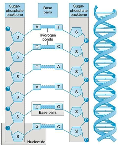 DNA Structure
Difference between DNA Profiling and Blood or Protein Tests
- Genetic Makeup vs. Genetic Products: DNA tests reveal the genetic makeup of an individual, while blood or protein tests indicate the genetic products present in the body.
- Avoiding Dominance and Recessiveness Issues: DNA methods bypass the complications of genetic dominance or recessiveness. For instance, blood group AA or Aa cannot be distinguished through blood tests, but DNA analysis can differentiate between them.
- Individual Identification at Crime Scenes: DNA does not combine, allowing for the identification of the number of individuals present at a crime scene based on their DNA contributions. This is crucial in forensic investigations to determine the presence of multiple individuals.
Disadvantages of DNA Analysis
- DNA analysis depends on the availability of nucleated cells. It cannot be conducted on biological samples lacking these cells or where they are in low numbers.
- Examples of such samples include:
- Semen without spermatozoa, as seen in vasectomised males
- Hair shafts, which do not contain nucleated cells
The Basics of Molecular Biology
Forensic genetics involves the use of molecular biology techniques to analyze DNA for legal purposes. A fundamental understanding of molecular biology, particularly the structure of DNA and the organization of the human genome, is essential to comprehend the principles behind forensic genetics.
Human Genome Overview
- The human genome comprises approximately 3 billion base pairs (bp) organized into 23 pairs of chromosomes.
- Each human cell contains diploid cells with two copies of each chromosome, while gametes (ova and sperm) are haploid, containing one copy of each chromosome.
- During fertilization, one chromosome from each parent is inherited, resulting in a diploid zygote.
Structure of DNA
- DNA consists of coding and non-coding regions. About 5% of the genome contains protein-coding genes, which are crucial for normal biological functions.
- Genes are made up of exons (functional parts) and introns (non-functional segments).
Polymorphic Markers and Genotyping
- Polymorphic markers are found in regions of the genome that appear to have little functional significance. These markers, located at specific loci, exist in different sequence forms, known as alleles.
- Individuals typically have two alleles for a locus, one inherited from each parent.
- If the alleles are the same, the individual is a homozygote. if they differ, the person is a heterozygote.
- The combination of alleles at a locus defines the genotype of an individual at that locus. Different genotypes across various loci make up a person’s genetic profile.
Genetic Variation and Forensic Identification
- Genetic variation ensures that no two individuals are genetically identical, except for monozygotic twins.
- Forensic genetics typically involves analyzing specific, highly polymorphic loci rather than the entire genome to distinguish between individuals.
- Tandemly repeated DNA sequences are commonly used for forensic identification due to their high variability in the number of repeats among individuals.
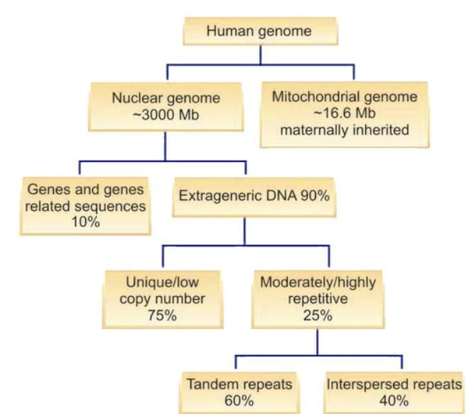 A schematic diagram showing the organisation of the human genome
Extraction, Detection and Visualization of DNA
- The process of extracting DNA has become relatively straightforward.
- DNA is obtained from nucleated cells by breaking them open using osmotic pressure.
- Initially, cellular proteins are precipitated, resulting in a clear liquid that contains DNA.
- DNA is then precipitated using isopropanol, followed by purification with ethanol and dilution in a buffer solution or pure water.
- There are many commercial kits available for DNA extraction, which are easy to use by following the manufacturer’s instructions.
- After DNA is extracted, it needs to be visualized, which is traditionally done using agarose gel or polyacrylamide gel electrophoresis with a buffer system.
- For high DNA concentrations, ethidium bromide, a fluorescent dye that binds to DNA bases, is used to make DNA visible on an agarose gel.
- For low DNA concentrations, a polyacrylamide gel is used, and DNA can be visualized with ethidium bromide staining.
- Over time, various other stains have been developed for DNA detection, but for very small DNA amounts, more effective methods were needed.
- The breakthrough involved using fluorescent dyes that attach to DNA segments, which require advanced equipment like a charge-coupled device (CCD) camera for detection.
- This camera, part of automated DNA sequencers, captures the fluorescence of DNA labelled with a dye when exposed to a laser, and the images are processed into an electropherogram.
- This method is now standard in automated DNA sequencers.
- Recent advancements have improved the medium for electrophoresis, with modern DNA sequencers performing electrophoresis in a thin capillary, allowing faster separation of DNA markers without the need for gels.
Tandemly Repetitive DNA
Tandemly repetitive DNA consists of segments of DNA arranged in a specific sequence that repeats multiple times. For example, a sequence like GGGCCCTTAA may be repeated many times. Depending on their size, tandemly repetitive DNA can be classified into three groups: (a) Microsatellites, (b) Minisatellites, and (c) Satellite DNA.
(a) Microsatellites
- Microsatellite polymorphisms consist of short tandemly repeating units of a specific sequence, known as a core repeat, typically ranging from 1 to 6 base pairs (bp) long. These are also referred to as short tandem repeats (STRs).
- STRs are highly variable regions found throughout the genome and are used in various applications, including forensic analysis and genetic mapping.
- The variability in STRs arises from differences in the number of core repeats present in individuals. For example, one person may have a repeat of AATG 10 times (AATG10), while another may have it 12 times (AATG12).
(b) Minisatellites
- Minisatellite polymorphisms are similar to microsatellites but involve longer tandemly repeating units, with core repeats typically ranging from 16 to 80 bp long. These are also known as variable number of tandem repeats (VNTRs).
- Highly variable regions associated with minisatellites have been identified near genes such as human insulin, alpha-globin, and the c-Ha-ras-1 oncogene. The variation in these regions is due to differences in the number of core repeats.
- In 1985, Prof. Sir Alec Jeffreys discovered a hypervariable minisatellite using the multilocus probe technique (MLP) and suggested its application for human identification.
- The multilocus system developed by Jeffreys demonstrated high discriminatory power due to the numerous alleles present at each locus. However, MLP profiles were difficult to interpret and standardize.
- To address this, several single locus probe systems (SLPs) were developed, which detect two alleles in a heterozygous individual—one inherited from each parent. This approach became the preferred method for DNA profiling.
- The process of DNA profiling using RFLP technology was time-consuming and required a large quantity of DNA, involving steps like gel electrophoresis, Southern blotting, and membrane probing.
- RFLP was not suitable for automation, limiting its use in many laboratories. This method is now considered outdated, requiring at least 50 ng of intact high molecular weight DNA, whereas PCR-based testing can utilize as little as 500 pg of DNA.
(c) Satellite DNA
- Satellite DNA consists of large tandemly repeating units of a specific sequence, typically ranging from several hundred base pairs to several kilobases in length.
- Satellite DNA is found in specific regions of the genome, such as centromeres and telomeres, and plays a role in chromosome structure and function.
- The variation in satellite DNA is attributed to differences in the number of repeat units present in individuals. For instance, one individual may have a repeat unit of 500 bp repeated 20 times in a centromeric region (CEN20), while another may have it repeated 25 times (CEN25).
Polymerase Chain Reaction (PCR)
- PCR, or Polymerase Chain Reaction, is a method developed in 1985 that significantly advanced the field of molecular genetics. It is a simple yet powerful technique used to make millions of copies of a specific DNA sequence.
- DNA replication occurs naturally in cells, and this process involves the unwinding of the two strands of DNA. For replication to take place, these strands must separate to allow the formation of a new strand.
- The initial step in PCR is called denaturation, where the DNA is heated to a high temperature to break the hydrogen bonds holding the strands together. This process creates single strands of DNA that serve as templates for the new strands.
- After denaturation, new nucleotides are added to the single strands by enzymes, leading to the synthesis of new DNA strands.
- PCR is an in vitro technique that targets a specific DNA sequence, defined by synthetic oligonucleotide primers that match the ends of the sequence.
- The PCR process involves several key steps:
- DNA segments are replicated using an enzyme, special buffers, synthetic nucleotides, and primers.
- Primers are short DNA sequences that bind to the denatured DNA at specific locations.
- The DNA is initially heated to near boiling to denature it, then cooled to allow the primers to attach to their complementary sequences on the DNA.
- The temperature is increased again to enable the Taq enzyme to add nucleotides to the growing DNA strand after the primer.
- This cycle of denaturation, annealing, and extension is repeated multiple times (usually 30 to 50 cycles), resulting in the rapid production of millions of copies of the target DNA segment within a few hours.
- The amplified DNA, known as amplicons, is produced in such large quantities that it can be used for various analyses.
- PCR is particularly useful in forensic applications where DNA samples may be small, degraded, or contaminated.
- The reactions are carried out in specialized equipment called thermal cyclers.
- PCR quickly gained recognition in forensic genetics for its ability to generate substantial amounts of DNA from minimal samples, such as saliva on a postage stamp.
- However, VNTRs had very long alleles, making amplification difficult.
- The scientific community then identified smaller minisatellite loci, which have alleles 9-15 bp long.
- These are known as amplified fragment length polymorphisms (AFLPs) and consist of short core repeat units.
- Notable examples include DIS80 and ApoB loci.
- Unlike VNTR loci, most AFLPs could be successfully amplified.
Microsatellites
- Microsatellites, also known as short tandem repeats (STRs), are a prevalent form of DNA variation found in the human genome.
- These variations are spaced approximately every 300 to 500 kilobases throughout the genome.
- Each repeat unit within a microsatellite ranges from 1 to 6 base pairs in length and exhibits a high degree of variability.
- Microsatellites can be easily amplified using a technique called polymerase chain reaction (PCR), resulting in products of varying sizes depending on the specific STR being targeted.
- Due to their variability and abundance, STRs are extensively used as markers in forensic DNA profiling.
- Currently, STRs form the foundation of forensic DNA analysis, and we will delve into this topic in greater detail.
- This discussion will include an exploration of Y chromosome-specific STRs and the analysis of mitochondrial DNA in forensic applications.
Short Tandem Repeat (STR) DNA Profiling
STRs have become essential tools for human identification in forensic science. Research has shown that the chosen human STR loci do not produce nonspecific reactions with microorganisms or various substrates, making them highly effective for identifying human DNA. These STR loci can be accurately sized using both fluorescent and non-fluorescent methods, such as silver staining of polyacrylamide gels.
Multiplex PCR
- One of the biggest advantages of using STRs is the ability to amplify multiple STRs simultaneously in a single PCR process.
- This is achieved by adding more than one primer pair, each targeting different DNA sequences.
- Each primer pair is responsible for amplifying a specific STR.
- This technique is known as multiplex PCR.
- Although optimizing this reaction can be challenging, once perfected, it significantly increases throughput.
- Multiplex PCR allows for the extraction of substantial information from very small DNA samples, which is often the case in forensic evidence.
- Various institutions, including the Forensic Science Services (FSS) in the UK, played a role in developing the first multiplex PCR kits, which included four STR loci, such as THO1.
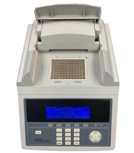 A modern thermal cycler capable of performing 96 PCRs at a time
CODIS STR Loci
- TPOX
- CSF1PO
- D5S818
- D13S317
- D16S539
- THO1
- D18S51
- D7S820
- VWA
- FGA
- D3S1358
- D8S1179
- D21S11
- Amelogenin
SGM+ Loci Used by FSS UK.
- D3S1358
- VWA
- D16S539
- D2S1338
- D8S1179
- D21S11
- D18S51
- D19S433
- THO1
- FGA
- Amelogenin
The match probability ( the chance that a DNA profile, meaning a genotype made up of all four STR loci, matches that of a random person in the population ) was 1 in 10,000.
- The FSS then introduced its second generation multiplex (SGM. , which included six STR loci: THO1, FGA, D8S1179, D18S51, and D21S11.
- SGM offered a match probability of 1 in 50 million, which was acceptable for the UK population.
- However, larger populations, such as those in the US and India, required a multiplex kit with greater discrimination capability.
- Two companies, PE Applied Biosystems and Promega, developed their own multiplex kits with high discrimination power.
In the USA, the main loci used in the CODIS system consist of 13 loci. All multiplex STR kits also feature an STR amelogenin for gender identification. The amelogenin locus is located on both the X and Y chromosomes, but they are not in the same positions because of a 6 bp deletion on the X chromosome. Consequently, the PCR produces two products ( or bands or peaks ) for males and only one product ( band or peak ) for females.
Classification of STRs
STRs can be classified according to the length of the repeat unit. The different types are:
- Di-nucleotide STRs (2 nucleotides)
- Tri-nucleotide STRs (3 nucleotides)
- Tetra-nucleotide STRs (4 nucleotides)
- Penta-nucleotide STRs (5 nucleotides)
While tetranucleotide STRs are common, the forensic community uses various types depending on specific needs and contexts. An important classification considers the configuration of alleles, which is crucial for establishing a universally accepted nomenclature.
Types of STRs
- The alleles of STRs are mostly easily distinguished from one another.
- The sizes can be measured in different ways.
Mechanisms of STR Mutation
The forensic importance of STRs (Short Tandem Repeats) lies in their high level of polymorphism, which results from mutations in genomic sequences, leading to the creation of different alleles at a specific locus. Typically, mutations that occur during DNA replication are corrected by enzymatic repair processes. However, mutations can also occur in regions of the genome with numerous simple repetitive sequences due to a process known as “slipped strand mispairing, ” which is the primary mechanism behind these mutations.
Types of STRs
- Simple STRs: These consist of a single repeating sequence. For instance, a STR locus may have several alleles, each differing only in the number of repeats while the sequence remains constant.
- Simple STRs with non-consensus alleles: An example is the STR locus HUMTHO1, where Allele 3 is [AATG] 3 and Allele 4 is [AATG] 4.
- Compound STRs with non-consensus alleles: For example, at the STR locus vWA, Allele 16 is TCTA [TCTG] 4[TCTA] 11TCCATCTA, and Allele 17 is TCTA [TCTG] 3[TCTA] 12TCCATCTA.
- Complex repeat STRs: Consider the STR locus D21S11, where Allele 25 is [TCTA] 4 [TCTG] 3 [TCTA] 3 TA [TCTA] 3 TCA [TCTA] 2 TCCA TA [TCTA] 10, and Allele 26 is [TCTA] 4 [TCTG] 6 [TCTA] 3 TA [TCTA] 3 TCA [TCTA] 2 TCCA TA [TCTA] 8.
- Hypervariable repeat STRs: An example is SE33, where certain loci not only exhibit different repeat regions but also various arrangements, as seen in SE33.
Techniques for STR Resolution
- Electrophoresis is a fundamental technique used to detect STR alleles.
- While agarose gel electrophoresis with ethidium bromide staining can separate STR alleles, polyacrylamide gel electrophoresis (PAGE) offers superior separation.
- In smaller laboratories, STRs can be identified using the silver staining method, which enables clear discrimination of alleles without the need for expensive automated DNA sequencers.
Challenges Faced by Forensic Laboratories
- Forensic laboratories often deal with a high volume of cases, necessitating rapid processing of samples to facilitate criminal or civil investigations.
- This demand has led to the need for automation and simplified methods for detecting STR alleles.
STR Analysis and Automation
Introduction: STR (Short Tandem Repeat) analysis is a powerful tool used in forensic science and DNA profiling. It involves the examination of specific regions in the DNA where short sequences are repeated. Automation has significantly improved the efficiency and accuracy of STR analysis.
History of Automation in STR Analysis
- In 1986, instrumentation was developed for the automatic analysis of DNA sequences using fluorescent dyes. This marked the beginning of automated systems in DNA analysis.
- Automated systems like ABI GeneScan® became capable of storing and tabulating electrophoretic data as alleles moved through a gel matrix and passed a laser detection window.
- The use of four distinct fluorescent dyes allowed for the tagging of different loci primers, enabling multiplexing of loci even with overlapping product sizes. This improved control with internal size standards.
Advantages of Automated STR Analysis
- Automatic Allele Sizing: Automated systems could achieve allele sizing by running an internal size standard with each sample.
- Normalisation of Electrophoretic Mobility: Variations in electrophoretic mobility from lane to lane and gel to gel were automatically normalised, addressing potential inconsistencies in allele sizing.
- Sensitivity: Initial studies demonstrated the sensitivity of automated methods compared to manual staining techniques, highlighting the benefits of automation.
- Reproducibility, Accuracy, and Precision: The automated system exhibited the necessary reproducibility, accuracy, and precision for forensic applications.
- Detection of Mixed Samples: Casework validation studies showed that mixed samples could be detected and accurately interpreted using automated STR analysis.
- Discrimination Power: STR analysis provided results with greater discrimination power compared to single locus probe analysis, HLA DQA1, and traditional blood grouping systems.
- Multiplexing Capability: Multiplexing allowed the amplification of multiple STR loci in a single tube PCR, with kits available to amplify 16 STR loci simultaneously, increasing throughput and discrimination power significantly.
Use of Allelic Ladders
- An allelic ladder is a crucial tool in DNA analysis, comprising multiple alleles from a specific Short Tandem Repeat (STR) system. This ladder serves as a reference guide for identifying and sizing alleles in DNA samples.
- The allelic ladder functions similarly to a ruler, allowing researchers to measure the size of alleles in a sample by comparing them to the known sizes of the alleles in the ladder. These known sizes are based on alleles that have already been sequenced.
- Utilizing sequenced allelic ladders has been shown to enhance the accuracy of allele sizing. Therefore, it is consistently recommended to use sequenced allelic ladders for allele designation in DNA analysis.
- Forensic standards for DNA analysis are often more stringent than those in typical laboratory settings. As a result, internal standards have been developed and are routinely added to samples before they are loaded onto gels or capillaries for analysis.
- These commercial internal standards are tagged with a red dye and contain DNA fragments of standard sizes. The software used by sequencers sizes alleles based on this internal ladder, ensuring accurate measurements.
- DNA analysts play a vital role in the analysis process by comparing the results of DNA samples with the allelic ladder that is run alongside each batch of samples. This comparison helps in verifying the accuracy of allele sizing and identification.
- In experiments, specific windows are set for allele sizes, and these windows are fed into software programs such as Genotyper™. This approach, known as the absolute window method, is used to identify alleles based on established size ranges.
- The absolute window method is also applicable for comparing crime scene samples with control samples, aiding in forensic investigations.
Non-specific Amplification Peaks
- Automated DNA sequencers, known for their sensitivity, detect all products generated during a PCR assay, presenting them as peaks on an electropherogram.
- These peaks encompass nonspecific peaks, stutter peaks, and allelic peaks.
- Nonspecific peaks are typically less than 5% the size of the allelic peak or exhibit an unusual shape, making them easily identifiable.
- Stutter peaks arise from enzyme slippage during the PCR reaction. While they cannot be entirely eliminated, they manifest as a smaller peak, approximately 15% the size of the allelic peak, and are one repeat smaller than the actual peak.
- Stutters play a crucial role in pinpointing the true peak but can complicate interpretation, particularly in di-nucleotide STR analysis.
- In mixture analysis, stutters have the potential to amplify the peaks of the minor component, posing challenges in identifying the minor peak.
|
71 docs|3 tests
|
FAQs on Forensic DNA Profiling - 1 Chapter Notes - Forensic Medicine and Toxicology (FMT) - NEET PG
| 1. What are the main advantages of using DNA for identification in forensic science? |  |
| 2. What are the disadvantages associated with DNA profiling? | |
| 3. How does the Polymerase Chain Reaction (PCR) work in the context of DNA analysis? | |
| 4. What are microsatellites and how are they used in forensic DNA profiling? | |
| 5. What role do allelic ladders play in STR analysis? | |


Sample Paper
,Exam
,Forensic DNA Profiling - 1 Chapter Notes | Forensic Medicine and Toxicology (FMT) - NEET PG
,shortcuts and tricks
,practice quizzes
,ppt
,Summary
,Objective type Questions
,MCQs
,mock tests for examination
,Viva Questions
,Important questions
,Free
,study material
,past year papers
,Forensic DNA Profiling - 1 Chapter Notes | Forensic Medicine and Toxicology (FMT) - NEET PG
,Extra Questions
,Semester Notes
,video lectures
,Previous Year Questions with Solutions
,Forensic DNA Profiling - 1 Chapter Notes | Forensic Medicine and Toxicology (FMT) - NEET PG
;


Chapter Notes: Forensic DNA Profiling - 1 Free PDF Download
Importance of Chapter Notes: Forensic DNA Profiling - 1
Chapter Notes: Forensic DNA Profiling - 1
Chapter Notes: Forensic DNA Profiling - 1 NEET PG Questions
Study Chapter Notes: Forensic DNA Profiling - 1 on the App
|
© EduRev
|
Education Revolution
|


|


