Animal Tissues | Science Class 9 PDF Download

The living organisms are either unicellular [eg. - Bacteria, Diatoms, Yeasts, Potozoans] or multicellular [eg. Man, Lion , Dog]. Each unicellular organism is able to perform all their vital activities like digestion, respiration, excretion, reproduction.
The multicellular organism, on the other hand, is composed of a millions of different types of cells. All the cells of a multicellular organism do not perform all functions of the body, rather they undergo differentiation and each type of cell becomes specialized for a limited number of specific functions. For examples, in human beings:-
- Muscle cells cluster together to perform contraction and relaxation to cause movements.
- Nerve cells or neurons coordinate to carry messages.
- Blood flows to transport oxygen, food, hormones and waste materials.
Utility of tissues in multicellular organisms:
With the increasing degree of multicellularity in living beings, it became difficult for each cell to efficiently perform all the physiological functions of the body. Hence, nature assigned specialized function to different group of cells called tissues. Thus, the utility of tissues in multicellular organisms is to perform specific functions of the body.
 Tissue
- Bichat introduced the term 'tissue'.
- Mayer introduced the term 'Histology'. [Study of tissue is called histology]
- Marcello Malpighi is the 'Founder of Histology'.
- The term 'epithelium' was introduced by Ruysch.
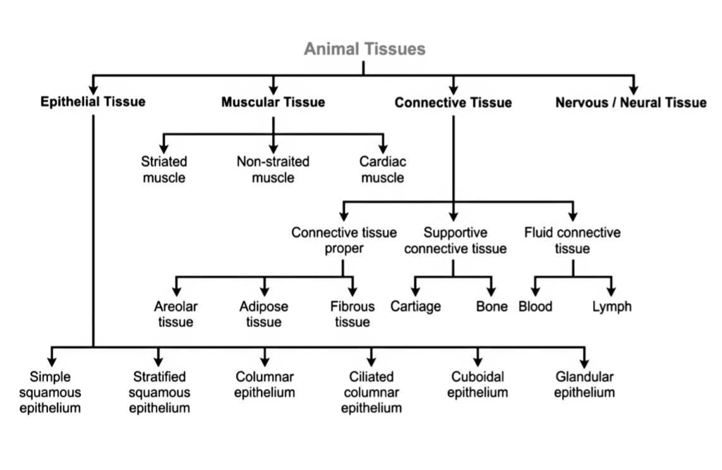
Types of Animal Tissue
Based on the location and function, the animal tissue are classified into four types
Epithelial Tissue
Word epithelium is composed of two words Epi-upon, Thelio-grows. (Means - A tissue which grows upon another tissue is called epithelium).
Nature
1. It is the simplest tissue. It is the protective tissue of animal's body.
2. It covers most organs and cavities within the body.
3. It also form a barrier to keep different body systems separate.
4. Epithelium cells are closely packed, so there is very little inter-cellular spacesare present between the cells. Due to absence or less of intercellular spaces blood vessels, lymph vessels and capallaries are unable to pierce this tissue, so blood circulation is absent in epithelium. Hence cells depend for their nutrients up on the underlying connective tissue.
5. It always rest upon underlying connective tissue.
6. At the junction of the (Epithelial tissue and connective tissue) layer is present which is called of basement membrane, which is formed of mucopolysaccharides and collagen fibrils.
7. Epithelial tissue has great regeneration power because meristematic cells can divide to replace old and dead cells.
The skin & lining of buccal cavity, blood vessels, alveoli (of lungs) and kidney tubules are made of epithelial tissue.
The tissue which evolved first in animal kingdom and appears first during embryological developement is the epithelial tissue.
Points for Competitive Exams
Epithelial tissue may be given different names in the different organs of body.
1. Endothelium: It lines cavity of heart, blood vessels and lymph vessels.
2. Mesothelium: It is peritoneum which forms outer most covering of body organs in coelomic cavity.
(Body cavity is called as coelom. Peritoneum is the covering of all visceral organs.)
3. Pericardium: It forms outer covering of the heart.
4. Pleura: This is covering over the lungs.
5. Germinal epithelium: This occurs in the gonads-testis and ovary.
6. Pigemented epithelium: It contains pigments.
7. Glandular epithelium: It forms glands.
8. Sensory epithelium: It occurs in sense organs.
General Functions of Epithelial Tissue
1. Protection: Epithelia protect the underlying cells from mechanical and chemical injuries and bacterial or viral infection.
2. Acts as Barriers: It acts as selective barriers.
3. Absorption: Helps in absorption of water and nutrients.
4. Elimination: Helps in elimination of waste products.
5. Secretion: Some epithelial tissues secrete secretion, such as sweat, saliva, mucus, enzymes, etc.
6. Respiration: Epithelia of alveoli of lungs exchange oxygen and carbon dioxide between blood and inhaled air.
7. Exoskeleton: It produces exoskeleton structures, such as scales, feathers, hair, nails, claws, horns and hoofs.
8. Regeneration: This tissue facilitates rapid healing of wounds by its regeneration power.
Types of Epithelial Tissues
(Depending upon the shape & function of the cells)
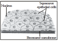
(a) Squamous epithelium:Squamous epithelium is made up of thin, flat, disc-like, polygonal or irregular-shaped cells with round and flat nucleus. Adjacent cells fit together to form a compact structure which gives an impression like tiles on a pavement or floor.
- The plasma membrane is wavy in these cells when they form lining of blood vessels, lymph vessels and in coelomic epithelium hence, such epithelium is called tessellated epithelium.
- Simple squamous epithelium is given different names on the basis of different position in the body. When it form lining the cavity of heart, blood vessels and lymph vessels, it is called endothelium. Coelomic cavity is lined with coelomic epithelium or mesothelium, when it form covering around visceral organs it is called peritoneum and while lining bone marrow it is called endosteum.
The squamous epithelium is of two kinds:
- Simple squamous epithelium: It is made up of single layer of flat cells. It forms the delicate lining of cavities [nose, pericardium, alveoli etc.] and of blood vessels.
- Stratified squamous epithelium: Composed of more than one layer of squamous cells. It is present where thick covers are required, e.g., surface of the skin and oral cavity, oesophagus, etc. This epithelium is water proof and highly resistant to mechanical injury.
Functions
(i) It protects the internal organs of body from mechanical injury, desiccation, entry of germs, chemicals & drying.
(ii) It forms a selectively permeable surface through which filtration occurs.
(iii) In certain organs, it also facilitates diffusion of gases.
(b) Cuboidal Epithelium: Cuboidal epithelium is composed of cube-like cells of almost equal height and width. The cells appear square-like in vertical section but their free surface seems to be hexagonal.
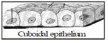
Place of Occurrence
Cuboidal epithelium is present in kidney tubules, salivary glands, sweat glands, pancreatic duct, thyroid follicles, etc.
It is also present in the germinal epithelium of testes and ovaries.
Functions
(i) It helps in absorption, excretion & secretion.
(ii) It also provides mechanical support.
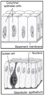
(c) Columnar epithelium: Columnar epithelium consists of tall or pillar-like cells. The basal part of the cell which rests on the basement membrane bears oval nucleus. The free end of the cell has large number of minute finger-like projections calledmicrovilli or brush border. Microvilli increase the absorptive surface. Most of the columnar epithelia are simple, i.e., one cell thick but stratified columnar epithelium with more than one layer of cells also exist.
- The tissue specialized for secretion is called glandular tissue. Glands are derived from folding of glandular epithelium. Cells of glandular tissue have nucleus and cytoplasm containing zymogen granules. These cells secrete mucus, hormones, enzymes or saliva. Cells of glandular epithelium are cuboidal or columnar in shape.
Place of Occurrence
The columnar epithelium lines the inner surface of stomach, intestine and gall bladder. It also occurs in salivary glands, sweat glands, oviduct, etc.
Functions
- Absorption: Absorption of digested food (Stomach, Intestine)
- Secretion: Mucus by goblet cells or mucus membrane.
(d) Ciliated epithelium: It is made up of tall cells with cytoplasmic hair like ciliaat free ends. The cells may be cuboidal or columnar, and hence, also called ciliated cuboidal epithelium or ciliated columnar epithelium.
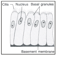
Place of Occurrence
The ciliated cuboidal epithelium is found in sperm ducts (vas deferens).
The ciliated columnar epithelium forms the lining of trachea (wind pipe), fallopian tube (oviducts), lungs (bronchi), nasal passage, kidney tubules, etc.
Functions
(i) The rhythmic, concerted beating of the cilia moves solid particles [eg. mucus, ova] in one direction through ducts.
(ii) It causes movement of ovum and zygote towards uterus.
(iii) It helps in removing unwanted particles from trachea.
Pseudostratified Epithelium
Some times columnar epithelium has cells of different sizes. Besides column like tall cells, some cells are small called basal cells which do not reach upto the margin. Due to different size of cells nuclei appear to be present in more than one layers. Although it is single layer of cells but it appears to be multilayered and is called pseudostratified epithelium. It occurs in the lining of trachea, bronchi, vas deferens, urethra, epididymis and pharynx.
Connective tissue originates from embryonic mesoderm. Hertwig (1883) gave the term mesenchyme for adult tissues derived from mesoderm which fills space between ectoderm and endoderm. Hence, connective tissue is also sometimes considered mesenchyme.
The connective tissue is specialised to connect and anchore various body organs. As such it can connect bones to each other, muscles to bones, bind tissues and give support to various parts of body by forming packing around organs so that they donot get displaced by body movements.The main functions of connective tissue are binding, supporting & packing together different organs of the body.The cells of connective tissue are living, separated from each other [i.e. loosely spaced] and are very less in number.
Homogeneous, gel-like intercellular substance called medium or matrix. This matrix may be jelly-like, fluid or dense [as blood] and solid [as in bone and cartilage] or fibrous in nature and binds other tissues. The nature of the matrix decides the function of tissue.
components of connective tissue
There are three components present in all the connective tissues:
(i) Intercellular medium (ii) Connective tissue cells. (iii) Fibres.
1. Connective tissue contains the following types of cells:
(a) Fibroblasts: They form ground substance and fibres [eg. collagen]
(b) Adipose cells: They store fats in their vacuoles.
(c) Macrophages: They may be free living or fixed phagocytes [ Leucocytes or WBC's] They are involved in the destruction and removal of invading bacteria, foreign bodies & damaged cells from tissues.
(d) Mast cells: They secrete substances such as heparin [anticoagulant], histamin[Vasodilator - dilation of blood vessels] serotonin [Vaso-constrictor-constriction of blood vessels].
They promote inflammation of the infected area.
(e) Immunocytes: These include cells such as lymphocytes and plasma cells both producing antibodies for the immune response.
2. Protein fibres of matrix: Matrix of connective tissue is secreted by the component cells. It chemically contains GAG's [Glycosaminoglycons or Mucopolysaccharides]
(a) White fibres of collagen
(b) Yellow fibres of elastin
(c) Reticular fibres of reticulin
General Functions of Connective Tissue
(i) Storage: Certain connective tissue like adipose tissue store fats.
(ii) Supports: Skeletal connective tissue like bones and cartilage provide the body with a supporting skeletal framework.
(iii) Transport: Fluid connective tissues such as blood and lymph transport various material in the body.
(iv) Defence and scavenging: Plasma cells synthesize antibodies, macrophages, lymphocytes, which ingest foreign matter and harmful bacteria.
(v) Shock absorber: The jelly like ground substances of connective tissue acts as shock absorber around some organs like eyeballs and kidney.
(vi) Formation of blood corpuscles: The bone marrow produces blood cells.
(vii) Packing material: Areolar tissue act as packing material in various organs.
(viii) Repair: Collagen fibre of connective tissue help in repairing of injured tissues.
Types of Connective Tissue
(a) Areolar [loose] connective tissue.
(b) Dense regular connective tissue.
(c) Adipose tissue
(d) Skeletal tissue
(e) Fluid connective tissue.
(a) Areolar [loose] connective tissue:
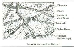
Nature
It is a loose and cellular connective tissue. It is the most abundant of all types of connective tissues. It has large amount of matrix. Its matrix consists of two kinds of fibres -
(i) White collagen fibres
(ii) Yellow elastic fibres or elastin.
Occurrence
It is simplest & most widely distributed connective tissue. It joins skin to muscles, fills spaces inside organs and is found around muscles, bone marrow, blood vessels & nerves.
Functions
(i) It acts as a supporting & packing tissue between organs lying in the body cavity.
(ii) It helps in repair of tissues after an injury.
(iii) It also helps in combating foreign toxins.
(iv) It fixes skin to underlying muscles.
(v) It provides rapid diffusion of oxygen and nutrients from blood vessels.
(b) Dense regular connective tissue:
It is a fibrous connective tissue which is characterized by systematically and densely packed fibres and cells. Dense regular connective tissue is the principal component of tendons & ligaments.
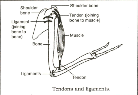
(i) Tendons: These are cord like, strong, inelastic, structures that join skeletal muscles to bones. It has great strength but its flexibility is limited. It is made up by collagen fibres.
(ii) Ligaments: They are elastic structures which connect bones to bones. It is highly elastic and has great strength but contains very little matrix. It is made up of both collagen and elastin fibres.
Ligaments strengthen the joints of body and they permit normal movement but prevent over flexing or over-extension. Sprain is caused by excessive pulling [stretching] of ligaments.
| Sr. No. | Characters Tendons Ligaments | ||
| 1 | Nature | Tough and non-elastic | Strong and elastic |
| 2 | Structure | Made up of white collagen fibrous tissues. | Made up of yellow fibrous tissue and white collagen fibrous tissue |
| 3 | Arrangement of fibroblasts | Present in rows between fibres | Scattered in matrix in between the bundles of white fibres. |
| 4 | Function | Join muscle to bone | Join bone to bone |
(c) Adipose tissue: It consists of large number of oval and rounded adipose cells [Adipocytes] filled with fat globules.Adipose cells may contain single large fat droplet [white adipose tissue] or several tiny droplets [Brown adipose tissue] Besides adipocytes, adipose tissue also contains fibroblasts, macrophages, collagen and elastic fibres.
| Competition window |
Adipose tissue occurs in different parts of body and forms about 15% of our body weight. It forms cushions around kidney and heart and it also occurs in yellow bone marrow. It mainly occurs as subcutaneous fat layer under skin called penniculus adiposus. In whale and elephant blubber is a thick adipose layer. Hump of camel, thick tail of merino sheep and fat bodies of frog represent adipose tissue. It is very important component of skin in mammals living in polar regions. Adipose tissue is fat depot in the body. It stores fat and releases it for energy production, whenever needed in the body. Stored fat is generally of two types: white (or yellow) fat and brown fat.Generally white fat occurs in the body. |
Functions
(i) Adipose tissue acts as food reservoir by storing fat.
(ii) This tissue is found below the skin, between internal organs and in the yellow bone marrow.
(iii) It acts as an insulator and regulates body temperature.
(iv) Animals living in cold climates have a lot of this tissue to protect them from the cold.
(d) Skeletal tissue: Skeletal tissue forms the rigid skeleton which supports the vertebrate body, helps in locomotion and provides protection to many vital organs. It is mesodermal in origin. There are two types of skeletal tissues :-
(i) Cartilage (ii) Bone.
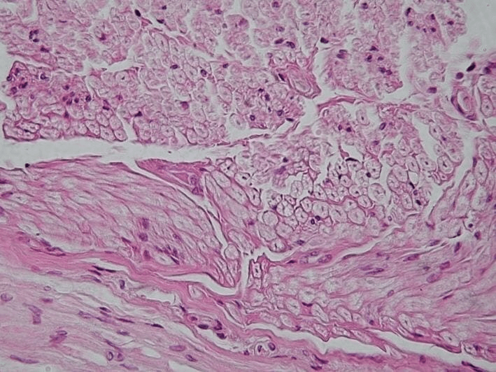
Cartilage
Cartilage is a special type of connective tissue which forms the soft endoskeleton of the body. It consists of extensive ground substance or matrix calledchondrin. Matrix is composed of proteins and sugars and because of the presence of calcium salts becomes slightly hardened. It also contains network of white collagen fibres and yellow elastic fibres. Nerves and blood vessels do not penetrate into chondrin.
The cartilage cells called chondrocytes are present in groups of 2, 3 or 4 in fluid filled cavities called lacunae
Types of Cartilages
On the basis of composition of matrix, amount and nature of fibres cartilages are of four types:
(i) Hyaline cartilage (ii) White fibrous cartilage (iii) Yellow elastic cartilage. (iv) Calcified cartilage.
Occurrence
This tissue occurs in very few parts of the body. In humans, the cartilage occurs at the ends of long bones, the pinnae of ears, the ends of nose, in the walls of respiratory ducts, etc. In sharks and rays, the entire skeleton is cartilage.
Functions
1. Cartilage provides support and flexibility to the body parts.
2. It smoothens bone surfaces at joints.
Bone
- Bone is hardest tissue of the body. It forms endoskeleton to give firm support to the muscles.
- Like other connective tissues, it consists of intercellular material (matrix) and cells (Osteocytes).
- The matrix is composed of about 30% organic materials (Ossein protein) and about 70% inorganic materials (Mainly phosphates and carbonates of calcium and magnesium). These inorganic salts are responsible for hardness of the bone.
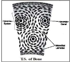
- The matrix of bone is arranged in the form of thin concentric rings calledlamellae.
- In between the lamellae, the bone cells (osteoblasts) are present in fluid filled cavities called lacunae, which have fine extensions called canaliculi.
- In long bones of mammals, the lamellae are arranged around a haversian canal. The Haversian canal contains blood vessels, nerves and lymphatic canals. Haversian canals along with concentric rings of lacunae and osteocytes is calledHaversian system. Its function is transportation of nutrients and oxygen.
Functions
(i) Bones form hard endoskeleton which give shape and support to the body.
(ii) Bones protect vital organs of the body, such as brain, spinal cord, lungs, etc.
(iii) Bones provide skeletal support to the body.
(iv) Bone marrow is the centre of blood cell formation in vertebrates.
(v) Bone attaches the muscles.
| IMPORTANT POINTS FOR COMPETITIVE EXAMS |
|
(e) Fluid Connective Tissue
It is a special type of connective tissue which maintains link among different parts of the body. It receives materials from certain parts of the body and transports them to the other parts
It constitutes the transport system of animals.
It consists of two basic components - blood and lymph.
Blood
Blood is a mobile connective tissue. It measures about 5-5.5 litres in an adult human being. It is slightly alkaline with a pH value of 7.4.It consists of an aquous (watery) mixture of substances in solution (blood plasma)in which are suspended different types of free floating cells (blood corpuscles).Plasma constitutes about 55% of blood volume while corpuscles constitute 45%.
Blood Plasma
It is a pale straw-coloured fluid matrix or medium consisting of about 90% water and 10% mixture of different types of molecules that enter the blood at various locations. These substances include - proteins (soluble proteins such as albumins, globulins and fibrinogen), glucose, amino acids, lipids, vitamins, urea, uric acids, enzymes and hormones.
Blood corpuscles
(i) Red Blood Corpuscles (RBC) or Erythrocytes
(ii) White Blood Corpuscles (WBC) or Leucocytes
(iii) Platelets or Thrombocytes
RBC
(i) In mammals, RBCs are small, circular, biconcave & discs shaped and lack nuclei when mature.
(ii) There are about five million red blood cells per mm3 of blood.
(iii) Their most important character is the presence of an iron protein,haemoglobin. The presence of haemoglobin gives the blood its red colour.
(iv) They are manufactured in bone marrow. Their lifespan in human beings is about 120 days, after which they are destroyed in liver.
The RBCs constitute about 99% of blood corpuscles. Erythrocytes occur only in vertebrate blood and red colour of blood is due to erythrocytes.
Smallest RBCs occur in musk deer (Tragulus). During maturation, cell organelles of RBC like nucleus, mitochondria, Golgi body and centrosome become disappear. Hence surface area of mature RBC increases. It can accommodate more haemoglobin and can carry more O2.
WBC
(i) These are rounded or amoeboid, nucleated, colourless cells.
(ii) WBCs are formed in red bone marrow, spleen, thymus and lymph nodes.
(iii) They are capable of amoeboid movement and play an important role in the body's defence mechanism.
(iv) The white blood corpuslces belong to two main categories: Phagocytes (carry out the function of body defence by engulfing pathogen) and Immunocytes (they are responsible for immunity and carry out immune responses by producing antibodies).
Phagocytes are further divided into two types: Granulocytes (having cytoplasmic granules) and Agranulocytes (having non-granular cytoplasm)
Granulocytes: On the basis of staining these are of three types:
(a) Eosinophils (stained with acidic dyes)
(b) Basophils (stained with basic dyes)
(c) Neutrophils (stained with neutral dyes).
Agranulocytes: It includes Monocytes and Lymphocytes.
Functions of blood:
(i) It transports nutrients, hormones and vitamins to the tissues and carries excretory products from the tissues to the excretory organs.
(ii) The RBC's of blood helps in the transport of respiratory gases, oxygen & CO2.
(iii) The WBCs fight with diseases by producing antibodies and engulfing the germs.
(iv) Blood platelets helps in the clotting of blood.
(v) Blood helps in thermoregulation, water balance and maintenance of pH of body.
Lymph
Lymph is actually filtered blood which is similar to blood in composition except that it is devoid of RBC, platelets and some blood protein. WBC are present in abundance in lymph. Due to the absence of haemoglobin, lymph is colourless.
Functions of Lymph
(i) Helps in the transport of nutrients. Nutrients that filter out from blood capillaries into lymph are transported back by lymph into blood through heart.
(ii) Helps in the transportation of fat absorbed from intestine to the venous blood.
(iii) Keeps the tissues and organs of the body moist.
(iv) Lymphatic organs (lymph nodes, spleen) produce lymphocytes which in turn produce antibodies to strengthen the immune system of the body.
Ques. Distinguish between the following :
(a) Cartilage and bone on the basis of matrix.
(b) Blood and lymph on the basis of components.
Ans. (a) Matrix of cartilage may or may not have calcium salts whereas calcium salts, mainly calcium phosphates, are allways present in the matrix of bone.
(b) Blood consists of plasma, erythrocytes, leucocytes and platelets whereas lymph consists of plasma and leucocytes.
Ques. What will happen if stratified squamous epithelium lines the alveoli of lungs?
Ans. The permeability of alveoli of lungs will be affected so that it will not be able to perform the function of absorption and transportation of substance and selective permeability of alveoli wall will be affected.
| Review questions |
1. Define connective tissue ? 2. Name the common type of connective tissue of animal's body ? 3. Why is blood called a connective tissue ? 4. What is the function of areolar tissue ? 5. What is the name of bone cell ? 6. Write the name of various types of WBC's. 7. Name the cells which are responsible for fibres information. 8. Name the chemicals which are formed against antigens in our body. |
Muscle Tissues
Muscular tissue is distinguished from other tissues by its unique ability to contract & relax and thereby perform mechanical work. It is responsible for movement of body organs and locomotion of body.
General structure
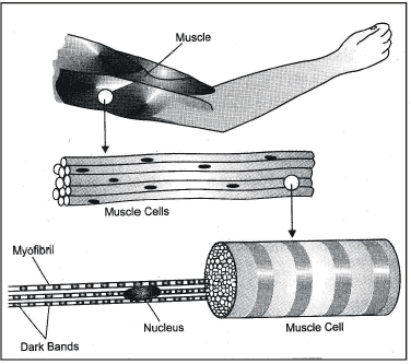
The structural unit of muscle tissue is the muscle cells which because of its elongated shape is also called muscle fibre.
The contractility is due to the presence of contractile proteins (Actin & Myosin) in the muscle fibre.
The plasma membrane of muscle cells is called sarcolemma and endoplasmic reticulum of muscle cell is called sarcoplasmic reticulum.
General functions of Muscular Tissue
1. It supports the bones and other organs of the body.
2. Muscles cause peristalsis of gut, heart beat, production of sound, etc.
3. Muscles cause movements of body parts and locomotion of the animals.
4. Facial expression also depends on muscles.
5. Contraction of muscles causes delivery of a baby.
(A) Unstriated Muscle (Smooth Muscle)
Characteristics
These are called smooth or unstriated muscles because they do not show any stripes of striations across the muscle fibres. Each cell (or fibre) is long, narrow spindle shaped with pointed ends and has only one nucleus(uninucleate) situated in the centre. These fibres are generally shorter than the striated muscle fibres.
Place of Occurrence
Unstriped muscles are found in the wall of alimentary canal (stomach and intestine), urinary bladder, blood vessels, lungs, etc.
Functions
These muscles cause slow and prolonged contraction which is involuntary, i.e., not under the control of individual's will. These are under the control of autonomous nervous system. These muscles help in peristalsis of alimentary canal, urinary tract, blood vessels, etc., and contraction of other visceral organs (not heart).
(B) Striated Muscle or Skeletal Muscle
Charcteristics
The striated muscles form more than 80% of the mass of soft tissues in a vertebrate body. They are attached to the bones by tendons and help in the movement of external body parts. Therefore, they are also called skeletal muscles. The contraction and relaxation of these muscles are under the control of the animal's will. They are, therefore, also called the voluntary muscles. The muscle fibres show alternate dark and light stripes (striations or bands), hence they are called striated muscles.
The striated muscle consists of long, narrow, cylindrical, unbranched fibres (cells) with blunt ends (non-tapering ends). Each fibre is enclosed in a thin but distinct plasma membrane, called sarcolemma. The cell contains many elongated, flattened nuclei characteristically located towards the periphery near the sarcolemma. The multinucleate condition of the fibre results from cell fusion.
Place of Occurrence
Striped muscles are found in limbs, body wall, tongue, pharynx, face, neck, initial part of oesophagus, etc.
Functions
Striped muscles produce rapid and powerful contractions which help in the movement of limbs and consequently cause locomotion. They are also helpful in the movement of other body parts which are in voluntary control of the individual.
(C) Cardiac Muscles
Cardiac muscles are the muscles of heart. They are involuntary in action. Cardiac muscles possess characteristics of both striped as well as unstriped muscles, resembling striped muscles structurally and unstriped muscles functionally.
Their muscle fibres are uninucleate, branched. The branches of adjacent fibres join to form a network. Each muscle fibre contains a centrally located nucleus. Sarcoplasm(Cytoplasm of muscle cell is called Sarcoplasm) bears contractile, longitudinal myofibrils which give the cardiac muscles a striated appearance in the form of dark cross bands calledintercalated disc.
Place of Occurrence
Wall of heart(Myocardium).
Characteristics
(i) Striations Present Absent Present
(ii) Shape of the cells Cylindrical Spindle shaped Cylindrical
(iii) Branches Not branched Not branched Branched
(iv) Number of nucleus Many Single Single
(v) Position of Nucleus Peripheral Peripheral Central
(vi) Intercalated discs Absent Absent Present
(vii) Mode of contraction Voluntary Involuntary Involuntary
(viii) Speed of contraction Fast Slow Fast
(ix) Length of fibres 0.02 mm to 0.5 mm 0.01 to 30 cm 85 to 100 mm (very short)
[longest muscles]
Nervous Tissue
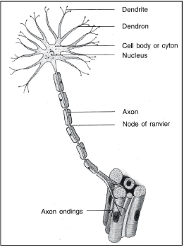
The nervous tissue, contains densely packed cells called nerve cells or neurons, is present in the brain, spinal cord and nerves. The neurons are specialised for conduction of nerve impulses. They receive stimuli from within or outside the body and conduct impulses (signals) which travel from one neuron to another neuron.
Each neuron has following 2 parts:
1. Cyton or cell body: Contains a central nucleus and cytoplasm with characteristic deeply stained particles called Nissl's granules [i.e. clumps of ribosomes]
2. Cell Processes
(A) Dendrites: These may be one to many, generally short and branched cytoplasmic processes. Dendrites areafferent processes because they receive impulse from receptor or other neuron and bring it to cyton.
(B) Axon: It is single generally long efferent processwhich conducts impulse away from cyton to other neuron.
Longest cell in body is neuron because axon can be more than one metre long. Axon has uniform thickness but it has terminal thin branches called telodendria. Terminal end buttons or synaptic knobs occur at the end of telodendria.
Competition Window
German neurologist Franz Nissl (1860-1919) first described Nissl granules in nerve cell, these are formed of rough ER and Ribosomes.
Synapses are junction between two adjoining neurons.
Nissl granules disappear during fatigue and injury to nerve cell and reappear after rest.
Types of Neuron: Based on number and nature of process arising from cyton the neurons are of different types:
(a) Multipolar neuron: It has many dendrites and one axon.
(b) Bipolar neuron: A neuron having one dendron and one axon is called bipolar. They generally occur in sensory layers like olfactory epithelium.
(c) Unipolar neuron: It has single process as axon but dendrite is absent.
(d) Pseudounipolar neuron: Such neuron has single fibre arising from cyton which bifurcates into one dendron and one axon.
(e) Nonpolar or apolar neuron: These neurons have many fibres but they are not distinguished into dendrites and axon. Each fibre can receive impulse towards cyton or can conduct impulse away from cyton.
|
84 videos|543 docs|60 tests
|
FAQs on Animal Tissues - Science Class 9
| 1. What are the four types of animal tissues? |  |
| 2. What is the function of epithelial tissue in animals? | |
| 3. How does connective tissue contribute to animal bodies? | |
| 4. What is the role of muscle tissue in animal physiology? | |
| 5. How does nervous tissue enable communication in animals? | |


Semester Notes
,mock tests for examination
,past year papers
,Exam
,practice quizzes
,study material
,Free
,MCQs
,shortcuts and tricks
,Important questions
,Animal Tissues | Science Class 9
,Extra Questions
,ppt
,Previous Year Questions with Solutions
,Summary
,Animal Tissues | Science Class 9
,Objective type Questions
,Viva Questions
,video lectures
,Sample Paper
,Animal Tissues | Science Class 9
;


Animal Tissues Free PDF Download
Importance of Animal Tissues
Animal Tissues Notes
Animal Tissues Class 9 Questions
Study Animal Tissues on the App
|
© EduRev
|
Education Revolution
|


|


