Digestive System, Chapter Notes, Class 11,Biology PDF Download

INTRODUCTION
To perform various functions of the body energy is required, which is obtained from food. The process of conversion of complex food material in to simple and diffusible forms by hydrolysis is termed as Digestion
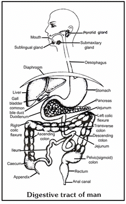
The alimentary canal is tubular structure which extends from mouth to anus. It develops from ectoderm & endoderm.
- Ectoderm – up to hard palate
- Endoderm – from soft palate to rectum
- Ectoderm – from anal canal to Anus
The alimentary canal is divided into following parts:
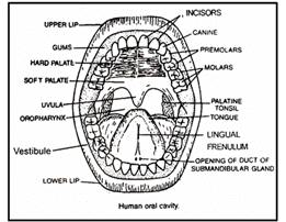
(i) Mouth and Buccopharygeal cavity
(ii) Oesophagus
(iii) Stomach
(i) Mouth and Buccopharyngeal Cavity: Mouth is a horizontal transverse slit like aperture which is surrounded by upper and lower lip. Orbicualaris oris voluntary muscles. are found in lips. Sebaceous glands are found on the outer part of lip. Serous glands are found on the inner part of lip. Serous glands is the modification of mucus glands. Its secretory substance is watery.
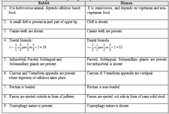
In Rabbits a small cleft is found in the middle part of upper lip, such type of lip is called as Hare lip
- Buccal vestibule: It is a peripheral part which, present between the gums and cheeks where the food is stored temporarily for some time
- Oral cavity: It is inner & central part which, is surrounded by upper and lower Jaw. Lined by stratified squamous epithelium. Upper Jaw is Fixed and Lower jaw is Movable.
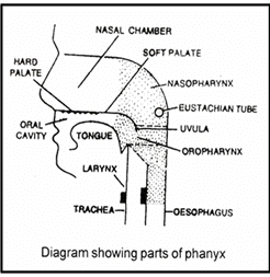
The roof of oral cavity is called as Palate. This palate is horizontal partition which situated between oral cavity and nasal chamber
Palate is differentiated into two parts
(i) Hard Palate: It is the anterior part of the palate. It is made up of Maxilla and palatine bones in humans. But in Rabbit it is made of Premaxilla, maxilla, palatine bone.
On the ventral surface of the hard palate, some projection or transverse ridges are present which are called palatine rugae. These rugae prevent slip out of the food from buccal cavity during mastication. These rugae are well developed in carnivorous animals.
In rabbits, one pair opening of Nasopalatine duct is present at the anterior part of hard palate, these connect the buccal cavity to the Nasal passage. In Rabbits some olfactory receptors are also found in nasopalatine ducts which are called as Jacobson's organ. It makes them aware of the smell of food while chewing.
(ii) Soft Palate: It is the posterior part of palate. It is made up of involuntary muscle, fibrous connective tissues and mucous epithelium. (Stratified squamous epithelium)
The posterior part of soft palate becomes out grow and hangs down in the form of finger like process called as Uvula or Velum palati One pair of large lymph node is present on the posterolateral surface of soft palate, called as Palatine tonsil or Tonsils Soft palate is situated in the pharynx and is divided into two parts. Upper and dorsal part of pharynx is called as Nasopharynx which is related to the nasal chamber. The lower and ventral part of pharynx is called oropharynx which is related to the oral cavity. One pair of openings of the Eustachian tube is present in the nasopharynx. This Eustachian tube is related to the middle ear.
TONGUE
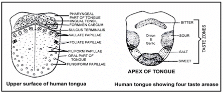
On the floor of oral cavity a muscular, flat, fleshy plate like structure is present which is called tongue. The anterior part of tongue is free while posterior part of Tongue is connected to the Hyoid bone. The surface of tongue is connected to the floor of buccal cavity through a very flexible membrane/ligamentous fold called as frenulum linguae On the dorsal surface of tongue, it is divided into two unequal parts by a V shaped sulcus, called as sulcus terminalis. The two limbs of the 'V' meet at a median pit named Foramen Caecum.
It is divided into two parts
1. Pharyngeal or Lymphoid Part: It is the posterior 1/3 part of the tongue. Many small lymph nodes are present in this part which are called Lingual tonsil.
2. Oral or papillary Part: It is anterior 2/3 part of tongue. Four types of papillae are found in this part in which gustatory or taste receptors are present.
(i) Fungiform Papillae: It is pink coloured, small & spherical in shape. It is found on the entire surface of tongue but Their maximum concentration at the anterior tip part of tongue. It is attached to tongue with the help of small pedicle. It provides pink colour to the tongue.
(ii) Filiform Papillae (Conical papillae): They are thread like, white coloured & conical in shape. They are also found on the entire surface of tongue. They are most numerous.
(iii) Foliate Papillae: They are found on the mid lateral surface of tongue. They are vestigial in the human. Their structures is leaf like present in rabbit and other mammals.(iv) Circumvallate papillae: They are large spherical shape papillae which are found near to sulcus terminalis. They are least in number (approx 8 to 12)
Two type of muscles are present in tongue
(i) Extrinsic muscle
- It is found on the outer and superficial part of the tongue.
- It helps in outward and inward movement of tongue.
(ii) Intrinsic muscle
- It is situated in the deep part of tongue.
- It help in the change of shape of tongue
TEETH
Teeth are ecto mesodermal in origin. Major portion of teeth arises from Dermis. Part of tooth present outside the gums only is derived from ectoderm or Epidermis (Enamel part).
In human teeth of upper jaw are attached to the maxilla bone. While teeth of lower jaw are attached to Mandible bone. But in rabbit upper incisors are attached to premaxilla. While upper pre molars and molars attached to the maxilla bone. While lower teeth are attached to dentary bone.
Structure of Teeth
There are a three parts of the tooth
(i) Crown: It is the outer part of the tooth, exposed outside gums
(ii) Neck: It is the middle part of the tooth which is embedded inside the gums.
(iii) Root: It is the part of tooth that is inserted inside the socket of jaw bone. (Alveoli)
The crown part of the tooth is made up of a very hard substance called the Enamel. It is the hardest material of animal kingdom.
(iv) Enamel is ectodermal. It is secreted by Ameloblast cells of the ectoderm. It has maximum amount of inorganic salt (96%) in it, Inorganic salt are mainly found in the form of phosphate and carbonate of Ca, Mg, Na and K. 3% of water is found in the enamel. Along with the keratin & ossein protein (1%) are also found in teeth.
(v) Ossein is a protein of bones. Remaining part of tooth develops from mesoderm of embryo.
Dentine is the main part of tooth. Approximately 69% inorganic salts are present in dentine and 65% are present in cement. (62% inorganic salts are present in bones.)
Dentine surrounds a cavity called pulp-cavity. This cavity contains soft connective tissue, blood capillaries, nerve fibres. Pulp cavity is necessary for the nutrition and survival of the teeth. At the base of pulp-cavity an aperture is present . Through this aperture, blood capillaries and nerve fibres enter inside the teeth. This aperture is called apical-foramen. A special type of cells form the lining of the pulp-cavity called the Odontoblast cells. These cells are the dentine secreting cells. Cytoplasmic process of odontoblasts are embedded into dentine in the form of fine tubule . These processes are called canaliculi. These canaliculi secretes dentine. The teeth continue to grow till the odontoblast cells remain active. In adults, the pulp-cavity shrinks and the odontoblasts become inactive so the teeth stops to grow. The cement layer is made up of the melanocytes cells. Between the root and the bones of the teeth a periodontal membrane is present.
(vi) In Rabbit and rat the pulp-cavity of the incisor remains wide throughout their life, so these teeth grow continuously throughout their life span.
(vii) If one incisor of Rabbit & rat is broken then the opposite incisor grows continuously, finally the animal can neither close the mouth nor gnaw the food. So the animal dies due to starving.
Four type of teeth found in mammals are:
1. Incisor: These are long, chisel like teeth for gnawing the food. They are more developed in gnawing animals e.g. lagomorphs, rodents, tusk of elephant are modification of upper Incisor. Tusk is used to protection from enemies, attack on enemies (not for feeding purpose)
2. Canines: These are sharp pointed teeth meant for tearing the food. Canines are most developed in carnivorous animals. canines are absent in herbivorous animals e.g. Rabbits do not have canines. In herbivores, the space of canines in gums is empty and this empty space is called diastema.
3. Premolar: These teeth are meant for chewing and crushing of food, they are triangular in shape.
4. Molars (Cheek teeth): These are also meant for chewing & crushing food. They are rectangular in shape. Premolar and molar help in the mastication of food. In human teeth of upper jaw are attached to the maxilla bone. While teeth of lower jaw are attached to the mandible bone.
(i) In animals, except Premolar and Last molar, all type of teeth appear twice in life. Teeth which appear during childhood are called milk teeth or temporary teeth. Due to the activity of osteoclast cells. These milk teeth are shed, off then permanent teeth appear.
(ii) When temporary molars shed, their socket are filled by premolar and new socket are formed for permanent molar. This occurs once in life time.
(iii) In frog, only upper jaw has teeth.
(iv) In Rabbit teeth of upper jaw are attached to the pre maxilla and maxilla bone, while teeth of lower jaw are attached to the dentary bone Hippocampus, tortoise and birds do not have teeth.
Type of Teeth
- Monophyodont: The teeth which appear only once in life e.g. Pre Molar & Last molar of man.
- Diphyodont: The teeth which appear twice in life e.g. Incisors, Canines, Molars of human.
- Polyphyodont: The teeth which appear more than twice in life. e.g. Fish, Amphibians.
- Thecodont: The teeth which are present in bony socket of jaw. e.g. Man & crocodile
- Pleurodont: The teeth which are present on the lateral side of jaw bone. e.g. Reptiles
- Acrodont: The teeth which are present on the terminal part of Jaw bone. eg. Fish, amphibian
- Heterodont: When the teeth are of different type in mammals on the basis of structure and function. e.g. Mammal.
- Homodont: Whether all teeth are of similar type in animal on the basis of structures and function e.g. Fish, Amphibians.
- Secodont: These are canine teeth of carnivorous animals. In this type of structure canine teeth become long and pointed which are bended towards the backward direction.
- Hypsodont (Smiling teeth): In this type of teeth the crown part is large root is either absent or small such as incisor and canine. These teeth are also called smiling teeth.
- Brachyodont (Cheek teeth): In this type of teeth crown part is small root is long such as premolar and molar
- Wisdom teeth: These are the last molar teeth of humans which appear in the age of 18 to 25 year.
LOPHS OR CUSPS
The upper surface of premolar & molar is broad and some small projections are present in the upper surface of premolar and molar. These projections are called Lophs or cusps. On the basis of structure of Lophs.
These teeth are of four types:
(i) Lophodont: In this type of teeth the lophs are large, wide and flat such as rabbit & elephant.
(ii) Bunodont: In this type of teeth. Lophs are small and spherical in shape , such as human.
(iii) Solenodonte: In this type of teeth the lophs are large and semilunar shape e.g. Ruminant animals (Cow, Buffalo).
(iv) Carnesial: in this type of teeth the lophs are long & pointed e.g. Carnivorous Animal.
Note
In humans, premolar teeth appear in the alveoli of molar teeth while permanent molar teeth are developed in new alveoli.
SALIVARY GLANDS
In mammals, 4 pair of salivary glands are present
1. Infra-orbital-glands: Gland is located just below the eye-orbit. The duct of these glands open in the upper-jaw near the 2nd molar teeth.
2. Parotid-glands(largest salivary glands): These glands are located just below the external auditory canal. Their duct is called Parotid duct/Stenson's duct which open in the upper jaw i.e. the Buccal-vestibule.
Whenever in human, these glands are infected by viruses this disease is called as Mumps. Due to this, the gland swells up.
3. Sub maxillary or submandibular glands: These are located at the junction of the upper and the lower jaw Their duct is called Wharton's duct (largest salivary duct). These ducts open in the lower jaw just behind the Incisor teeth.
4. Sublingual glands: These are the smallest salivary glands. These glands are found in the lower jaw. Many ducts arise from these glands called as the Ducts of Rivinus or also the Bartholin's ducts. These ducts open in the bucco-pharyngeal cavity on the ventral side of the tongue
- Aldosterone – increases the K+ and decreases the Na+ concentration in saliva.
- Aptyalism/xerostomia – Stoppage of secretion of saliva, fear, anxiety etc.
- Maximum saliva is secreted by the Submaxillary glands or Submandibular gland.
- Salivary glands are Exocrine glands. The secretion of salivary gland is termed as the saliva.
- In saliva, water, mucous, starch-digesting Ptyalin enzyme, Lysozyme and thiocyanates are present.
- Ptyalin is secreted only by parotid gland. Lysozyme and Thiocyanates mainly kill bacteria. They also check the growth of bacteria in bucco-pharyngeal cavity.
- In addition to it 5th pair of molar gland is found in Cat which is situated near to the upper molar teeth and also open near upper molar teeth.
WALDEYER'S RING
The lymphatic tissues of the pharynx and oral cavity are arranged in a ring like manner, which are collectively called Waldeyer's ring (= Waldeyer's lymphatic ring). The ring mainly consists of the following:
(i) Nasopharyngeal Tonsil (= Pharyngeal Tonsil): Refer to the nasopharynx. In children nasopharyngeal tonsil may become enlarged and referred as adenoids. The resulting swelling may be a cause of obstruction to normal breathing.
(ii) Tubul Tonsil: Refer to the nasopharynx.
(iii) Palatine Tonsils (= Faucial Tonsils): Refer to the oropharynx. The Palatine tonsils are often infected (tonsillitis) leading to sort throat. Such enlarged tonsils may become a focus of infection and their surgical removal (Tonsillectomy) becomes necessary.
(iv) Lingual Tonsil: They are situated on posterior part of tongue.
OESOPHAGUS
Two apertures are found in central part of Buccopharyngeal cavity
- Ventral or lower aperture is called Glottis which is related to the Larynx. Which is guarded by epiglottis
- The Dorsal and upper aperture is called Gullet which open into oesophagus.
- Oesophagus is simple uniform tube which runs downward and pierces the diaphragm and finally opens into stomach.
- Longitudinal folds are found on the inner surface of Oesophagus.
- In its lumen digestive glands are absent, only mucous glands are present here.
- Voluntary muscles are found on the upper 2/3 part of oesophagus while, involuntary muscles are found in lower 1/3 part of oesophagus.
- The length of oesophagus depends on length of neck so the longest Oesophagus is present in Giraffe.
STOMACH
It is situated on left side of abdominal cavity. It is the widest part of alimentary canal. It is a bag like muscular structure, J shaped in empty condition. The stomach is divided into three part (Fundus, Body, pylorus or antrum). It has two orifices (opening)
(i) Cardiac orifice: It is proximal aperture of stomach which is joined by the lower end of the oesophagus.
(ii) Pyloric orifice: It distal aperture of stomach which opens into the duodenum. Mucous membrane of the stomach is thick. In empty stomach numerous longitudinal folds are found called gastric rugae. They disappear when stomach is distended. Stomach is covered by layer of peritoneum, fat tissue and lymph tissue deposits on the peritoneum. Such type of peritoneum are called Omentum. Left curved surface of stomach is called greater omentum. Right curved surface of stomach is called lesser omentum.
GASTRIC GLANDS
These are numerous microscopic, tubular glands formed by the epithelium of the stomach. The following types of cells are present in the epithelium of the gastric glands.
(i) Chief cells or Peptic cells (=Zymogen cells) are usually basal in location and secrete gastric digestive enzymes as proenzymes or zymogens, pepsinogen and prorennin. The chief cells are also produce small amount of gastric amylase and gastric lipase. Gastric amylase action is inhibited by the highly acid condition. Gastric lipase contributes little to digestion of fat. Pro rennin is secreted in young mammals. It is not secreted in adult mammals.
(ii) Oxyntic cells: (=Parietal cells) are large and are most numerous on the side walls of the gastric glands. They are called oxyntic cells because they stain strongly with eosin dye. They are called parietal cells as they lie against the basement membrane. They secrete hydrochloric acid and Castle intrinsic factor.
(iii) Mucous cells: (= Goblet cells) are present through out the surface epithelium and secrete mucus. The epithelium of gastric glands also has the following two parts of cells.
G-cells. Argentaffin cells produce serotonin (its precursor is 5-hydroxy-tryptamine, 5-HT), somatostatin and histamine . Gastrin cells (G-cells) are present in the pyloric region and secrete and store the hormone Gastrin. Serotonin is vasoconstrictor and stimulates the smooth muscles. Somatostatin suppresses the release of hormones from the digestive tract. Histamine dilates the walls of blood vessels (vasodilator). Gastrin stimulates the gastric glands to release the gastric juice.
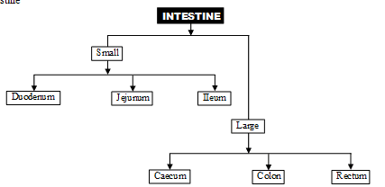
It is divided into two part
(i) Small Intestine: Small intestine is differentiated in to three part
(i) Duodenum (ii) Jejunum (iii) Ileum
First part is duodenum, it is 25 cm long, c-shaped in humans and has opening of hepatopancreatic duct (bile duct + pancreatic duct)
A small swelling is present at the opening of hepatopancreatic duct and is called 'Ampulla of Vater' or hepatopancreatic ampulla and the opening is regulated by sphincter of oddi.
Next parts of small intestine are jejunum and ileum. The wall of intestine has thin layers of longitudinal and circular muscles. Mucosa has folds plicae circular e (folds of Kerckring or Valvulae conniventes) and villi towards lumen of the intestine. Epithelial cells lining the villi have microvilli which further increase the absorptive area. Intestinal glands or Crypts of Lieberkuhn have epithelial cells (secrete mucus), Paneth cells (secrete digestive enzymes) and argentaffin cells (probably secrete hormones). In duodenum Brunner's glands are also present (located in submucosa) which secrete mucus. Diffused patches of lymphoid tissue are present through out the small intestine and are aggregated in ileum to form Peyer's patches.
(ii) Large intestine: It is 1.5 m long and consists of three part caecum, colon and rectum. A blind pouch of caecum is a vermiform appendix. These parts help in digestion of cellulose in herbivores. Wall of colon has sac like haustra. Histologically wall of colon has three bands of longitudinal muscles called taeniae coli. Another characteristics of colon surface is the presence of small fat filled projections called epiploic appendages.
The colon part is divisible into ascending, transverse, descending and sigmoid colon. Sigmoid colon is also called as pelvic colon .Ascending colon is the smallest and is without mesentery. Last part of rectum is anal canal having a strong sphincter. It opens outside by anus. In certain conditions (like persistent constipations) rectal veins can get distented or enlarged due to weakening of valves of it (varicosity). It leads to swollen areas called haemorrhoids.
HISTOLOGY OF ALIMENTARY CANAL
Wall of alimentary canal is made up of four layer (outer to inner)
(i) Serosa: It is the outermost layer of the alimentary canal , it is called tunica adventitia in oesophagus, which is made up of fibrous connective tissue. Except oesophagus, remaining part of alimentary canal in covered by serosa layer which is made up of visceral peritoneum while, tunica adventitia is made up of white fibrous connective tissue.
(ii) Muscularis Externa of mucularis coat: It is made up of two types of muscles. Outer muscle layer is made up of longitudinal muscle while inner layer is made up of circular muscle. Extra oblique muscles are found in stomach. Thickest muscular coat is found in stomach so maximum peristalsis are found in stomach least muscles are found in rectum so least peristalsis are found in rectum.
(iii) Sub mucosa: It is made up of loose connective tissue layer with blood lymph vessels and nerves.
(iv) Mucosa: It is the innermost layer of gut which contains the secretory and absorptive cells.
It is differentiated into 3 parts.
1. Outer part Called mucosa muscularis or muscularis interna
- It is made up to longitudinal and circular muscles.
- But these muscles are vestigial.
- They provide support to the folds of the alimentary canal.
2. Middle part
- It is made up of reticulate and fibrous connective tissue, dense network of blood capillaries are found in this part.
3. Innermost part
- In oesophagus this layer is made up of non keratinised stratified squamous epithelium.
- Except oesophagus this layer is single layer thick.
- This layer makes the lining of lumen of Alimentary canal.
- This layer is made up of columnar mucous epithelium.
- Folds of oesophagus are less developed
- This layer makes the folds of alimentary canal
- Folds of stomach are finger shaped.
- Folds of the small intestine are conical shaped called Villi.
- Small slit like space is found at the base of villi.
- These spaces are called crypts of Lieberkuhn
- Villi of Duodenum are small blunt.
- Villi of jejunum and Ileum are long and pointed.
- Maximum villi are found in Jejunum.
4. Brunner's Gland
- They are small spherical multicellular glands.
- They open into crypts of lieberkuhn with the help of fine tubules.
- These glands are found in the submucosa and mucosa of duodenum.
- They synthesize and secrete the non enzymatic secretion of intestinal juice.
5. Paneth Cells
- These cells are found in the mucosal layer of crypts of the lieberkuhn of jejunum.
- They are unicellular glands.
- They synthesize and secrets enzymes of intestinal juices.
- The secretory substances of brunner's glands and paneth cells are together known as intestinal juice or succus entericus.
6. Peyer's patches
- They are small lymph nodes which are found in the mucosa of the small intestine (Jejunum and Ileum more in number). They are also called intestinal tonsils and provide immunity.
7. Nerve supply
Two types of Nerve plexus are found in muscle of the alimentary canal. (These control muscle contraction).
(i) Auerbach's Nerve Plexus: This nerve plexus is found between longitudinal muscles and circular muscles.
(ii) Meissner's Nerve plexus: Found between circular muscles and sub mucosa but in stomach it is found between oblique muscle & submucosa.
LIVER
The human liver is made up of four lobe. Left lobe is small, the right proper lobe is large, two additional lobe quadrates and the caudate lobe are also found on the posterior side of the right proper lobe.
It develops from endoderm. (Weight 1.5 kg., both exocrine and endocrine
In humans it is found on the right side of the abdominal cavity, below the diaphragm.
The liver is the largest gland of the body.
Right and left liver lobe are separated from each other by the falciform ligament, (Fibrous connective tissue) Which is made up of a fold of peritoneum.
Right and left hepatic duct develop from the right and left liver lobe. Both these ducts combine to form a Common Hepatic duct.
Gall bladder is situated below the right lobe of the liver.
Cystic duct of gallbladder is connected to the common hepatic duct and forms a common bile duct which is also called ductus choledocus or common bite duct.
The liver is made up of numerous polygonal lobules. These lobules are covered by fibrous connective tissue, the covering layer is called Glisson's Capsule.
Each lobule consists of radial rows of hepatic cells, two rows of hepatic cells are combinedly called as hepatic cord. Each hepatic cord is lined by an endothelial layer.
In between the hepatic cord, a space is present called a hepatic sinusoid. These sinusoids are filled with blood. Sinusoids are lined by the endothelial cells mostly but a few macrophages cells are also present. These are called kupffer's cells. (Phagocyte cells)
The bile canaliculi run in between the two layers of hepatic cells in each hepatic cord. Hepatocytes (hepatic cells) pour bile into the canaliculi. Canaliculi opens into a branch of hepatic duct which is situated at the angular part of lobule in the Glisson's capsule.
All branches of the hepatic duct of right and left lobe are combined to form right and left Hepatic ducts which come out from the liver and form a common hepatic duct.
Hepatic artery and hepatic portal vein enter into liver and divide to form many branches. These branches are also found at the angular part of Glisson's capsule. Its fine branches open in to hepatic sinusoids. Branch of hepatic portal vein, branch of hepatic artery and branch of hepatic duct are collectively called as Portal triad. All hepatic sinusoids of one Glisson's capsule are open into central vein or intra lobular vein, all Central veins are combined and form one pair hepatic vein which, comes out from liver and opens into inferior vena cave.
Function of Liver
Most of the biochemical functions of the body are done by the liver.
1. Secretion & synthesis of bile: This is the main function of liver. Bile is yellowish-green, alkaline fluid. In bile juice, bile salts, sodium bicarbonate, glycocholate, taurocholate, bile pigments, cholesterol, Lecithin etc. are present. Bile salts help in emulsification of fats. Bile prevents the food from putrefaction. It kills the harmful bacteria.
2. Carbohydrate Metabolism: The main centre of carbohydrate metabolism is liver. Following steps are related with carbohydrate metabolism.
- Glycogenesis: The conversion and storage of extra amount of glucose into glycogen from the digested food is called glycogenesis. The main stored food in the liver is glycogen
- Glycogenolysis: The conversion of glycogen into glucose back when glucose level in blood falls down is called glycogenolysis.
- Gluconeogenesis: At the time of need, liver converts non-carbohydrate compounds (e.g. Amino acids. Fatty acids) into glucose. This conversion is called gluconeogenesis. This is the neo-formative process of glucose.
- Glyconeogenesis: Synthesis of glycogen from lactic acid (which comes from muscles) is called glyconeogenesis
3. Storage of fats: Liver stores fats in a small amount. Hepatic cell play an important role in fat metabolism. The storage of fats is increases in the liver of alcohol addict persons (Fatty liver). this storage of fats decreases the activity of liver. the damage of liver due to alcohol intake is called Alcoholic Liver cirrhosis.
4. Deamination and Urea formation: Deamination of amino acids is mainly done by liver (Amino acid ® NH3 separation of ammonia from the amino acids is done by the liver) Liver converts ammonia (obtained form deamination) into urea through orinithine cycle. So after the spoilage of liver, the ammonia level in the animal body is increased and the animal dies.
5. Purification of blood: The spleen and liver separate dead blood cells and bacteria from the blood. Kupffer cells in liver and phagocytes in spleen perform this function.
6. Synthesis of plasma proteins: Many types of proteins are present in blood plasma. Except gamma globulins all type of plasma proteins are synthesized in the liver.
7. Most of the blood clotting factors are synthesized in the liver.
8. Synthesis of heparin: Heparin is an anticoagulant (Mucopolysaccharide).
9. Some heparin is also formed by basophils, that are special type of white blood cells
10. Synthesis of Vitamin A: The liver converts the b-carotene into vitamin A : b- carotene is a photosynthetic pigment which is obtained from plants. It is abundantly found in carrot.
11. Liver stores vitamin A, D, E, K and B12
12. Storage of minerals: Liver stores iron in the form of ferritin. Liver also stores the, copper, zinc, cobalt, molybdenum etc Liver is a good source of iron.
13. Detoxification: In this process liver converts the toxic substances into non-toxic substances. The toxic substances are formed by metabolic activities of the body. e.g. Prussic acid is converted into neutral Potassium sulphocyanide (It is a non-toxic salt) by the liver.
14. Heamopoiesis: The formation of blood cells is called haemopoesis. In empbryonic stage R.B.C. and WBC are formed by liver.
15. Yolk synthesis – Most of the yolk is synthesized in liver.
16. Secretion of enzymes – Some enzymes are secreted by liver, participate in metabolism of proteins, fats and carbohydrates e.g. Dehydrogenase, cytochrome oxidase etc.
17. Prothrombin and fibrinogen proteins are also formed in hepatic cells. These help in blood clotting
18. Factors I, II, V, VII, IX and X are formed in liver, which are responsible for blood clotting.
PANCREAS (SWEET BREAD)
- Develop from endoderm.
- It is soft, lobulated and elongated organ..
- It is made up or numerous acini. Acini is a group of secretory cells surrounding a cavity.
- Each acini is lined by pyramidal shaped cells. These acinar cells secrete the enzyme of pancreatic juice.
- Each acini opens into pancreatic ductule. Many penacreatic ductule combine to from main pancreatic duct (duct of wirsung). The main Pancreatic duct is join with the bile duct to form the hepatopancreatic ampulla which opens into duodenum. The accessory Pancreatic duct (duct of santorini) opens into duodenum with separate openings located above the opening of main Pancreatic duct.
- Some group of endocrine cells also found in between group of acini called islets of Langer han's. These islets secrete insulin & glucagons hormone. So this gland is exocrine as well as endocrine. Its 99% part is exocrine while 1% part is endocrine (Heterocrine)
- In rabbit, bile duct and pancreatic duct both are separately open into Duodenum.
- Bile duct opens into proximal limb of duodenum and is controlled by sphincter choledocus.
- Pancreatic duct opens into distal limb of duodenum and is controlled by sphincter pancreaticus.
- In humans both bile duct and main pancreatic duct combine to form common duct called as Hepto-Pancreatic duct. The terminal end of common duct is swollen and is called as Ampulla of Vater or Hepato Pancreatic ampulla. Ampulla of Vater opens into middle part of Duodenum and is controlled by sphincter of Oddi while, bile duct is controlled by sphincter of Boyden
PHYSIOLOGY OF DIGESTION
1. Digestion in Oral Cavity: Food enters through mouth food is tasted in oral cavity and mixed with saliva, tongue mixes the food with saliva. This food with saliva is called bolus. This saliva (pH 6.8 – 7.0) contains water (99.5 %) and electrolytes (Na+, K+, Cl–, HCO3– , Thiocynate )
Chemical digestion
- In this type of digestion saliva act with food particles.
- Saliva contain 99.5 % water & 0.5 % salts.
- These salts are organic and inorganic type
- The main contents are Mucin, Lysozyme, Thiocynate and Ptyalin
(i) Mucin: It is a glycoprotein. It lubricates the food particles. It helps in the swallowing of food.
(ii) Lysozyme: It is an enzyme which kills the harmful bacteria. Due to this reason saliva is a antiseptic lotion.
(iii) Thiocynate: It is a special salt which kills the harmful bacteria. So it is called bacterioscidal salt.
(iv) Ptyalin: Ptyalin is found in human saliva, because human food is mainly made up of starch. Ptyalin digest only ripe and cooked starch. It does not digest the raw starch.
Note
Ptyalin is absent in saliva of rabbit and carnivorous animal, because food of rabbit is mainly made up of cellulose.
Bolus is pushed inward through the pharynx into the oesophagus. The tongue blocks the mouth. Soft palate close off the nasopharynx and larynx rises so that epiglottis bend and closes off the glottis food move downward into the oesophagus A traveling wave of contractions are called peristalsis pushes the Bolus (food) downward. Peristalsis is produced by involuntary contactions of circular muscles, which is preceded by a simultaneous contraction of the longitudinal muscle and relaxation of the circular muscle lining the gut. When a peristaltic wave reaches the end of the oesophagus. (Digestion or digestive enzymes are absent in Oesophagus) The sphincter opens allowing the passage of bolus food to the stomach. Gastroesophageal sphincter of the oesophagus and stomach normaly remains closed and does not allow contents of the stomach to move back.
- Secretion of saliva is mainly controlled by nervous type. Sympathetic nerve decreases the secretion of saliva while secretion of saliva increases by parasympathetic nerve.
- Secretion of saliva also controlled by reflex action e.g. smell of food, sight reflex etc.
2. Digestion of Food In Stomach
When the food enters into stomach G-cells secrete gastrin hormones which stimulate the secretion of gastric juice by gastric glands. Secretion of gastric juice is controlled by nerve, hormones and chemical substances.
Secretion of gastric juice is divided into 3 phases–
(i) Cephalic Phase: This phase is mediated by parasympathetic. It is the first of step of secretion. When person see the food then due to sight or optic reflex small amount of gastric juice secretes in the stomach.
(ii) Gastric phase: When food enter into stomach then gastric phase is started. When food particles strikes to the fundic part of stomach then small amount of gastric juice is secreted due to strike reflex action and distension. Gastric juice developes the peristalsis movement in the stomach. Due to peristalsis food particles are rubbed on mucosal layer of stomach. Due to rubbing process cells stimulates and secretes gastrin hormone. This hormone powerfully stimulate the gastric glands for secretion of gastric juice.
Some drinking substances also stimulates the secretion of gastric juice such a soup, alcohol, caffeine, histamine. These drinking substance and gastric juice stimulate the desire of appetite. So these substances are called Appetiser juice.
(iii) intestinal phase: When food reaches at the Ileum then mucosal layer of ileum secretes a chemical substance. Its nature is similar to the histamine or gastrin. This chemical substance goes into stomach through blood circulation where it stimulates the secretion of gastric juice. Its actual cause is yet unknown. But it is believed that this phase starts after 8–10 hour of taking of meal.
COMPOSITION OF GASTRIC JUICE
- Water = 99.5 %
- HCl = 0.2 – 0.3 %
- pH = 1.5 to 2.5 (very acidic)
- rest part = mucous water, HCl and gastric enzymes (Pepsinogen, Prorennin, Gastric Lipase Gastic amylase etc.)
Functions of HCl
- The main function of HCl is to convert inactive enzymes (zymogens) into active enzymes.
- Pepsinogen and Prorennin are inactive enzymes.
- It destroys harmfull the bacteria present in the food.
- HCl stops the action of saliva on food. In stomach, the medium is highly acidic.
- It dissolves the hard portions of the food and makes it soft.
- It releases the fat globules from tissue or cells which found in food
- HCl of gastric juice converts Fe+3 into Fe+2 which makes the absorption of iron possible.
DIGESTION BY RENNIN (CHYMOSIN)
Renning is active in the childhood stage of mammals only. It converts milk into curd like substance (clot the milk). Rennin, acts on milk protein casein. Casein is a soluble protein.
In the presence of Rennin, Casein gets converted into insoluble Calcium-paracaseinate, This process is termed as Curdling of milk. After becoming insoluble, milk can remain in the stomach for a loger time. Rennin is absent in human (clotting of milk is done by HCl in human).
Digestion by Pepsin
Inactive pepsinogen on getting proper pH conversts into active pepsin.
Peptidase – An enzyme which breakes the peptide bond. These peptidase are of two types.
(a) Exopeptidase: The peptidase enzyme which breaks the outer and marginal bond of polypeptide called exopeptidase. In this process amino acid and polypeptides are formed.
(b) Endopeptidase: The peptidase enzyme which breaks the inner peptide bond of large polypeptide and forms the small polypeptides such as peptone, proteoses and peptides.
- Pepsin is a strongest Endopeptidase. It breaks proteins into smaller molecules.
- In stomach, endopeptidases are found so only digestion of proteins can take properly in the stomach.
- Digestion by Gastric Lipase – It converts fats into fatty-acids and glycerol. It is secreted in a less amount
so less digestion of fats takes place here. - This lipase acts on emulsified fat and convert it into fatty acid & glycerol. 1% emulsified fat is already present in the food. Peristalsis. Continues during the process of digestion so the gastric-juice mixes properly with the food.
- Due to peristalsis the food is converted into a paste. This form which is thick. Acidic & semi digested in the stomach is called chyme.
- After short intervals, the pyloric valves on opening and closing so the chyme is enters the intestine in installments
DIGESTION OF FOOD IN DUODENUM
When food leaves the stomach through its pyloric end enters the duodenum it is called chyme (Acidic). The HCl of chyme stimulates the wall of duodenum to secrete hormones. It secretes various hormones–
- Hepatocrinin: It promotes the synthesis and secretion of Bile juice from liver.
- Cholecystokinin: It stimulates the liver and the gall bladder (mainly gall bladder) to secrete bile juice.
- Secretin: It is the most important hormone of digestive tract and also first discovered hormone. This hormone stimulates pancreas for synthesis and secretion of non enzymatic part of pancreatic juice. It also stimulates liver for secretion of bile juice and inhibit the gastric juice secretion in stomach and reduce rate of contraction of stomach.
- Pancreozymin: It stimulates the synthesis as well as secretion of enzymatic part of pancreatic juices.
- Secretin promotes the secretion of the non enzymetic part of the pancreatic-juice. While pancreozymin promotes the secretion of enzymatic part of the pancreatic juice.
- Duocrinin: It stimulates the Brunner's gland for synthesis and secretion of non-enzymatic part of intestinal juice.
- Enterocrinin: This hormone stimulated Paneth cells for synthesis and secretion of enzymatic part of intestinal juice.
- Villikinin: It stimulates the activity of villi.
- Enterogasterone: It inhibits the secretion of HCl in stomach.
- Gastric inhibitory polypeptide (GIP): It inhibits the secretion of gastrin hormone.
- Vasoactive intestinal peptide and somatostatin: They inhibits the motility of stomach
BILE-JUICE
In the proximal part of the duodenum bile-juice is secreted. The parenchyma cells of the liver produce bile-juice and it is stored in the Gall-bladder. Bile-juice does not contain any digestive enzyme. Therefore it is not a true digestive juice (Pseudodigestive-juice).
Composition of Bile-juice – Bile juice is a greenish (Biliverdin) yellow (Bilirubin) coloured alkaline fluid. Bile juice pH, 8.0 H2O 98 % Organic constituent are bile acid, bile pigment, cholesterol, Lecithin, inorganic constituents Na+, K+, HCO3– etc Bile-pigments are the excretory-substances of the liver.
Bile-salts are of two types:
(i) Inorganic salts – Bile-juice contains NaCl, Na2CO3, NaHCO3 etc in it. Inorganic salts neutralize the acidity of the food and make the medium basic. It is necessary for the medium to become basic because the pancreatic-juice enzymes can act only in basic medium.
(ii) Organic salts – Organic salt like Na-glycocholate and Na-taurocolate are found in Bile juice. The main function of these salts is the emulsification of fats. Because pancreatic Lipase can act only on emulsified fats.
Bile salts also help in the absorption of fat and fat-soluble vitamin (A, D, E, K) Bile salts combine with fats and these vitamins to form compounds called Micelles which are absorbed rapidly. Bile-sallts promote-peristalsis in the small-intestine. Bile-pigments, cholesterol and Lecithin are the excretory substances found in Bile-juice.
Gall Stone: Sometimes the passage inside the bile-duct gets blocked or becomes narrow, so the cholesterol gets deposited or precipitated in the gall-bladder. This is termed as the Gall-stone (Cholelithiasis)
Obstructive Jaundice: If the passage of bile is blocked then the amount of bilirubin increases in the blood. So the yellowish colouration of body like skin, cornea and nails appear yellow. Urine also becomes yellow.
PANCREATIC JUICE
Pancreozymin stimulates the acini and glandular cells so pancreatic juice are secreted. The pancreatic-juice is secreted by the exocrine cells of the pancreas. Pancreatic juice is highly odouriferous, colourless basic fluid which contains enzymes and salts.
Compostition of Pancreatic juice:
- Total amount in man = 500 – 800 ml/day
- Water = 98%,
- pH = 7.5– 8.3,
- Salts =2 %
- Pancreatic juice contains only inorganic-salts
The action of enzymes present in the pancreatic juice is as follows:
1. Pancreatic a-Amylase: Amylase or Amylopsin dissociates starch into Maltose. Majority of starch breaks up into the duodenum.
2. Trypsinogen and Chymotrypsinogen: The step of these enzymes is as follows–
- Tripsinogen Trypsin
- Chymotrypsinogen Chymotrypsin
- Enterokinase is secreted by the Duodenal mucosa.
Trypsin and chymotrypsin are Endopeptidase type of enzymes. They dissociate protein into peptones and proteoses. Majority of proteins are broken into the stomach and the remaining are broken into the duodenum.
3. Procarboxy Peptidase: These are also called zymogens. Trypsin convert it into active Carboxy-peptidase.
4. Large Peptides: Oligopeptide
5. Elastin: Oligopeptides
6. Fat Digesting enzyme: In pancreatic-juices various Fat-digesting enzymes are found which are collectively called steapsin.
- Pancreatic Lipase: It converts triglyceride into monoglyceride, fatty acid, glycerol
- Cholesterol esterase: It digest cholesterol esters. These esters are made up of cholesterol and fatty- acid Like- Lanolin, (cholesterol and Palmitic acid).
- Phospholipase: These digest phospholipids.
7. DNase and RNase – For digestion of DNA and RNA.
DIGESTION IN JEJUNUM AND ILEUM
These hormones stimulate the crypts of lieberkuhn to secrete Succus-entericus or intestinal juice. This succusentericus mainly contain water (99%) and digestive enzymes (< 1%). Intestinal juice act on food (chyle).
Succus- entericus mainly contains the following enzymes-
- Peptidase or Erepsin - This is a type of Exopeptidase. It converts oligopeptides into amino-acids Disaccharidases
- Sucrase – It is also known as invertase. It converts sucrose into glucose and fructose.
- Maltase – It convers maltose sugar into Glucose molecules. maltose Glucose + Glucose
- Lactase – This enzymes is found only in mammals. It converts milk sugar lactose into Glucose and Galactose.
- Lactose Glucose + Galactose
- Intestinal Lipase– This fat-digesting enzyme converts fats into Fatty-acids and Glycerol.
- Nucleotidase and Nucleosidase –These act in the following way–
- maximum digestion of carbohydrates is done in duodenum, but its digestion is completed in Jejunum.
DIGESTION IN CAECUM
In herbivores, the symbiotic bacteria and protozoans present in the caecum help in digestion of cellulose into glucose. So the digestion of cellulose takes place in caecum by the process of decompostion. This decomposition process is very slow. So very less amount of cellulose is digested at a time in caecum. In the last part of the large intestine faeces is temporarily stored.
Millions of microscopic folds or finger like projection are present in the lumen of gut which are called villi. These villi are supplied with a network of blood capillaries and Lymphatic vessels. Largest of which is central Lacteal. The cells that line the surface of villi numerous microscopic bristle like projections are called microvilli or brush border. These further increase the surface area for the absorption of the nutrients/digested food. On the surface of the mucous epithelium are billions of single cell mucous glands called mucous or goblet cells. These cells mainly secrete mucus that acts as a lubricant and protects the epithelial surface from damage and digestion.
Epithelial cells of small intestine showing absorption of nutrients.
(A) Absorption of a aminoacid.
(B) Absorption of monosaccharides.
(C) Absorption of fatty aicds.
Role of Some Major Gastrointestinal Peptide Hormones in Digestion
Intestinal wall
(Duodenum)
Food and strong acid in stomach and intestine
Pancreas, secretory cells and muscles of stomach, secretion of water and bicarbonate (NaHCO3), inhibition of gastric motility
Gastric Inhibitory Peptide (GIP)
Upper small intestine (Duodenum)
Monosaccharides and fats (fatty chyme) in duodenum
Gastric mucosa and muscles, inhibition of gastric secretion and mobility/motility (slowing food passage)
An Overview of the Action of Major Enzymes in Human
Salivary Juice (Salivary Gland)
Salivary amylase of Ptyalin
Mouth and stomach Buccal cavity
Starch
Disaccharides (few)
Gastric Juice (Stomach)
Pepsinogen : pepsin
Stomach
Proteins
Large peptides
The lining cells of the villi are columnar epithelial cells called enterocytes. On the surface of entrocytes, numerous microvilli are found, they increase the surface area of mucous membrane.
Absorption of Digested Food
The process through which the food stuff diffuses through the intestinal mucous membrane and reaches the blood, is termed as absorption. The process of absorption. The process of absorption in different parts of the alimentary takes place in the following manner.
- Absorption in Buccal-Cavity: No absorption of food takes place in the oropharynx and the oesophagus. Only some chemicals/medicines and alcohol are absorbed in oro-pharyngeal cavity.
- Absorption in stomach: In the stomach, absorption of water, some salts, alcohol and glucose takes place, complete absorption of alcohol takes place in the stomach.
- Absorption in duodenum: Iron & calcium ion are absorbed in the duodenum.
- Absorption in Jejunum: Maximum absorption take place in jejunum.
CARBOHYDRATE
The principal carbohydrate of our food is usually starch (from rice or wheat) which is broken down by the pancreatic amylase. Disaccharides are broken down to their monsaccharide by enzymes of the succus-entricus. Monosaccharides are absorbed via the capillary blood with in the villus to finally reach into portal vein. Absorption of glucose molecules occurs along with Na+ by active symport. Fructose is absorbed passively.
(i) Digestion and absorption of amino acid: All these proteins are exposed to pepsin, trypsin, chymotrypsin, carboxypeptidases etc and as a result they are converted into tri and dipetides or free amino acids. Amino acid are of two types L-amino acid & D-amino acid. The L-amino acids are absorbed by active process against the concentration gradient while D-amino acid are absorbed passively by diffusion. Di–and tripeptide enter the enterocytes where they are hydrolyzed to amino acids by dipeptidases and then absorbed via portal blood.
(ii) Digestion and absorption of Fat: One molecule of triglyceride is hydrolyzed into one molecule of monoglyceride and two molecule of fatty acids by pancreatic lipase. After hydrolysis, the bile salt, monoglyceride and the fatty acid together produce a complex called a mixed micelle. These are water soluble & enter in the enterocytes. Monoglyceride and fatty acid are resynthesized with in enterocyte to form a molecule of trigylyceride (TG).TG combines with a small amount of protein Phosphate and cholesterol and resultant complex is called chylomicron (150mm, white) Chylomicron enters the lacteal Fat soluble vitamins are absorbed along with dietary fat whereas water soluble vitamins are absorbed by passive diffusion. Vit. B12 is absorbed with intrinsic factor by forming a complex. In ileum Vit. B12 & Bile salt are absorbed. In colon only metabolic water is absorbed.
All lymph-capillaries coming out of the alimentary canal and unite to form Lymph-vessels. All lymph-vessels coming from the alimentary canal open into the Left Thoracic Lymph Duct. This duct now opens into the Left Subclavian vein. Through the blood, fats reaches the heart and from here it is distributed throughout the body.
Besides fats, other substances of the digested food like-sugars, amino-acids, vitamins, minerals-salts after being absorbed, enter the blood capillaries. All blood-capillaries coming out of the alimentary canal, join together to form the Hepatic portal vein. This vein takes the digested food material into the liver. From the liver, the Hepatic vein and the inferior or post – caval vein takes them to the heart. Heart distributes them throughout the body. Liver performs some necessary and important actions on the digestive food.
Maximum water absorption occurs from upper part of small intestine passively.
ABSORPTION IN COLON
Colon absorbs metabolic water form the undigested food. Due to Haustra the water-absorbing surface of colon increases and it efficiently increases absorption of water.
The excreta of rabbit is given out of the body in the form of small Pellets. The process of removal of undigested food from the body is termed as the Defaecation. The process of defaecation is involuntary in rabbit, thought it is voluntary in most animals. Symbiotic bacteria found in colon. Bacteria synthesis vitamin-K, B1 , B2 etc.
Undigested food goes into rectum where it gets converted into faeces contains – water and solid matter. Solid matter contains dead bacteria 30%, fat 10-12%, proteins 2-4% and others. These faeces ejected outside through anus.
- In the morning the excreta of rabbit is in the form of semi-solid pellets. It has more amount of undigested cellulose in it. Cellulose is a colloid substance, Colloid have the capacity to bind water on their surfaces, so complete absorption of water is not possible in intestine . To completely digest the cellulose rabbit again ingests the semi-solid excreta so again digestion of cellulose takes place in the caecum.
- In the evening the excreta of rabbit is in the form of solid, dry pellets. These have less amount of undigested cellulose in them. This nature of rabbit of eat is own excreta is termed as Coprophagy or Caecotrophy or also Pseudorumination. Double Circulation of food through the alimentary-canal is termed as Caecotrophy. Food of rabbit mainly consists of cellulose so this activity is necessary for rabbit.
- Brown colour of the excreta is due to 2 pigments-Sterobilin and Urobilin. Both of them are formed due to the degradation of Bilirubin. Foul smellof the excreta is due to Indole, Scatole and Typtophan. CH4 , NH3 , H2S. These are found in the colon due to the decomposition of undigested protein by bacteria. Pellets of rabbit don't have a foul smell because it has minimum amount of proteins in its diet. Carnivores have excess protein-rich diet so there is highly foul-smelling.
COMPOUND STOMACH
Gastric juice-secreted by Abomasum. So it is called true stomach. Inner surface of Rumen and Reticulum lined by keratinised epithelium. Symbiotic bacteria found in Rumen and Reticulum. Voluntary muscles are found in Rumen and Oesophagus. Hence reverse peristalsis are found in Rumen and oesophagus which is controlled by will power of animal. Omasum is absent in Camel and Deer.
(i) Calorific Value: The amount of heat liberated from complete combustion of 1 gm food in a bomb calorimeter (a closed metal chamber filled with O2) is its gross calorific value or gross energy value (G.C.V.). The actual amount of energy liberated in the human body due to combustion of 1 gm of food is the physiologic calorific value (P.C.V.) of food.
(ii) Assimilation: The use of absorbed digested food by the body is termed as assimilation. Amino-acids synthesise proteins, which in turn synthesis enzymes and new protoplasm. Glucose fatty-acids and glycerol on oxidation provide energy.
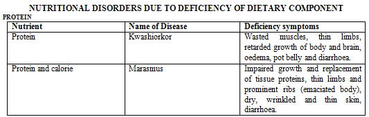
PROTEIN ENERGY MALNUTRITION
1. Kwashiorkor
- it is a protein deficiency disease.
- it commonly affects infants and children between 1 to 3 year of age.
- Symptoms: Underweight, stunted growth, poor brain development, loss of appetite, anaemia, oedema on lower leg and face.
- Cure: Proteins are necessary for growth, repair of tissue and for body defence therefore adequate amount of proteins must be present in the diet.
- Daily requirement: 1 gm protein per kg. body weight in adult. 2 gm protein per kg body weight in growing children.
- Sources in food – Cereals pulses, meat, fish, milk, groundnut, peas, leafy vegetables etc.
2. Marasmus
- It is caused by Protein-Energy-Malnutrition (PEM) or deficiency of protein and total food caloric value.
- Symptoms: Impairs physical growth, subcutaneous fat diseappears, ribs become very prominent, limbs become thin and skin becomes dry, thin & wrinkled. There is no oedema on leg and face but loss of weight occurs.
- Cure: Diet with adequate proteins and proper caloric value should be given to the infants.
- Source in food: Same as kwashiorkor.
3. Hypercholesterolemia: It is caused due to intake of excess of saturated fat such as butter, ghee, red meat, egg. Cholesterol level in blood rises abnormally (hypercholesterolemia) this may cause thrombosis and heart attack.
4. Obesity: It is caused by excessive intake of high caloric nutrients such as sugar, honey and saturated fat. Fat accumulates in the tissue. This may cause high blood pressure, diabetes and heart diseases. Regular exercise and taking of green leafy vegetable are recommended to such persons.
5. Hypervitaminosis: It is caused by excessive intake of vitamin. Such as excess of vitamin D causes deposition of calcium in soft tissue. Excess of vitamin A causes lack of appetite, itching rash etc. Hypervitaminosis of vitamin 'D'-Nausea, anorexia, hypocalcaemia, hyperphosphetemia, calcification of soft tissue.
6. Fluorosis: It is caused by excessive intake of fluorine. It is characterized by mottled (brownish discolouration) teeth.
7. Constipation: Because of show reaching of excreta into the large-intestine hard and dry excreta deposits in the colon.
8. Diarrhoea: Fast and rapid removal of excreta from the large-intestine is called Diarrhoea. It may be due to viral or bacterial infection in the intestine.
VITAMINS
- The study of vitamins is called as vitaminology.
- Vitamins were discovered by ''Lunin'' .
- The term ''Vitamin'' was given by ''Funk'' and ''Hopkins'' (B1 from unpolished rice – 1912)
- Vitamins are micronutrients, biological regulators and metabolic regulators (Vitamin theory)
- Vitamins are important to maintain health, but cannot synthesize in the body.
- Earliest known vitamin - vitamin 'C' (James Lind – Scottish naval surgeon – 1747.)
- Earliest extracted vitamin = Vitamin - B1
Vitamins are following types:
- Fat soluble
- Water soluble vitamin
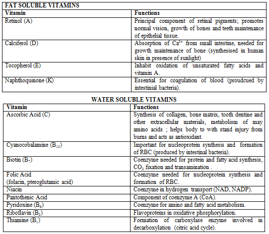
FAT – SOLUBLE VITAMIN
Vitamin- 'A' (Retinol)
- Can be synthesized in liver from yellow and red carotenoid pigment.
- It is also known as anti-infectious and anticancer vitamin.
- It is also known as anti-xeropthalmic vitamin.
Isomer of vitamin 'A' are:
- A1 – Retinol for vision.
- A2 – Dehydro retinol which is essential for epithelial lining of glands and tear production. It is essential for growth and epithelical cell division.
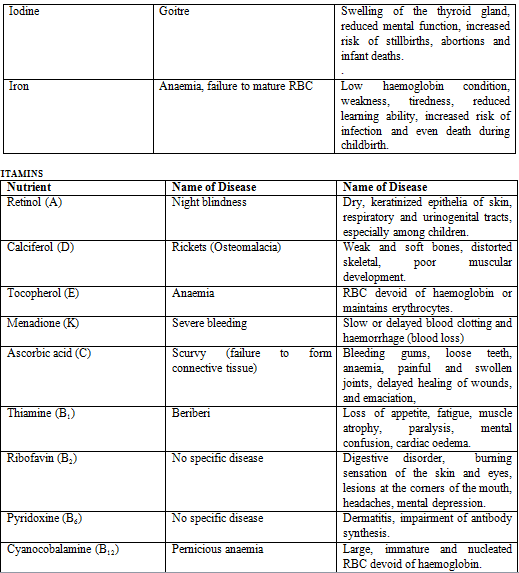
DEFICIENCY DISEASES
1. Night blindness
- Due to deficiency of A1
- It is also known as Nyctolopia (Inability to see in dark)
2. Xeropthalmia
- Due to deficiency of A2
- Tear formation is absent.
- In this disease conjunctiva & cornea become dry due to keratinization of conjunctiva & cornea .
- It is the main problem of blindness in children throughout the world.
- Source – Good source is carrot, other sources are Guava, papaya, mango, spinach etc.
3. Vitamin – ''D'' (Calciferol)
- It is also known as ''Sunshine vitamin'' or ''Anti Ricket'' vitamin.
- Synthesized in skin from cholesterol by UV light.
- It is necessary for bone & teeth.
- It regulates the absorption of calcium & phosphorous.
- It organize the calcium in bone and teeth.
WATER – SOLUBLE VITAMIN
(i) Vitamin B1 (Thiamine)
- It is also known as ''anti beri-beri'' factor or antineuritic factor.
- Beri-Beri affects peripheral nervous system, alimentary canal & cardiovascular system.
- It is essential for formation of coenzyme carboxylase in carbohydrate and amino acid metabolism.
- Defi disease: Beri-Beri, Wernicke's encephalopathy. Anorexia, constipation, weak-Heart and muscle atrophy.
- Source: Rice, wheat, egg and fish etc.
(ii) Vitamin B2 (Riboflavin)
- it is also known as vitamin ''G'' or lactoflavin or yellow enzyme.
- it is essential for formation of FMN & FAD.
- It maintain the oral epithelial lining.
- Defi. Disease – Cheliosis (Cracked lips at the corner of mouth) sore mouth and ulceration, digestive
disorder, Pellagra like, beri-beri like. - Source – Cow's milk, egg, liver, yeast etc.
(iii) Vitamin B3 (Niacin or Nicotinic acid)
- It is also known as ''antipellagra'' factor or vitamin PP (Pellagra preventing factor)
- It forms essential component of NAD & NADP.
- It maintains the epithelial lining of lumen of alimentary canal.
- Defi. Disease: Pellagra in human beings (diarrhoea, dermatitis, dementia) and black tongue (hyper pigmentation) disease in dogs.
- Source: Kidney, liver, milk, yeast, egg etc.
(iv) Vitamin B5 (Pantothenic acid)
- Greek work pantothen = everywhere
- It is also known as yeast factor or chick antidermatitis factor.
- It is help in formation of acetylcholine and co enzyme A. It regulated the secretion of steroid hormones.
- Defi. Disease: Burning feet syndrome, fatigue & paralysis of muscles.
- Source: Liver, meat, yeast, milk, egg, meat etc.
Vitamin B6 (Pyridoxine)
- Function as co-enzyme. It is also known rat antidermatitis factor.
- Defi disease – Dermatitis, Anaemia, nervousness.
- Source – Liver, meat, yeast, egg etc.
Vita B7 (Biotin)
- It is also known as vitamin 'H' or antiegg white injury factor (egg white contain avidin protein which is antagonist to vit. B7) – Dermatitis, hair loss, nervous symptom).
- It is essential for fat synthesis and energy production
- Defi. Disease – Dermatitis
- Source – Vegetables, yeast, wheat egg etc.
Vitamin B12 (Cyanocobalamine) – Extrinsic factor of castle
- It is also known as '' antipernicious anaemic'' factor or '' RBC maturing'' factor
- It promotes DNA synthesis & Maturation of RBCs.
- Defi. Disease – Pernicious anaemia.
- Source – meat, liver etc.
Folic Acid
- It is also known as folacin or Vitamin M.
- It is needed for formation of RBC & synthesis of DNA
- Deficiency disease: Anaemia.
- Source: green foliage of plant – cabbage, cauliflower.
Vitamin ' C' (Ascorbic Acid)
- It is also known as '' anti-scurvy'' or anti-viral, anti-cancer vitamin.
- It is necessary for healing of the wound and formation of collagen fibre
- Deficiency disease – Scurvy (deficient formation of collagen fibres).
- Source-Amla, Tomato, orange, Guava, Lemon (citrus fruit).
POINT TO REMEMBER
- Spoil hay of Sweet clover (melilotus indica) (Fodder and green manure) contains a substance called dicumarol. Dicumarol prevents the action of vitramin 'K'
- Non-secretion of HCl is called as achlorhydria condition.
- Chalogogues are substances which cause. The contraction of gall bladder
- Choloretic are substances which increase bile juice from liver.
- ''Achalasia Cardia'' condition is characterized by failure of cardiac sphincter to relax completely on swallowing causing accumulation of food in oesophagus and proximal oesophagus dialates.
- One pair of vomerine teeth is found in the palate of frog.
- Fangs are the poison teeth of snakes, these are the maxillary teeth.
- Upper incisor teeth are modified in tusk in elephant.
- Upper canine teeth are modified in tusk in walrus.
- Homodont type dentition are found in toothed whale.
- Enamel is absent in sloth and Armadillo.
- Salivary glands are absent in whale.
- The tongue is non-motile in whale.
- Gall bladder is absent in lemprey, whale, rat and horse.
- The main pancreatic duct is also known as duct of wirsung while accessory pancreatic duct is known as duct of santorini.
- Citrin is also known as vitamin 'P' and controls vascular permeability.
- Vitamin B17 – It is recently discovered anticancer vitamin.
- Vitamin Q – helps in blood clotting.
- Vita B15 – It is also known as pogonic acid, deficiency causes disorder in liver.
- Vitamin B6 also used in the treatment of tuberculosis.
- Thecodont teeth are also found in crocodile.
FAQs on Digestive System, Chapter Notes, Class 11,Biology
| 1. What is the function of the digestive system? |  |
| 2. What are the organs involved in the digestive system? | |
| 3. How does the digestive system work? | |
| 4. What are some common digestive system disorders? | |
| 5. How can you maintain a healthy digestive system? | |


Chapter Notes
,study material
,shortcuts and tricks
,Important questions
,Class 11
,Sample Paper
,ppt
,Viva Questions
,Digestive System
,Free
,Exam
,Extra Questions
,video lectures
,Biology
,practice quizzes
,Objective type Questions
,Class 11
,Class 11
,MCQs
,Digestive System
,past year papers
,Chapter Notes
,mock tests for examination
,Summary
,Biology
,Previous Year Questions with Solutions
,Digestive System
,Chapter Notes
,Semester Notes
,Biology
;


Digestive System, Chapter Notes, Class 11,Biology Free PDF Download
Importance of Digestive System, Chapter Notes, Class 11,Biology
Digestive System, Chapter Notes, Class 11,Biology
Digestive System, Chapter Notes, Class 11,Biology Class 11 Questions
Study Digestive System, Chapter Notes, Class 11,Biology on the App
|
© EduRev
|
Education Revolution
|


|


