Muscles, Chapter Notes, Class 11, Biology PDF Download

MUSCLES
INTRODUCTION
Study of muscles known as Myology.
Myology also known as Sarcology.
Development of muscles : -
Except muscles of Iris & cilliary body all muscles of body develop from mesoderm.
– Muscle of Iris, cilliary body & myoepithelial cell of sweat gland develop form Ectoderm. Conductivity & contractility are the two main characteristics of muscles.
Three types of muscles are found in the body.
- Voluntary or skeletal muscles.
(ii) Involuntary or smooth muscles.
(iii) Cardiac muscles.
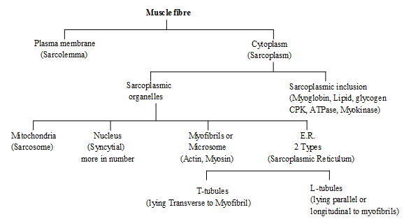
FORCE GENERATING PROTEIN
Actin –
The Actin or thin filament is a double helix made up of protein molecule called as. G-Actin.
(Globular actin) Many G-actin combined to form a filament like structure, which is called as filamentous-actin. G-actin contain a active site where myosin head is attached.
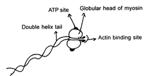
Myosin –
Each myosin molecule consist of a tail & a head. Tail is made up of two chains interwined with each other like double helix.
The myosin head has sites for attachments with (i) The actin filament (ii) ATP molecules.
Each myosin (thick) filament is also a polymerised protein. Many monomeric proteins called Meromyosins constitute one thick filament. Each meromyosin has two important parts, a globular head with a short arm and a tail, the former being called the heavy meromyosin (HMM) and the latter, the light meromyosin (LMM)> The HMM component, i.e.: the head and short arm projects outwards at regular filament and is known as cross arm. The globular head is an active ATPase enzyme and has binding sites for ATP active sites for actin.
REGULATING PROTEIN
Tropomyosin –
It is one type of contractile protein. In the relaxed state of the muscle situated in such a way, that the active sites remain covered by the Tropomyosin & attached at the terminal end of actin.
Troponin –
It is one type of protein which attached with one of ends of the tropomyosin molecules.
Troponin is made up of three subunit.
(a) Troponin I (b) Troponin T (c) Troponin C
(Inhibitory site) (Tropomyosin site) (Ca+2 binding site)
STRUCTURAL PROTEIN
Actinin –
It is one type of protein which found in Z-line.
PHYSIOLOGY OF MUSCLES CONTRACTION...
SLIDING FILAMENT THEORY
This theory is given by A.F. HUXLEY, H.E. HUXLEY & HANSEN
The junction of Nerve & muscle is called as neuromuscular junction.
Terminal branches of Axon of motor nerve is embedded into sarcolemma.
Its bulb like structure is called as motor end plate.
Sarcolemma invaginate inside & form a fimbriated structure which is called synaptic gutter or subneural cleft. The cell membrane of the bulbous terminals is called as the pre junctional membrane where as the cell membrane of muscle fibre which invaginates called post junctional membrane.
In motor end plate large number of vesicles & mitochondria are present. Each vesicle contains Ach in high
concentration. In post junctional membrane, Ach receptor are presented.
(Ach = Acetylcholine, it is a neurotransmitter chemical)
Neuromuscular Transmission –
– When motor nerve fibre stimulated it develops an Action potential (Resting potential 50 to 100 mV)
– AP reaches in the neuromuscular junction & goes to bulbous expansion of the nerve terminal.
– Than it increases permeability of Ca++ in the Pre junctional membrane & Ca++ enter from E.C.F. in to the cytosol of motor end plate by penetrating the prejunctional membrane.
– Ca++ ions causes bursting of the vesicles & releases the Ach.
– These Ach now cross the prejunctional membrane. via subneural cleft reach the post junctional membrane attach the Ach receptor also called as End plate receptor.
– End plate receptor stimulate & develop end plate potential by opening of Na+– K+ channels in post synaptic membrane.
– When end plate potential sufficiently higher than A.P. develop on sarcolemma & myofibril.
– Sarcolemma invaginates inside & form transvers & longuituidnal tubules which are also called as
T-tubul and L-tubule
– T-tubules are parallale = to Z-line whereas L-tubule is perpendicular to the Z-line.
– T-Tubules communicates with ECF.
– T & L system of tubules together called as endoplasmic reticulum.
– L-Tubules dilated on both side of T-Tubules this dilated part called cisterns.
– A.P. proceeds along the sarcolemma & A.P. contact with T-Tubules & further proceeds via T-tubules & enter with in muscle fibre & now this AP called as T-tubule potential.
– T-tubule potential come in close contact of L-tubules at region of the Triads (T + L-tubules).
– L-tubules in very rich source & store house of Ca++ ion in higher concentration release of Ca++ ion in large amount.
– Released Ca++ ion combine with troponin C.
– In Relaxed state tropomyosin covers the active site of actin.
– But troponin-C combines with Ca++ ion some physiochemical changes occur in tropomyosin & Tropomyosin move away from active site of actin.
– Myosin have strong tendency to bind the actin molecule & Actomyosin complex
– Myosin head attach on active site of actin with the help of cross bridges.
– Now the myosin head twists in the groove of the active site of actin-F. This causes movement of actin toward H-zone.
– Contraction is caused by overlapping of actin filament over myosin – sliding filament hypothesis.
– All the cross bridges move simultaneously in one direction so the actin filaments move vigorously towards H-zone.
– When cross bridge disrupted than myosin molecule detached & reattach the new active site of actin.
– After muscle contraction H-Zone disappears & length of sarcomere & I-band decreases by 20%. The length of A-band remains unchanged.
All process are reversible, at the time of relaxation Ca++ are goes into L-tubules.
Role of ATP –
(i) The Rotational movement of myosin head with in the groove.
(ii) Deattachment of myosin head form the actin.
Chemical reaction in Muscles :
1. ATP + H2O ADP + P1 + Energy (For contractile muscle)
2. Creatine phosphate + ADP Creatine + ATP (Muscle contraction )
3. Gycogen Lactic acid + Energy
4. 80% Lactic acid + Water Glycogen (Liver cell)
5. 20% Lactic acid + Oxygen CO2 + H2O + ATP (Liver cell)
6. Creatine + ATP Creatine phosphate + ADP (Resting Muscle)
PROPERTIES OF MUSCLES
Terminology
1. Origin – Fixed end of muscle (Proximal end)
Insertion – Distal end of muscle which is attach to bone (Movable end)
2. Excitability – Muscles responds to stimuli which can be nervous, chemical, electrical & thermal mechanical.
Conductivity – Stimulus acting in one region of muscle fibres propagated to all parts within no time.
Contractility – On being stimulated the muscle fibres contract & shorten followed by Relaxation.
3. Threshold stimulus – Intensity of stimulus below the threshold value does not produces contraction in muscle fibres is called.
Sub threshold Stimulus shambles stronger than threshold one is called suprathreshold stimulus.
4. All or none law – Response of muscle fibre is maximum whether the stimulus has threshold value or suprathreshold value. Response is absent when intensity is subthreshold. (Below threshold value)
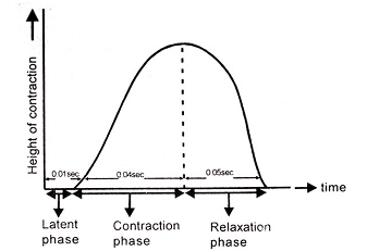
5. Muscle twitch – It is single isolated contraction of Muscle fibres due to single stimulus. Muscle curve or kymograph indicates three phases.
(a) Period of latent excitation (Latent period) Interval between the application of appropriate stimulus & initiation of contraction
It is 0.01 sec. in skeletal muscle. 3sec. in smooth muscle.
(b) Contraction phase – Duration for which muscle remain contracted state. It is 0.04 sec. in skeletal muscle. 20 sec. in smooth muscle.
(c) Relaxation phase – Interval for contracted muscle to regain its original/Relaxed state 0.05 sec. in skeletal muscle 23 sec. in smooth muscle.
6. Refractory Period – It is period between two twitched during this muscle does not respond to second stimulus after single twitch.
It is 0.002 – 0.005 second in skeletal muscles and 0.1 – 0.2 second in visceral muscles.
7. Summation of stimuli – Two subliminal stimuli Applied simultaneously get added up & Evoke the response
Muscle response = (1st stimulus +2nd stimulus ³ threshold value )
Subliminal subliminal
Howerver a muscles consist of large no. of muscle fibres with different threshold value so in intensity of stimulus increases contraction of muscle although individual fibres obey all/none law.
Summation of IInd stimulus during contration phase
8. Muscle tone – In relaxed muscles, a few fibres always undergoing contraction alternately so maintain the health of muscles. It is known as Muscles tone
9. Tetanic condition – It is sustained muscles contraction due to rapid series of impulse, during this relaxation of muscles does not take place.
10. Paralysis – Supply of motor nerve impulse completely cut off. So function of muscle contraction is stoped.
11. Shivering – Involuntary contraction of muscles to make body warm.
12. Muscle tension – force produced during contraction of muscle is known as muscle tension.
Isometric –
Contraction occur when a muscle is stimulated adequately but is prevented to shorten.
eg. applying too heavy load against the muscle so that the muscle but cannot lift the at all ext. work done is zero Isotonic –
When muscles is stimulated adequately & is allowed to shorten, then the contraction is called Isotonic some external work is done. Technically called a load is lifted
13. Antagonistic muscles–
They are pair of muscles which causes opposite movement at the same site when one muscle is contracting, the other is relaxes & viceversa.
e.g. – Biceps (flexor) & Triceps of arms (extensor)
14. Motor unit – Groups of mucles fibres supplied by single motor neuron. It is a functional unit of muscles because all the muscle fibres of motor unit contract & relax simultaneously.
15. Cori cyles – Lactic acid accumulated in muscles during sustained contraction. formed lactic acid transported in blood as blood lactate to liver where is changes into liver glycogen which is changed in to glucose.
Glycogen
16. Speed of – Skeletal muscle = 0.1 sec. per contraction per cycle
Cardiac muscle = 0.8 sec. per contraction per cycle
Smooth muscle = 46 sec. per contraction per cycle
17. 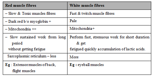
Eg : Extensor muscles of back,
flight muscles
Eg : eyeball muscles
18. Fatigue : Marathian athelets develops red fibers in thigh muscles due repeated contraction
Due to sustained contraction initially muscle give beneficial effects of contraction (warm ups) but after it ATP is exhausted & muscle is as state of permanent contraction & no relaxation because no fresh ATP Available for detachment of actomysosin complex.
– B/o 1. Accumulation of lactic acids
2. Consumption of stores glycogen. ATP, CTP (Creatinine phosphate)
Infatigue–
(i) Increase latent period and phase of relaxation
(ii) decrease height of contraction
19. Rigor Mortis
After death fresh supply of ATP. become impossible so one the local store of ATP molecule are exhausted. Due to non availability of ATP/C.P. deattachment of myosin from actin cannot take place resulting in permanent state of contraction of muscle. This phenomenon is called rigor mortis. This condition helps fixation of the hour of death.
20. E.D.T.A (Ethylane diamine tetra acetic acid) injected inside muscle combined with Ca+ and stops contraction
21. Muscle and nerve exitability is reduced by K+.
22. During muscle contraction chemical energy changed into mechanical energy.
23. Over stretching of tendon is called sprain.
MUSCLE TYPES ON BASIS OF MOVEMENTS
1. Flexor = Fore arm move in upward direction. (Bend)
Bending of part over one another Eg. biceps brachii
Extensor – Fore arm move in downward direction. Straighting of bending part
Eg. Triceps
2. Adductor - Toward body axis. Towards the body
Lattissimus dorsi brings the arms towards body
Abductor-upper & lower limb move away from body axis. Away from the body (midline) deltoids.
3. Pronator – Palm state in down. Rotate downward eg pronater teres
Supinator – Palm state in upward Rotate upward eg brachioradialis
4. Dilation – Diameter increases, widening of Iris (radial muscle of iris)
Constrictor – Diameter decreases, Closing an aperture–sphincter ani closes anus
5. Depressor – Lower Jaw move in downward direction. Lowering part depressor mandibularis
Elevator–Lower Jaw move in downward direction.Raising the part eg. Massetar.
6. Median Rotation – Upper & lower limb rotate in inward direction.
Lateral Rotation – Outward direction rotation
7. Inversion when sole of foot turn toward body axis.
Eversion - Away from body axis
Aryeiglotticus muscle is called Hilton muscle.
Gastrocenemius muscle present in shank.
Sartorius the longest muscle of body
Gluteus maximus (Buttock muscles) – Largest muscle of body
Stapedius – smallest muscle of body.
In Human beings 639 muscle are found 634 muscle are paired and 5 muscle are unpaired. 400 muscles are striated & most of muscles are found in back region & number of back muscles are 180. Jaw muscles are strongest. Longest smooth muscles is present in present in uterus of pregnant lady.
FAQs on Muscles, Chapter Notes, Class 11, Biology
| 1. What are muscles and what is their role in the human body? |  |
| 2. How many types of muscles are there in the human body? | |
| 3. How do muscles contract and relax? | |
| 4. What are the major functions of skeletal muscles? | |
| 5. How can muscles be kept healthy and strong? | |


Chapter Notes
,shortcuts and tricks
,Objective type Questions
,MCQs
,Biology
,Viva Questions
,Semester Notes
,Muscles
,Free
,ppt
,mock tests for examination
,Exam
,video lectures
,Muscles
,Sample Paper
,Previous Year Questions with Solutions
,Biology
,Class 11
,Chapter Notes
,Important questions
,Class 11
,Extra Questions
,study material
,Biology
,practice quizzes
,Class 11
,past year papers
,Summary
,Muscles
,Chapter Notes
;


Muscles, Chapter Notes, Class 11, Biology Free PDF Download
Importance of Muscles, Chapter Notes, Class 11, Biology
Muscles, Chapter Notes, Class 11, Biology
Muscles, Chapter Notes, Class 11, Biology Class 11 Questions
Study Muscles, Chapter Notes, Class 11, Biology on the App
|
© EduRev
|
Education Revolution
|


|


