TOOLS AND TECHNIQUE IN CYTOLOGY( Part - 1) - Notes, Zoology; Class 11 PDF Download

TOOLS AND TECHNIQUE IN CYTOLOGY
STAINING :
| Stain | Final colour | Suitable for |
1 | Schultzs's solution | rYellow→ | lignin, Cutin, Suberin & Starch |
2 | (Chlor-Zinciodine) | ^-Blue/Violet → | Cellulose |
3 | Sudan IV | Scarlet red | Suberin |
4 | Sudan black and Red | Black/Red | lipid |
5 | Haematoxylene | Red | Nuclear stain "I Ctell Division |
6 | Acetocarmine | Pink | Nuclear stain |
7 | Feulgen' s stain | Red/Purple | DNA |
8 | Osmium tetraoxide | Black | Eats, Stain for electron microscopy |
9 | Toludine blue | Blue | RNA |
10 | Saffranine | Red | lignin |
11 | PeriodicacidSchiff (PAS) | Red | Polysaccharides |
12 | Aniline blue (Cotton blue) | Blue | Fungal hyphae |
13 | Eosin | Pink, Red | Cytoplasm, Cellulose |
14 | Janus green | Greenish blue | Mitochondria |
15 | Methylene blue | Blue | Nucleolus |
16 | Phloroglucinol | Red | lignin |
17 | Aurantia | Yellow | Mitochondria |
18 | Brazlin | Red | DNA |
19 | Para Rosailin | Purple | DNA |
20 | Iodine solution | Blue, Red | Starch, Glycogen |
21 | Crystal Violet | Violet | Bacteria |
22 | Azure B | Red | RNA |
Vital Stains → Stains the cell organelles without killing the cell are called asvital stains. Normally cell becomes dead after staining but vital stains keep the cell alive.Janus Green,Neutral red, Methylene blue, malachite Green.
FEULGEN REACTION
Feulgen reaction was discovered byFeulgen andRosen beck (1924). Feulgen reaction specifically stains DNA . Feulgen reaction involves two steps –
(i) Acid hydrolysis of material :-It removes the purine at the level of thepurine-deoxyribose glycosidic bond of DNA thereby unmasking the aldehyde group of deoxyribose.
(ii) The free aldehyde group react withSchiff's reagent. The Schiff's reagent is prepared by treatingbasic fuschin withsulfurous acid.
MICROSCOPY
- Microscopy means the use of microscope for studying the cells.
MAGNIFICATION POWER :
- Maximum magnification power of light compound microscope is 2000.
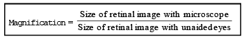
| Magnification = power of objective lens × power of eye piece lens |
In light compound microscope maximum power of objective lens is 100 X and maximum power of eye piece lens is 20 X. Thus the maximum magnification achieved is 100 × 20 = 2000
- Resolving power of microscope refers to the smallest distance between two objects which can be identified as separate images. Resolving power of human eye is 100 mm. l Resolving power of light microscope is 0.25mm to 0.30mm.
- Resolving power of microscope can be calculated with the help of the Abbe equation.
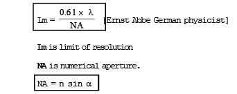
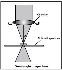
- n is referactive index of medium and sin a is the sine of the half angle of light entering the objective lens from the specimen.
- l is the wavelength of light used to illuminate the object.
- Limit of resolution is inversely proportional to resolving power. So when Lm becomes smaller, the resolution increases.
- Resolving power is inversely related to wavelength of light. Therefore, the highest resolution is obtained with the light of shortest wavelength i.e. (450 – 500 nm) light at theblue end of the visible spectrum.
- The angle of light that can enter a lens depends on the refractive index of the medium as well as upon the object itself.
- Refractive index of air is 1.00. Since sin µ can not be higher than 1 (µ = 90° and sin 90°=1), no lens working in air can have a numerical aperature more than 1. The semi angle of aperture for the best objective lens is 70° (sin 70° = 0.94), Therefore, the only way to increase the numerical aperture is to increase the refractive index by using an oil(cedar wood oil, olive oil) which has a refractive index identical to that of a glass.
Generally the value of NA for oil immersion lens is 1.4.
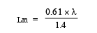
Therefore, resolution of microscope is roughly one half of the wavelength of illuminating light.
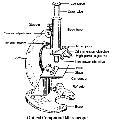
LIGHT COMPOUND MICROSCOPE
- Light compound microscope was first invented by Z. Janssen and H. Janssen (1590)
- Wilson (1710) improved the microsope of Janssen and Janssen and presented it in the form of present day Laboratory Microscope.
- Normally light compound microscope is bright field microscope. In a bright field microscope the image is formed when the light is transmitted through the specimen.
- The specimen being denser and more opaque than its surroundings, absorbs some of this light and the rest of the light is transmitted directly upto the eye piece. As a result, the image produced by the specimen will be darker than the surrounding brightly illuminated field.
Use :- The bright field microscope is a multipurpose instrument and can be used for live unstained materials as well as preserved and stained materials.
DARK FIELD MICROSCOPE OR ULTRA MICROSCOPE
- Dark field microscope developed by Zsigmondy (1905).
- In dark field microsocpe, the object is illuminated by oblique beam of light. The resulting image is illuminated brightly against the dark background.
- The most effective advantage of Dark field microscope is that, the nuclei, mitochondria and vacuoles are readily detected.
PHASE CONTRAST MICROSCOPE
- It was developed by Zernicke (1935)
- This microscope multiplies the small differences among the phases or refractive indices of different cellular constituents.
- It converts the differences of refractive index into differences in brightness with the help of a transparent phase plate situated at the back focal plane of objective lens.
- So different structures of cell appear in different brightness.
- Phase contrast microscope does not require staining of material so cell can be observed in living state.
- This microscope is useful to study functional aspect of cell like cell division.
INTERFERENCE MICROSCOPE
- It was developed by Merten et. al.
- It is one modification of phase contrast microscope. Here, parts of cells with different thickness and refractive index appear in contrasting colours so as to provide clear observation.
- In this microscope, the light rays are split into two beams before passing through the object by two prisms that create contrasting colours in a image. One beam passes through the object and undergoes a phase change. The second beam does not pass through the object and remains unchanged.
- The two beams are brought together above the object.
- This microscope can determine the presence of several light absorbing substances like nucleic acid, proteins, lipids etc.
- By using this microscope, it is possible to find out the dry weight of macromolecules like DNA, RNA and proteins.
FLUORESCENT MICROSCOPE
- It was developed by Coons (1945).
- It employs invisible ultraviolet rays instead of oridinary light for illumination. It is built on the principle that certain substances absorb short wave ultra-violet radiations falling on them, get excited and emit them back but in the form of longer wave length of visible light. This phenomenon is called Fluorescence.
- Some cellular components like chlorophylls, riboflavin, vitamin A, collagen fibers etc. have the property of autofluorensce. They are therefore, visible under the fluorescent microscope.
- Other cellular components (e-g. cellulose, starch, glycogen, lipids, proteins) can be made visible under the fluorescent microscope with the help of special dyes called fluorochromes (e.g. Acridine orange, coriphosphine). The phenomenon of fluorescence with the help of fluorochromes is called secondary fluorescence. l Fluorescent microscope is useful in detecting the microbes present inside the infected tissue.
Some Fluorochrome :
(i) Fluorescein – (emitting green light when excited with blue light)
(ii) Rhodamine – (emitting deep red light when excited with green yellow light),
(iii) quinine sulphate
(iv) auramine
FAQs on TOOLS AND TECHNIQUE IN CYTOLOGY( Part - 1) - Notes, Zoology; Class 11
| 1. What are some commonly used tools in cytopathology? |  |
| 2. What techniques are used in cytopathology to analyze cellular samples? | |
| 3. What is the role of cytology brushes in cytopathology? | |
| 4. How does fine needle aspiration (FNA) work in cytopathology? | |
| 5. What is a cell block kit used for in cytopathology? | |


Previous Year Questions with Solutions
,MCQs
,TOOLS AND TECHNIQUE IN CYTOLOGY( Part - 1) - Notes
,TOOLS AND TECHNIQUE IN CYTOLOGY( Part - 1) - Notes
,ppt
,study material
,Free
,Sample Paper
,Viva Questions
,Extra Questions
,shortcuts and tricks
,Important questions
,Summary
,Exam
,Zoology; Class 11
,Zoology; Class 11
,Zoology; Class 11
,past year papers
,Objective type Questions
,TOOLS AND TECHNIQUE IN CYTOLOGY( Part - 1) - Notes
,video lectures
,Semester Notes
,practice quizzes
,mock tests for examination
;


TOOLS AND TECHNIQUE IN CYTOLOGY( Part - 1) - Notes, Zoology; Class 11 Free PDF Download
Importance of TOOLS AND TECHNIQUE IN CYTOLOGY( Part - 1) - Notes, Zoology; Class 11
TOOLS AND TECHNIQUE IN CYTOLOGY( Part - 1) - Notes, Zoology; Class 11
TOOLS AND TECHNIQUE IN CYTOLOGY( Part - 1) - Notes, Zoology; Class 11 Class 11 Questions
Study TOOLS AND TECHNIQUE IN CYTOLOGY( Part - 1) - Notes, Zoology; Class 11 on the App
|
© EduRev
|
Education Revolution
|


|


