JAMB Exam > JAMB Notes > Biology for JAMB > Alimentary Canal: Stomach, Small Intestine & Large Intestine
Alimentary Canal: Stomach, Small Intestine & Large Intestine | Biology for JAMB PDF Download
| Table of contents |

|
| Stomach |

|
| Gastric Glands |

|
| Intestine |

|
| Histology of Alimentary Canal |

|
Stomach
- It is situated on the left side of the abdominal cavity. It is the widest part of the alimentary canal. It is a bag like muscular structure, J shaped in empty condition.
- The stomach is divided into three parts as Fundus, Body, Pylorus or Antrum.
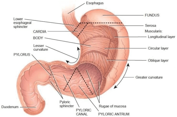 Anterior View of Regions of Stomach
It has two orifices (opening):
(i) Cardiac Orifice
- It is a proximal aperture of the stomach that is joined by the lower end of the oesophagus.
(ii) Pyloric Orifice
- It is a distal aperture of the stomach that opens into the duodenum.
- The stomach is covered by a layer of peritoneum, fat tissue and lymph tissue deposits on the peritoneum. Such a type of peritoneum is called Omentum.
- The left curved surface of the stomach is called the greater omentum. The right curved surface of the stomach is called the lesser omentum.
- The mucous membrane of the stomach is thick. In an empty stomach, numerous longitudinal folds are found called gastric rugae. They disappear when the stomach is distended.
Gastric Glands
These are numerous microscopic, tubular glands formed by the epithelium of the stomach.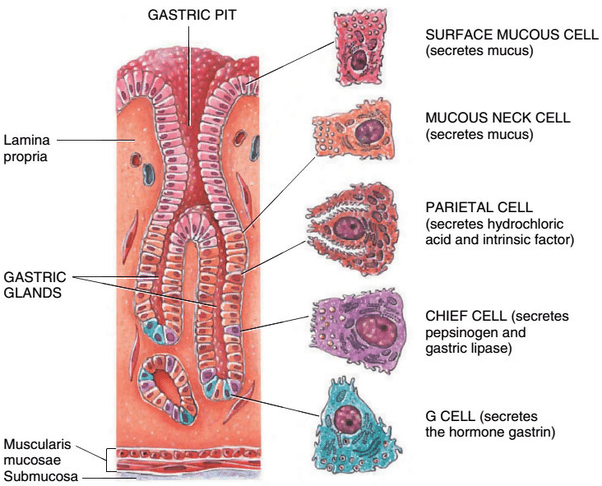 Sectional View of Gastric GlandsThe following types of cells are present in the epithelium of the gastric glands:
(i) Chief cells or Peptic cells (Zymogen cells)
- Chief cells are usually basal in location and secrete gastric digestive enzymes as proenzymes or zymogens, pepsinogen and prorenin.
- The chief cells also produce a small amount of gastric amylase and gastric lipase. Gastric amylase action is inhibited by the highly acid condition.
- Gastric lipase contributes little to the digestion of fat. Prorennin is secreted in young mammals. It is not secreted in adult mammals.
(ii) Oxyntic cells (Parietal cells)
- Oxyntic cells are large and are most numerous on the sidewalls of the gastric glands. They are called oxyntic cells because they stain strongly with eosin dye.
- They are called parietal cells as they lie against the basement membrane. They secrete hydrochloric acid and Castle intrinsic factor.
(iii) Mucous cells (Goblet cells)
- Goblet cells are present throughout the surface epithelium and secrete mucus. The epithelium of gastric glands also has the following two parts of cells.
- Argentaffin cells produce serotonin (its precursor is 5-hydroxy-tryptamine, 5-HT), somatostatin and histamine. Gastrin cells (G-cells) are present in the pyloric region and secrete and store the hormone Gastrin.
- Serotonin is a vasoconstrictor and stimulates the smooth muscles. Somatostatin suppresses the release of hormones from the digestive tract.
- Histamine dilates the walls of blood vessels (vasodilator). Gastrin stimulates the gastric glands to release the gastric juice.
Intestine
It is divided into two parts:
- Small Intestine
- Large Intestine
1. Small Intestine
- The small intestine is differentiated into three parts:
(i) Duodenum
(ii) Jejunum
(iii) Ileum (2m)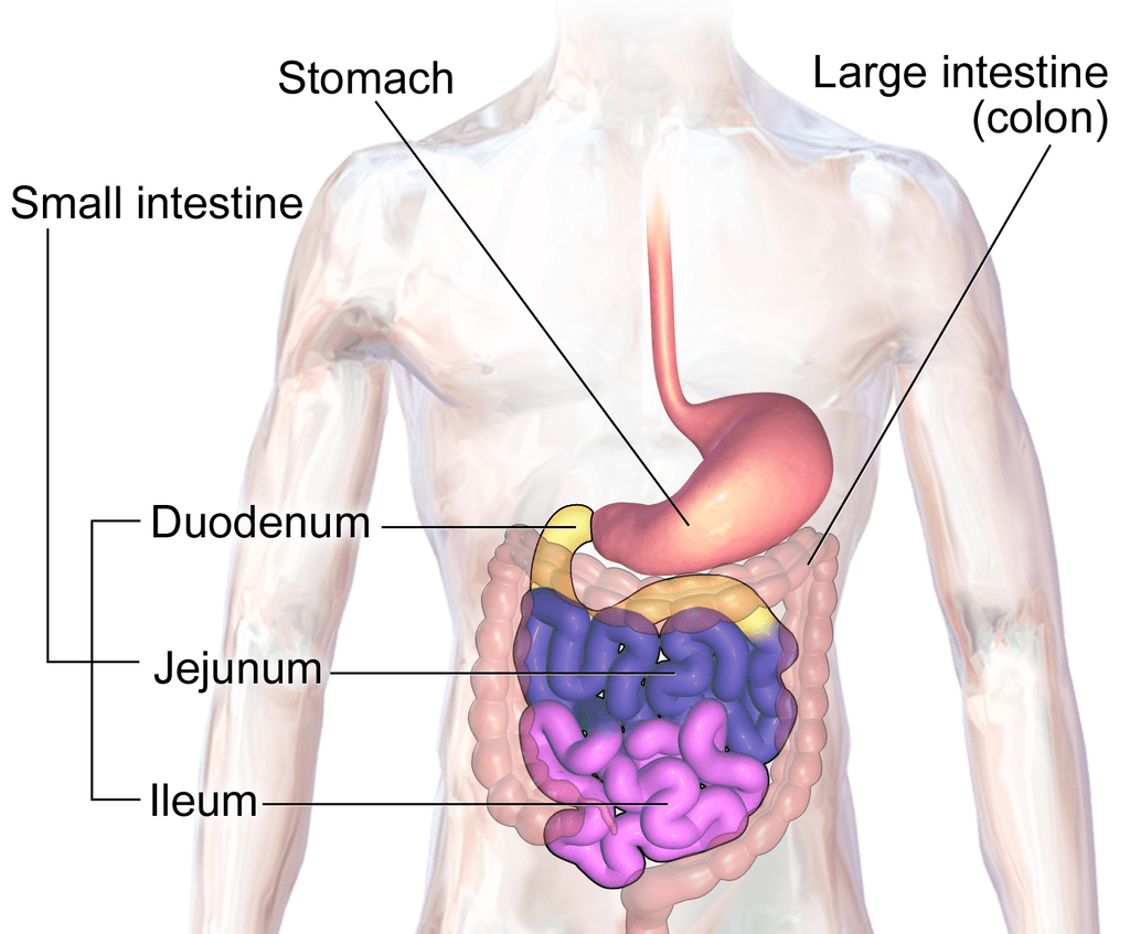 Anatomy of Small Intestine
- Duodenum is the retroperitoneal and the initial part of the small intestine. Duodenum is the shortest, widest and the fixed part of the small intestine. It is C/U-shaped.
- For absorption of digested food, a very large surface area is required.
Therefore some adaptations are present here:
(a) Great length of the intestine.
(b) The presence of permanent deep folds in the mucosa is called plicae circularlis, valvulae conniventae or valves of kerckring, along with villi and microvilli.
2. Large Intestine
- The large intestine (larger in diameter) is differentiated into three parts as caecum, colon and rectum.
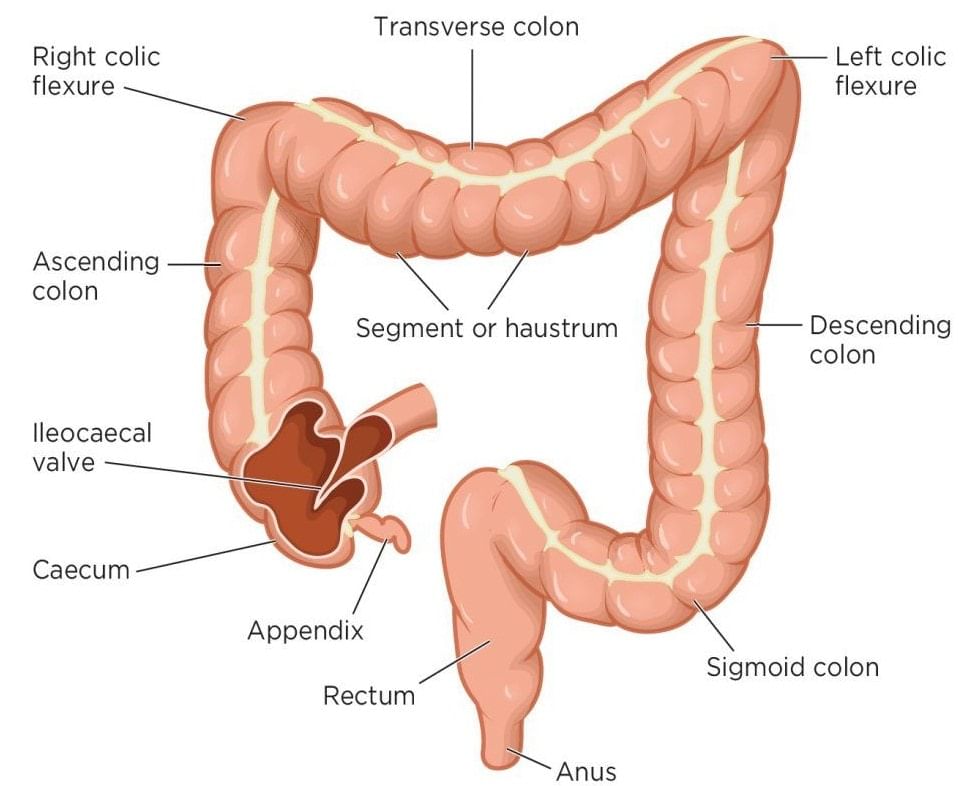 Anatomy of Large Intestine
 - In rabbit, the dilated last terminal part of the ileum is called sacculus rotundus. It is connected with the caecum through an ileocaecal valve.
- The lower end of the ileum opens on the Posteromedial aspect of the ileocaecal junction. The Ileocaecal opening is guarded by the ileocaecal valve. The caecum is a large blind sac. It is situated in the right Iliac fossa. It is 6 cm long and 7.5 cm broad.
- About 2 cm below the ileocaecal orifice, a worm-like structure arises from the caecum called the vermiform appendix. Its length varies from 2 to 20 cm. It is a vestigial organ. The caecum is well developed in rabbit and other mammals but is vestigial in human.
- In Rabbit, some symbiotic bacteria such as Clostridium, cello-bioparus and symbiotic protozoa such as Antidinium are found in the vermiform appendix. These symbiotic organisms secrete the cellulase enzyme which digests the cellulose in the caecum.
➢ Colon
- The caecum continues in the colon, which is the middle part of the large intestine.
- The longitudinal muscle coat forms three ribbon-like bands called Taeniae coli. Due to the presence of taeniae, a pouch-like structure develops in the Lumen of the colon called Haustra.
➢ Rectum
- This colon then continues in a uniform tube called the rectum (storage chamber for faeces).
- The rectum opens into a small bag like structure called anal-canal.
- In a rabbit, the rectum is a beaded shape in appearance.
➢ Anal-Canal
- The anal canal opens outside by the anus. The anus is controlled by an anal sphincter.
- Two types of anal sphincter are found at the opening of the anus.
- Two types of sphincter muscles are found in the Anal canal:
(i) Internal Anal sphincter → Involuntary
(ii) External Anal sphincter → Voluntary
Histology of Alimentary Canal
The wall of the alimentary canal is made up of four-layers (outer to inner):
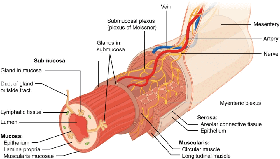 Three Dimensional View of Layers of Alimentary Canal
1. Serosa
- It is the outermost layer of the alimentary canal, it is called tunica adventitia in the oesophagus, which is made up of fibrous connective tissue.
- Except for the oesophagus, the remaining part of the alimentary canal is covered by the serosa layer which is made up of the visceral peritoneum while tunica adventitia is made up of white fibrous connective tissue.
2. Muscularis Externa or Mucularis Coat
- It is made up of two types of muscle outer muscle layer is made up of longitudinal muscle while the inner layer is made up of circular muscle.
- Extra oblique muscles are found in the stomach.
- The thickest muscular coat is found in the stomach so maximum peristalsis is found in the stomach least muscles are found in the rectum so the least peristalsis is found in the rectum.
3. Submucosa
- It is made up of a loose connective tissue layer with blood lymph vessels and nerves.
4. Mucosa
- It is the innermost layer of the gut that contains the secretory and absorptive cells.
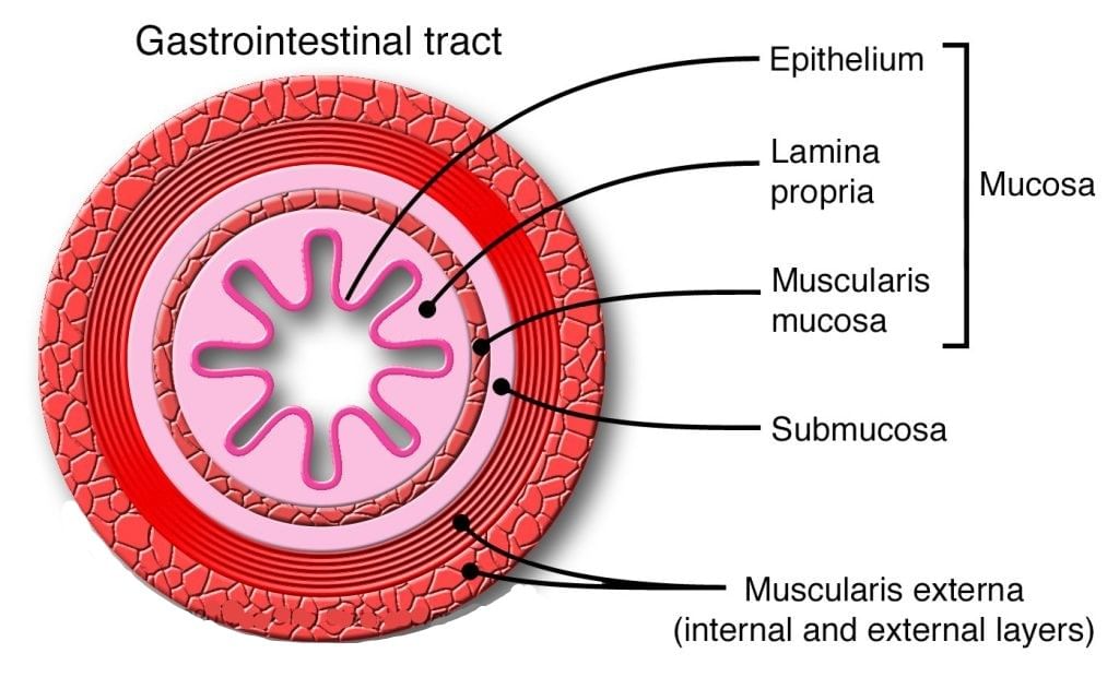 Different Parts of Mucosa Layer
It is differentiated into 3 parts:
(i) Outer Part
- It is called mucosa muscularis or muscularis interna.
- It is made up of longitudinal and circular muscles.
- But these muscles are vestigial.
- They provide support to the folds of the alimentary canal.
(ii) Middle Part
- It is called the lamina propria.
- It is made up of reticulate and fibrous connective tissue, a dense network of blood capillaries are found in this part.
(iii) Innermost Part
- It is called the mucosal layer.
- In the oesophagus, this layer is made up of non keratinised stratified squamous epithelium.
- Except for the oesophagus, this layer is a single layer thick.
- This layer makes the lining of the lumen of the Alimentary canal.
- This layer is made up of columnar mucous epithelium.
- Folds of the oesophagus are less developed.
- This layer makes the folds of the alimentary canal.
- Folds of the stomach are finger-shaped.
- The folds of the small intestine are conical shaped called Villi.
- A small slit-like space is found at the base of the Villi.
- These spaces are called crypts of Lieberkuhn.
- Villi of the Duodenum are small blunt.
- Villi of Jejunum and Ileum are long and pointed.
- Maximum villi are found in Jejunum.
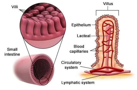 Villi in Small Intestine
➢ Brunner's Gland
- They are small spherical multicellular glands.
- They open into crypts of lieberkuhn with the help of fine tubules.
- These glands are found in the submucosa and mucosa of the duodenum.
- They synthesize and secrete the non-enzymatic secretion of intestinal juice.
➢ Paneth Cells
- These cells are found in the mucosal layer of crypts of lieberkuhn of jejunum.
- They are unicellular gland.
- They synthesize and secrets enzymes of intestinal juices.
- The secretory substances of Brunner's glands and paneth cells are combinedly called intestinal juice or succus entericus.
➢ Peyer's Patches
- They are small lymph nodes that are found in the mucosa of the small intestine (Jejunum and Ileum more in number). They are also called intestinal tonsils and provide immunity.
➢ Nerve Supply
- Two types of Nerve plexus are found in the muscle of the alimentary canal. These control muscle contraction. These are:
(i) Auerbach's Nerve Plexus: This nerve plexus is found between longitudinal muscles and circular muscles.
(ii) Meissner's Nerve Plexus: Found between circular muscles and submucosa but in the stomach, it is found between oblique muscle & submucosa.
The document Alimentary Canal: Stomach, Small Intestine & Large Intestine | Biology for JAMB is a part of the JAMB Course Biology for JAMB.
All you need of JAMB at this link: JAMB
|
221 videos|172 docs|126 tests
|
FAQs on Alimentary Canal: Stomach, Small Intestine & Large Intestine - Biology for JAMB
| 1. What is the function of the gastric glands in the stomach? |  |
Ans. The gastric glands in the stomach secrete gastric juice, which contains enzymes and hydrochloric acid. These substances help in the digestion of food, especially proteins. The enzymes break down proteins into smaller peptides, while the hydrochloric acid creates an acidic environment that activates the enzymes and kills any harmful bacteria present in the food.
| 2. How does the histology of the small intestine contribute to its function in the digestive system? |  |
Ans. The histology of the small intestine is characterized by the presence of numerous finger-like projections called villi and microvilli. These structures increase the surface area of the small intestine, allowing for efficient absorption of nutrients. The villi contain blood vessels and lacteals, which absorb nutrients and transport them to the bloodstream. Additionally, the presence of specialized cells in the small intestine, such as enterocytes and goblet cells, further aids in the digestion and absorption of nutrients.
| 3. What are the main differences between the small intestine and the large intestine in terms of their histology? |  |
Ans. The small intestine and the large intestine differ in terms of their histology. The small intestine has a larger surface area due to the presence of villi and microvilli, while the large intestine has a smoother inner surface. Additionally, the small intestine has a thicker muscular wall compared to the large intestine. The small intestine has a higher density of goblet cells, which secrete mucus, while the large intestine has a higher density of mucus-secreting cells called colonocytes. Furthermore, the small intestine has a higher concentration of enzymes for digestion, while the large intestine primarily absorbs water and electrolytes.
| 4. How does the stomach contribute to the digestive process? |  |
Ans. The stomach plays a crucial role in the digestive process. It receives food from the esophagus and churns it with the help of its muscular walls. The gastric glands in the stomach secrete gastric juice, which contains digestive enzymes and hydrochloric acid. These substances break down proteins into smaller peptides and create an acidic environment that aids in the digestion of food and kills harmful bacteria. The stomach also regulates the release of food into the small intestine, where further digestion and absorption occur.
| 5. What are the main functions of the alimentary canal? |  |
Ans. The alimentary canal, which includes the stomach, small intestine, and large intestine, has several important functions in the digestive system. It is responsible for the ingestion of food, where food enters the mouth and travels through the esophagus into the stomach. The alimentary canal also facilitates the mechanical and chemical digestion of food, breaking it down into smaller particles and molecules that can be absorbed by the body. Additionally, the alimentary canal absorbs water, electrolytes, and nutrients from the digested food and eliminates waste products through defecation.
Related Searches


