NEET PG Exam  >  NEET PG Tests  >  Test: Anemia and RBC Defects - NEET PG MCQ

Test: Anemia and RBC Defects - NEET PG MCQ


Test Description

30 Questions MCQ Test - Test: Anemia and RBC Defects

Test: Anemia and RBC Defects for NEET PG 2025 is part of NEET PG preparation. The Test: Anemia and RBC Defects questions and answers have been prepared according to the NEET PG exam syllabus.The Test: Anemia and RBC Defects MCQs are made for NEET PG 2025 Exam. Find important definitions, questions, notes, meanings, examples, exercises, MCQs and online tests for Test: Anemia and RBC Defects below.
Solutions of Test: Anemia and RBC Defects questions in English are available as part of our course for NEET PG & Test: Anemia and RBC Defects solutions in Hindi for NEET PG course. Download more important topics, notes, lectures and mock test series for NEET PG Exam by signing up for free. Attempt Test: Anemia and RBC Defects | 47 questions in 50 minutes | Mock test for NEET PG preparation | Free important questions MCQ to study for NEET PG Exam | Download free PDF with solutions
Test: Anemia and RBC Defects - Question 1

A 25-year-old female presents to your OPD 1 year post­partum. She was treated for iron deficiency anemia when she was pregnant. Now she is pale and haemoglobin value is 5 gm%. Her reticulocyte count is 9%. Calculate her corrected reticulocyte count. (AIIMS May 2017)

Detailed Solution for Test: Anemia and RBC Defects - Question 1

Reticulocyte is defined as RBC containing 2 or more dots of precipitated RBC
Corrected reticulocyte count =  Reticulocyte count x ((Hemoblobin in gm%)/15)
Hence corrected reticulocyte count = (9x5)/15 = 3%.

Test: Anemia and RBC Defects - Question 2

All are causes of Coombs positive haemolytic anaemia except? (AIIMS May 2016)

Detailed Solution for Test: Anemia and RBC Defects - Question 2
  • Aplastic anemia is due to marrow defect where pancytopenia is seen.
  • Haemolytic disease of new-born occurs due to Rh incompatibility.
  • Alpha-methyl-dopa results in warm antibody mediated haemolytic anaemia
  • Mismatched blood transfusion is due to type II hypersensitivity reaction leading to haemolysis significant enough to cause acute tubular necrosis.
Test: Anemia and RBC Defects - Question 3

Severe aplastic anemia is defined as bone marrow cellularity less than _____? (Recent Question 2016-17)

Detailed Solution for Test: Anemia and RBC Defects - Question 3

TABLE Classification of Aplastic anemia Based on severity of Pancytopenia

  • Severe aplastic anemia (SAA)
  • Bone marrow cellularity < 25%
  • Two of three peripheral blood criteria;
    • Absolute neutrophil count < 500/mm3
    • Platelet count < 20,000/mm3
    • Reticulocyte count <60,000/mm3 or < 1% corrected reticulocyte count
  • Very severe aplastic anemia (VSAA)
  • Same as SAA with absolute neutrophil count < 200/mm3
  • Nonsevere (moderate) aplastic anemia
  • Bone marrow cellularity < 25%
  • Peripheral blood cytopenias do not fulfil criteria for SAA
Test: Anemia and RBC Defects - Question 4

All are essential for diagnosis of Aplastic anemia except? (Recent Question 2016-17)

Detailed Solution for Test: Anemia and RBC Defects - Question 4
  • The bone marrow is usually readily aspirated but dilute on smear, and the fatty biopsy specimen may be grossly pale on withdrawal; a “dry tap" instead suggests fibrosis or myeloph-thisis.
  • In severe aplasia the smear of the aspirated specimen shows only red cells, residual lymphocytes, and stromal cells; the biopsy (which should be >1 cm in length) is superior for determination of cellularity and shows mainly fat under the microscope, with hematopoietic cells occupying <25% of the morrow space; in the most serious cases the biopsy is virtually 100% fat.
  • Cells bearing the CD34 antigen, a marker of early hem atopoietic cells, are greatly diminished, and in functional studies, committed and primitive progenitor cells are virtually absent; in vitro assays have suggested that the stem cell pool is reduced to <1% of normal in severe disease at the time of presentation.
Test: Anemia and RBC Defects - Question 5

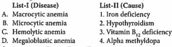
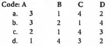
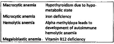
Match List-I with List-II and select the correct answer using the code given below the lists: (UPSC 2015)

Detailed Solution for Test: Anemia and RBC Defects - Question 5

Test: Anemia and RBC Defects - Question 6

Which one of the following is NOT associated with microcytic hypochromic anemia? (APPG 2015)

Detailed Solution for Test: Anemia and RBC Defects - Question 6

Causes of Microcytic hypochromic anemia:

  1. Sideroblastic anemia (INH, Lead poisoning)
  2. Iron deficiency anemia
  3. Thalassemia
  4. Anemia of chronic disease
Test: Anemia and RBC Defects - Question 7

All of the following cause microcytic hypochronic anemia, except? (Bihar PG 2015)

Detailed Solution for Test: Anemia and RBC Defects - Question 7

The causes of microcytic hypochromic anemia is
S = Sideroblastic Anemia
I = Iron deficiency Anemia
T = Thalassemia
A = Anemia of chronic disease

Test: Anemia and RBC Defects - Question 8

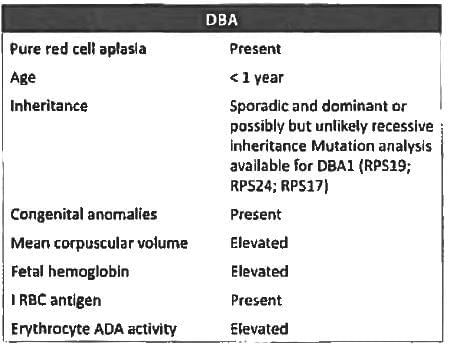
Diamond Blackfann anaemia is associated with? (JIPMER Nov 2015)

Detailed Solution for Test: Anemia and RBC Defects - Question 8

Diamond blackfann syndrome is an inherited bone marrow failure syndrome leading to pure red ceil aplasia. The diagnostic criteria are:

  • Normochromic, usually macrocytic anaemia
  • Reticulocytopenia
  • Normocellular bone marrow with selective deficiency of erythroid precursors
  • Normal or only slightly decreased granulocyte count
  • Normal or slightly increased platelet count

Test: Anemia and RBC Defects - Question 9

A 45-year-old lady diagnosed to have anemia Hb-7.8, MCV = 70 and serum ferritin = 8 ng/dl. After 1 month of iron therapy with adequate dose Hemoglobin is still 8 gm%. What is the most probable cause for treatment failure is?  (JIPMER Nov 2014)

Detailed Solution for Test: Anemia and RBC Defects - Question 9

The patient has presence of microcytic anemia with low serum ferritin. Oral iron was started but hemoglobin has increased by 0.2 gm% only. The most probable cause of the same is Non compliance

  1. Intolerance to oral iron leading to patient not taking medicine or avoiding for most of the days
  2. Consumption of less dose of iron which is consumed post meals

Responses to Iron Therapy

  1. The response to iron therapy varies depending on the erythropoietin stimulus and the rate of absorption.
  2. The reticulocyte count should begin to increase within 4-7 days after initiation of therapy and peak at 7-10 days.
  3. A useful test in the clinic to determine the patient’s ability to absorb iron is the iron tolerance test two iron tablets are given to the patient on an empty stomach and the serum iron is measured serially over the subsequent 2 hours.
  4. Normal absorption will result in an increase in the serum iron of at least 100 g/dl.
Test: Anemia and RBC Defects - Question 10

In cobalamin deficiency which is not seen? (Recent Question 2015-I6)

Detailed Solution for Test: Anemia and RBC Defects - Question 10

B12 deficiency is characterised by;

  • Loss of vibratory and position sense (Rhomberg sign)
  • Abnormal gait
  • Dementia
  • Impotence
  • Loss of bladder and bowel control
  • Macrocytic anemia
Test: Anemia and RBC Defects - Question 11

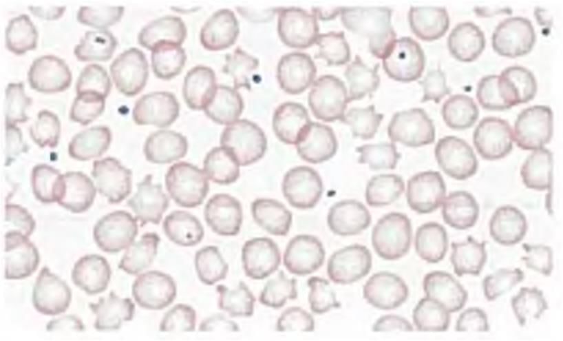
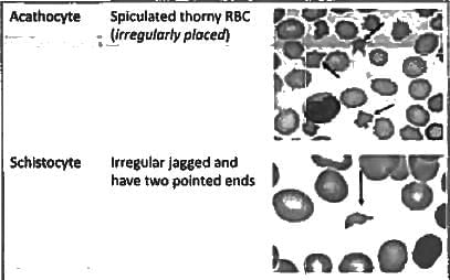
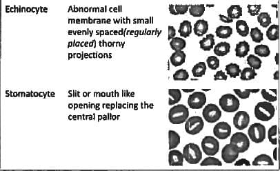
Poikilocytosis and anisocytosis is seen in? (Recent Question 2015-16)

Detailed Solution for Test: Anemia and RBC Defects - Question 11
  • Initially the anemia is normocytic normochromic and subsequently hypochromic and microcytic. There is mild to moderate anisacytosis( variation in size) and poikilocytosis (Variation and shape).
  • The long, thin elliptocytes of iron deficiency are sometimes referred to as pencil cells.
  • Target cells are uncommon and anisochromasia is characteristic. There are two features that can be useful in making the distinction from lhalassaemia trait.
  • Subsequently there is a fall in the MCV and the MCH and, when it is measured by a sensitive technique, a fall in the MCHC as well.
Test: Anemia and RBC Defects - Question 12

Blood transfusion associated acute lung injury occurs due to? (Recent Question 2015-16)

Detailed Solution for Test: Anemia and RBC Defects - Question 12
  • Transfusion-Related Acute Lung Injury (TRALI) is a syndrome characterized by acute respiratory distress following transfusion.
  • TRALI usually results from the donor plasma that contains high-titer anti-HLA antibodies that bind recipient leukocytes.
  • The leukocytes aggregate in the pulmonary vasculature and release mediators that increase capillary permeability.
  • Testing the donor's plasma for anti-HLA antibodies can support this diagnosis.
  • The implicated donors are frequently multiparous women, and transfusion of their plasma component should be avoided.
  • All plasma-containing blood products have been implicated including rare reports of IVIG and cryoprecipitate.
  • Symptoms of TRALI typically develop during, or within 6 hours of a transfusion. Patients present with the rapid onset of dyspnea and tachypnea. There may be associated fever, cyanosis, and hypotension and clinical exam reveals non-cardiogenic pulmonary edema.
Test: Anemia and RBC Defects - Question 13

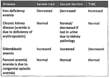
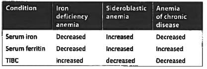
Low serum iron and low serum ferritin is seen in: (Recent Question 2015-16)

Detailed Solution for Test: Anemia and RBC Defects - Question 13

Test: Anemia and RBC Defects - Question 14

All are true about cross-matching of blood except: (Recent Question 2015-16)

Detailed Solution for Test: Anemia and RBC Defects - Question 14

Cross-matching blood is performed prior to a blood transfusion in order to determine if the donors blood is compatible with the blood of an intended recipient, or to identify matches for organ transplantation.

Test: Anemia and RBC Defects - Question 15

The image shows presence of ?

Detailed Solution for Test: Anemia and RBC Defects - Question 15

Test: Anemia and RBC Defects - Question 16

All the following are suggestive of iron-deficiency anemia except: (Recent Pattern 2014-15)

Detailed Solution for Test: Anemia and RBC Defects - Question 16
  • Iron-deficiency Anemia is one of the most prevalent forms of malnutrition.
  • Initially, a state of negative iron balance occurs during which iron stores become slowly depleted.
  • Serum ferritin may decrease, and the presence of stainable iron on bone marrow preparation decreases. When iron stores are depleted serum iron begins to fall Total iron- binding capacity (TIBC) starts to increase, reflecting the presence of circulating unbound transferrin. When the transferrin saturation falls to 15% to 20%, hemoglobin synthesis is impaired. The peripheral blood smear reveals the presence of microcytic and hypochromic red blood cells.
  • Clinically, these patients exhibit the usual signs of anemia, which are fatigue, pallor, and reduced exercise capacity.
  • Cheilosis and koilonychia are signs of advanced tissue iron deficiency. Some patients may experience pica, a desire to ingest certain materials, such as ice (pagophagia) and clay (geophagia).
  • Reticulocytes may also become hypochromic. Reticulocyte numbers are reduced relative to the level of anemia, reflecting a hypoproduction anemia secondary to iron deficiency.
Test: Anemia and RBC Defects - Question 17

Hemolytic anemia are associated with all of the following, except: (Recent Pattern 2014-15)

Detailed Solution for Test: Anemia and RBC Defects - Question 17
  • Haptoglobin is an α-globin which binds specifically to globin of hemoglobin. Hemolytic anemia leads to intravascular hemolysis that leads to hemoglobinuria.
  • Haemolytic anemia is characterized by hemoglobinuria, hemosiderinuria, increased reticulocyte count, unconjugated hyper-bilirubinaemia, increased plasma haemoglobin and LDH, polychromatophilia and bone marrow erythroid hyperplasia.
Test: Anemia and RBC Defects - Question 18

A patient with microcytic hypochromic anemia, Hb-9%, serum iron is 20 g/dl, ferritin level 800 ng/ ml, transferrin percentage saturation is 64. What is possible diagnosis: (Recent Pattern 2014-15)

Detailed Solution for Test: Anemia and RBC Defects - Question 18

Interpretation of values given:
Low serum iron = Normal (50-170 g/dl)
High serum ferritin = Normal (30-400 ng/ml)
High transferrin saturation = Normal (30-50%)
Iron deficiency anemia

  • Can be easily ruled out as serum ferritin is low in Iron deficiency anemia.

Hemochromatosis

  • Can be easily ruled out as hemochromatosis is characterized by increased serum iron level.

Atransferrinemia

  • Transferrin is an iron carrying protein that transmits iron to the erythroblasts.
  • Atransferrinemia or hypotransferrinemia results in reduced delivery of iron to erythroblasts and development of iron deficient anemia. This explains low serum iron.
  • This leads to massive but futile iron absorption and whatever LITTLE transferrin is there is saturated. This explains High serum transferrin. The difference from DMT 1 mutation is that since the iron absorption is defective transferrin saturation is low.
  • Iron is deposited in the visceral organs rather than in the bone marrow.

DMT 1 mutation

  • DMT 1 is a transmembrane protein involved in dietary non heme iron uptake at the brush border of duodenal enterocytcs and also plays crucial role in iron utilization at the endosomal membrane of the erythroid precursors, Divalent metal transporter 1 (DMT 1) is a duodenal apical iron transporter encoded by the SLC11A2 gene.
  • In DMT 1 mutation the iron absorption in the duodenum continues because the absorption of heme iron is not affected.
  • The mutation primarily affects iron utilization and not absorption. Iron utilization in erythroid precursors is disturbed leading to severe iron deficiency anemia.


Genetic forms of Iron deficiency anemia

  • Iron deficiency anemia is an acquired disease. It is generally associated with low cost diet and bleeding.
  • Recent advances in iron metabolism led to the recognition of new entities of iron deficiency anemia in nonbleeding and “high cost diet" nourished individuals. These are known as genetic forms of iron deficiency anemia.
  • Apparently rare these genetic forms of iron deficiency anemia should be recognized by hematologists as they are refractory to classical oral or intravenous iron administration.

These includes:

  • Mutations in gene encoding DMT 1
  • Mutations in gene encoding glutaredoxin 5.
  • Hypotransferrinemia or Atransferriemia
  • Deficiency of ceruloplasmin 
  • IRIDA (Iron Refractory, Iron Deficiency Anemia)
Test: Anemia and RBC Defects - Question 19

Seen in chronic inflammatory anemia is: (Recent Pattern 2014-15)

Detailed Solution for Test: Anemia and RBC Defects - Question 19
  1. In anemia of chronic disease, a product liberated by liver hepicidin, inhibits release of iron from the stores.
  2. Hence the stores will get increased while serum iron will fall.
  3. TIBC always shows an inverse relation to serum ferritin.
Test: Anemia and RBC Defects - Question 20

Reticulocyte count in Hemolytic jaundice is greater than? (Recent Pattern 2014-15)

Detailed Solution for Test: Anemia and RBC Defects - Question 20
  • Normally, reticulocytes are red cells that have been recently released from the bone marrow. They are identified by staining with a supravital dye that precipitates the ribosomal RNA. These precipitates appear as blue or black punctate spots. This residual RNA is metabolized over the first 24-36 h of the reticulocytes life span in circulation.
  • Normally, the reticulocyte count ranges from 1 to 2% and reflects the daily replacement of 0.8-1.0% of the circulating red cell population. A corrected reticulocyte count provides a reliable measure of red cell production.
Test: Anemia and RBC Defects - Question 21

Coombs negative hemolytic anemia is seen in: (Recent Pattern 2014-15)

Detailed Solution for Test: Anemia and RBC Defects - Question 21

Coombs negative hemolytic anemia is associated with several conditions. Let's break down the details:

  • Micro-angiopathic hemolytic anemia: This type is characterised by damage to small blood vessels, leading to red blood cell destruction, but it is not typically linked to Coombs negative hemolytic anemia.
  • SLE (Systemic Lupus Erythematosus): This autoimmune disease can cause various types of anemia, including hemolytic anemia, but it is usually Coombs positive.
  • CLL (Chronic Lymphocytic Leukaemia): CLL can lead to immune-related anemia, yet it is not specifically known for causing Coombs negative hemolytic anemia.
  • Rh incompatibility: This condition occurs when an Rh-negative mother is pregnant with an Rh-positive baby, leading to hemolytic disease of the newborn, which is often Coombs positive.

In summary, the condition most likely associated with Coombs negative hemolytic anemia is not clearly represented here, but it is often seen in certain types of autoimmune disorders or conditions not related to direct blood group incompatibility.

Test: Anemia and RBC Defects - Question 22

Coomb's positive hemolytic anemia associated with: (Recent Pattern 2014-15)

Detailed Solution for Test: Anemia and RBC Defects - Question 22

Connective tissue disorders associated with autoimmune hemolytic anemia (Associated with IgG autoantibody warm autoantibody) Common

  • Systemic lupus Erythematosus

Rare

  • Polyarteritis nodosa
  • Rheumatoid arthritis
  • Sjogrens syndrome
  • Scleroderma
Test: Anemia and RBC Defects - Question 23

A patient with previously normal hemoglobin suffered a sudden massive acute hemorrhage. He is most likely to show all of the following except: (Recent Pattern 2014-15)

Detailed Solution for Test: Anemia and RBC Defects - Question 23

In acute blood loss, the platelets and circulating granulocytes increase within few hours. Immature WBC’s may also be seen. Increase leucocytes and platelets are normal body response to stress. The body continuously strives to maintain homeostasis.
Packed Cell Volume

  • The body adjusts to severe hemorrhage by expanding the circulating volume at the expense of the extravascular fluid leading to low PCV
  • A hematocrit that is done immediately after a haemorrhage usually does not show the extent of RBC loss because at the time of haemorrhage plasma and red blood cells are lost in equal proportions.
  • However, within several hours after haemorrhage, plasma volume begins to increase due to shift of interstitial fluid into the vascular space.
  • Red blood cells, however, cannot be replaced quickly as the bone marrow takes approximately ten days to produce mature red blood cells.
  • As a result PCV done several hours after bleeding episode will show a more accurate picture of the hematocrit which is a Decreased Packed Cell Volume.
Test: Anemia and RBC Defects - Question 24

Spur cell anemia is seen in: (Recent Pattern 2014-15)

Detailed Solution for Test: Anemia and RBC Defects - Question 24

A mild hypoproliferative anemia may develop in patients with chronic liver disease from nearly any cause. The peripheral blood smear may show spur cells and stomatocytes from the accumulation of excess cholesterol in the membrane from a deficiency of lecithin-cholesterol acyltransferase.

Test: Anemia and RBC Defects - Question 25

Low serum haptogloblin in hemolysis is masked by: (Recent Pattern 2014-15)

Detailed Solution for Test: Anemia and RBC Defects - Question 25
  • Haptoglobin binding of free haemoglobin results in removal of the complex from the circulation.
  • Reduced levels occur in haemolysis (particularly intravascular), megaloblastic anaemia (red cell turnover in marrow) following blood transfusion, massive tissue haemorrhage, and hepatocellular disease.
  • Congenital absence of haptoglobin has also been recogn ised.
  • High levels may be seen in pregnancy, with steroid or oestrogen therapy, in conditions associated with an acute phase response, and in biliary obstruction.
  • Pre-existent high levels may mask haemolysis.
Test: Anemia and RBC Defects - Question 26

Which causes aplastic anemia? (Recent Pattern 2014-15)

Detailed Solution for Test: Anemia and RBC Defects - Question 26

Viruses leading to aplestic anemia

  1. EBV
  2. Hepatitis (non-A, non-B, non-C)
  3. HIV - I
  4. Parvovirus B19 (aplastic CRISIS)
Test: Anemia and RBC Defects - Question 27

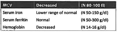
A young female has the following lab values: Hemoglobin = 9.8 gm%, MCV = 70 serum iron = 60, serum ferritin = 100, the diagnosis is: (Recent Pattern 2014-15)

Detailed Solution for Test: Anemia and RBC Defects - Question 27

Interpretation of parameters provided in question

  • Thalassemia trait is not diagnosed on the basis of serum iron indices, instead, definitive diagnosis of thalassemia trait is based on hemoglobin electrophoresis." HbA2 levels > 3.6 - 8% are considered diagnostic for thalassemia trait.
  • Serum iron level is increased in thalassemia trait because of increased intestinal absorption of iron. Choice A is ruled out
  • Due to low iron but normal ferritin iron deficiency is ruled out as serum ferritin is first to reduce in iron deficiency anemia. Choice B is ruled out.
  • Since MCV is low it is a microcytic anemia. Choice C is ruled out
  • This is likely a case of anemia of chronic disease in which serum ferritin is normal to increased while the serum iron level is usually on the lower side.
Test: Anemia and RBC Defects - Question 28

Warm-antibody immune-hemolytic anemia is seen in all except: (Recent Pattern 2014-I5)

Detailed Solution for Test: Anemia and RBC Defects - Question 28

Warm antibody hemolytic anemia is the most common form of autoimmune hemolytic anemia (AIHA); it is more common among women. Autoantibodies in warm antibody hemolytic anemia generally react at temperatures > 37” C. The autoantibodies may occur.

  • Spontaneously
  • In association with certain disorders (SLE, lymphoma, chronic lymphocytic leukemia)
  • After use of certain drugs which stimulate production of autoantibodies against Rh antigens (α-methyldopa-type of AIHA). Other drugs stimulate production of autoantibodies against the antibiotic-RBC-membrane complex as part of a transient hapten mechanism; the hapten maybe stable (e.g., high-dose penicillin, cephalosporins).
  • In warm antibody hemolytic anemia, hemolysis occurs primarily in the spleen. It is often severe andean be fatal. Most of the autoantibodies in warm antibody hemolytic anemia are IgG. Most are pan-agglutinins and have limited specificity.
Test: Anemia and RBC Defects - Question 29

Anemia of chronic disease is associated with: (Recent Pattern 2014-15)

Detailed Solution for Test: Anemia and RBC Defects - Question 29

Test: Anemia and RBC Defects - Question 30

Which of the following causes of Anemia is associated with a Hypoplastic marrow? (Recent Pattern 2014-15)

Detailed Solution for Test: Anemia and RBC Defects - Question 30

Pancytopenia with hypo-cellular marrow

  1. Acquired aplastic anemia
  2. Constitutional aplastic anemia (Fanconi's anemia, dyskeratosis congenita)
  3. Some myelodysplasia
  4. Rare aleukemic leukemia
  5. Acute lymphoid leukemia
  6. Lymphomas originating from the bone marrow
View more questions
Information about Test: Anemia and RBC Defects Page
In this test you can find the Exam questions for Test: Anemia and RBC Defects solved & explained in the simplest way possible. Besides giving Questions and answers for Test: Anemia and RBC Defects, EduRev gives you an ample number of Online tests for practice
Download as PDF