Software Development Exam > Software Development Questions > The following techniques can be used to see p...
Start Learning for Free
The following techniques can be used to see proteins in cells:
A. To directly see under a fluorescence microscope, express the gene (which codes for the aforementioned protein) as a fusion with the green fluorescence protein (GFP).
B. express the gene (which codes for the aforementioned protein) in a fusion with the lac Z gene, and then directly observe with a phase-contrast bright-field microscope.
C. The stated protein could be visualized in a fluorescent microscope using an antibody that has been fluorescence-tagged.
D. Explicitly observe the protein under a scanning electron microscope after overexpressing it.
Which of the following methods you would choose to visualize a protein in a living cell?
- a)A only
- b)B and C only
- c)A and D only
- d)D only
Correct answer is option 'A'. Can you explain this answer?
Verified Answer
The following techniques can be used to see proteins in cells: A. To d...
Concept:
- Ligand binding to cell membrane receptors sets off a series of protein interactions that convey the nuances of ligand identity to the cell interior.
- The information may be encoded in conformational changes, the interaction kinetics and, in the case of multichain immunoreceptors, by chain rearrangements.
- The signals may be modulated by dynamic compartmentalization of the cell membrane, cellular architecture, motility, and activation—all of which are difficult to reconstitute for studies of receptor signaling in vitro.
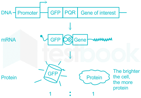
- GFP (green fluorescently labelled protein)and its variants have been used in organisms from bacteria and yeast to mice and human cells. One of the most common uses of GFP is in promoter and protein fusion constructs.
Explanation:
Fig 1: Quantifying of proteins in living cells

- Promoter fusions with GFP can document patterns of gene expression.
- Protein fusions are useful in determining the subcellular localization of a protein of interest and whether that localization changes during development, with different growth conditions, or in different genetic backgrounds.
- Protein interactions in general and receptor signaling in particular can be studied in living cells by different fluorescence imaging techniques.
- Particularly versatile are methods that exploit Förster resonance energy transfer (FRET), which is exquisitely sensitive to the nanometer-range proximity and orientation between fluorophores.
- Fluorescence correlation microscopy (FCM) can provide complementary information about the stoichiometry and diffusion kinetics of large complexes, while bimolecular fluorescence complementation (BiFC) and other complementation techniques can capture transient interactions.
Statement A: To directly see under a fluorescence microscope, express the gene (which codes for the aforementioned protein) as a fusion with the green fluorescence protein (GFP).
- Consider the explanation above thus this option is true, as promoter fusions with GFP can document patterns of gene expression
Statement B:
express the gene (which codes for the aforementioned protein) in a fusion with the lac Z gene, and then directly observe with a phase-contrast bright-field microscope.
- As we need to explore many genes and not just lac Z.
- thus this statement is not true.
Statement C:
The stated protein could be visualized in a fluorescent microscope using an antibody that has been fluorescence-tagged.
- Consider the explanation above thus this option is not true, as we need to localize proteins inside the living system and not invitro.
Statement D:
Explicitly observe the protein under a scanning electron microscope after overexpressing it.
- Consider the explanation above thus this option is not true, as we need to localize proteins inside the living system and not invitro.
hence the correct answer is option 1
Most Upvoted Answer
The following techniques can be used to see proteins in cells: A. To d...
Explanation:
Method A:
- This method involves expressing the gene coding for the protein of interest as a fusion with the green fluorescence protein (GFP).
- The fusion protein will fluoresce under a fluorescence microscope, allowing direct visualization of the protein in living cells.
- This technique is widely used in cell biology to observe protein localization and dynamics in real-time.
Method B and C:
- Method B involves expressing the gene as a fusion with the lac Z gene and observing it under a phase-contrast bright-field microscope. This method may not provide the specificity and sensitivity of direct fluorescence visualization.
- Method C involves using a fluorescence-tagged antibody to visualize the protein under a fluorescence microscope. While this method is commonly used for fixed cells, it may not be suitable for real-time imaging in living cells.
Method D:
- Method D involves overexpressing the protein and observing it under a scanning electron microscope. This method is not suitable for visualizing proteins in living cells as the preparation for electron microscopy usually involves fixation and dehydration, which would not preserve the cellular environment.
Conclusion:
- Among the given methods, method A (expressing the protein as a fusion with GFP and visualizing it under a fluorescence microscope) is the most appropriate for visualizing proteins in living cells due to its specificity, sensitivity, and suitability for real-time imaging.

|
Explore Courses for Software Development exam
|

|
The following techniques can be used to see proteins in cells: A. To directly see under a fluorescence microscope, express the gene (which codes for the aforementioned protein) as a fusion with the green fluorescence protein (GFP). B. express the gene (which codes for the aforementioned protein) in a fusion with the lac Z gene, and then directly observe with a phase-contrast bright-field microscope. C. The stated protein could be visualized in a fluorescent microscope using an antibody that has been fluorescence-tagged. D. Explicitly observe the protein under a scanning electron microscope after overexpressing it. Which of the following methods you would choose to visualize a protein in a living cell?a)A onlyb)B and C onlyc)A and D onlyd)D onlyCorrect answer is option 'A'. Can you explain this answer?
Question Description
The following techniques can be used to see proteins in cells: A. To directly see under a fluorescence microscope, express the gene (which codes for the aforementioned protein) as a fusion with the green fluorescence protein (GFP). B. express the gene (which codes for the aforementioned protein) in a fusion with the lac Z gene, and then directly observe with a phase-contrast bright-field microscope. C. The stated protein could be visualized in a fluorescent microscope using an antibody that has been fluorescence-tagged. D. Explicitly observe the protein under a scanning electron microscope after overexpressing it. Which of the following methods you would choose to visualize a protein in a living cell?a)A onlyb)B and C onlyc)A and D onlyd)D onlyCorrect answer is option 'A'. Can you explain this answer? for Software Development 2025 is part of Software Development preparation. The Question and answers have been prepared according to the Software Development exam syllabus. Information about The following techniques can be used to see proteins in cells: A. To directly see under a fluorescence microscope, express the gene (which codes for the aforementioned protein) as a fusion with the green fluorescence protein (GFP). B. express the gene (which codes for the aforementioned protein) in a fusion with the lac Z gene, and then directly observe with a phase-contrast bright-field microscope. C. The stated protein could be visualized in a fluorescent microscope using an antibody that has been fluorescence-tagged. D. Explicitly observe the protein under a scanning electron microscope after overexpressing it. Which of the following methods you would choose to visualize a protein in a living cell?a)A onlyb)B and C onlyc)A and D onlyd)D onlyCorrect answer is option 'A'. Can you explain this answer? covers all topics & solutions for Software Development 2025 Exam. Find important definitions, questions, meanings, examples, exercises and tests below for The following techniques can be used to see proteins in cells: A. To directly see under a fluorescence microscope, express the gene (which codes for the aforementioned protein) as a fusion with the green fluorescence protein (GFP). B. express the gene (which codes for the aforementioned protein) in a fusion with the lac Z gene, and then directly observe with a phase-contrast bright-field microscope. C. The stated protein could be visualized in a fluorescent microscope using an antibody that has been fluorescence-tagged. D. Explicitly observe the protein under a scanning electron microscope after overexpressing it. Which of the following methods you would choose to visualize a protein in a living cell?a)A onlyb)B and C onlyc)A and D onlyd)D onlyCorrect answer is option 'A'. Can you explain this answer?.
The following techniques can be used to see proteins in cells: A. To directly see under a fluorescence microscope, express the gene (which codes for the aforementioned protein) as a fusion with the green fluorescence protein (GFP). B. express the gene (which codes for the aforementioned protein) in a fusion with the lac Z gene, and then directly observe with a phase-contrast bright-field microscope. C. The stated protein could be visualized in a fluorescent microscope using an antibody that has been fluorescence-tagged. D. Explicitly observe the protein under a scanning electron microscope after overexpressing it. Which of the following methods you would choose to visualize a protein in a living cell?a)A onlyb)B and C onlyc)A and D onlyd)D onlyCorrect answer is option 'A'. Can you explain this answer? for Software Development 2025 is part of Software Development preparation. The Question and answers have been prepared according to the Software Development exam syllabus. Information about The following techniques can be used to see proteins in cells: A. To directly see under a fluorescence microscope, express the gene (which codes for the aforementioned protein) as a fusion with the green fluorescence protein (GFP). B. express the gene (which codes for the aforementioned protein) in a fusion with the lac Z gene, and then directly observe with a phase-contrast bright-field microscope. C. The stated protein could be visualized in a fluorescent microscope using an antibody that has been fluorescence-tagged. D. Explicitly observe the protein under a scanning electron microscope after overexpressing it. Which of the following methods you would choose to visualize a protein in a living cell?a)A onlyb)B and C onlyc)A and D onlyd)D onlyCorrect answer is option 'A'. Can you explain this answer? covers all topics & solutions for Software Development 2025 Exam. Find important definitions, questions, meanings, examples, exercises and tests below for The following techniques can be used to see proteins in cells: A. To directly see under a fluorescence microscope, express the gene (which codes for the aforementioned protein) as a fusion with the green fluorescence protein (GFP). B. express the gene (which codes for the aforementioned protein) in a fusion with the lac Z gene, and then directly observe with a phase-contrast bright-field microscope. C. The stated protein could be visualized in a fluorescent microscope using an antibody that has been fluorescence-tagged. D. Explicitly observe the protein under a scanning electron microscope after overexpressing it. Which of the following methods you would choose to visualize a protein in a living cell?a)A onlyb)B and C onlyc)A and D onlyd)D onlyCorrect answer is option 'A'. Can you explain this answer?.
Solutions for The following techniques can be used to see proteins in cells: A. To directly see under a fluorescence microscope, express the gene (which codes for the aforementioned protein) as a fusion with the green fluorescence protein (GFP). B. express the gene (which codes for the aforementioned protein) in a fusion with the lac Z gene, and then directly observe with a phase-contrast bright-field microscope. C. The stated protein could be visualized in a fluorescent microscope using an antibody that has been fluorescence-tagged. D. Explicitly observe the protein under a scanning electron microscope after overexpressing it. Which of the following methods you would choose to visualize a protein in a living cell?a)A onlyb)B and C onlyc)A and D onlyd)D onlyCorrect answer is option 'A'. Can you explain this answer? in English & in Hindi are available as part of our courses for Software Development.
Download more important topics, notes, lectures and mock test series for Software Development Exam by signing up for free.
Here you can find the meaning of The following techniques can be used to see proteins in cells: A. To directly see under a fluorescence microscope, express the gene (which codes for the aforementioned protein) as a fusion with the green fluorescence protein (GFP). B. express the gene (which codes for the aforementioned protein) in a fusion with the lac Z gene, and then directly observe with a phase-contrast bright-field microscope. C. The stated protein could be visualized in a fluorescent microscope using an antibody that has been fluorescence-tagged. D. Explicitly observe the protein under a scanning electron microscope after overexpressing it. Which of the following methods you would choose to visualize a protein in a living cell?a)A onlyb)B and C onlyc)A and D onlyd)D onlyCorrect answer is option 'A'. Can you explain this answer? defined & explained in the simplest way possible. Besides giving the explanation of
The following techniques can be used to see proteins in cells: A. To directly see under a fluorescence microscope, express the gene (which codes for the aforementioned protein) as a fusion with the green fluorescence protein (GFP). B. express the gene (which codes for the aforementioned protein) in a fusion with the lac Z gene, and then directly observe with a phase-contrast bright-field microscope. C. The stated protein could be visualized in a fluorescent microscope using an antibody that has been fluorescence-tagged. D. Explicitly observe the protein under a scanning electron microscope after overexpressing it. Which of the following methods you would choose to visualize a protein in a living cell?a)A onlyb)B and C onlyc)A and D onlyd)D onlyCorrect answer is option 'A'. Can you explain this answer?, a detailed solution for The following techniques can be used to see proteins in cells: A. To directly see under a fluorescence microscope, express the gene (which codes for the aforementioned protein) as a fusion with the green fluorescence protein (GFP). B. express the gene (which codes for the aforementioned protein) in a fusion with the lac Z gene, and then directly observe with a phase-contrast bright-field microscope. C. The stated protein could be visualized in a fluorescent microscope using an antibody that has been fluorescence-tagged. D. Explicitly observe the protein under a scanning electron microscope after overexpressing it. Which of the following methods you would choose to visualize a protein in a living cell?a)A onlyb)B and C onlyc)A and D onlyd)D onlyCorrect answer is option 'A'. Can you explain this answer? has been provided alongside types of The following techniques can be used to see proteins in cells: A. To directly see under a fluorescence microscope, express the gene (which codes for the aforementioned protein) as a fusion with the green fluorescence protein (GFP). B. express the gene (which codes for the aforementioned protein) in a fusion with the lac Z gene, and then directly observe with a phase-contrast bright-field microscope. C. The stated protein could be visualized in a fluorescent microscope using an antibody that has been fluorescence-tagged. D. Explicitly observe the protein under a scanning electron microscope after overexpressing it. Which of the following methods you would choose to visualize a protein in a living cell?a)A onlyb)B and C onlyc)A and D onlyd)D onlyCorrect answer is option 'A'. Can you explain this answer? theory, EduRev gives you an
ample number of questions to practice The following techniques can be used to see proteins in cells: A. To directly see under a fluorescence microscope, express the gene (which codes for the aforementioned protein) as a fusion with the green fluorescence protein (GFP). B. express the gene (which codes for the aforementioned protein) in a fusion with the lac Z gene, and then directly observe with a phase-contrast bright-field microscope. C. The stated protein could be visualized in a fluorescent microscope using an antibody that has been fluorescence-tagged. D. Explicitly observe the protein under a scanning electron microscope after overexpressing it. Which of the following methods you would choose to visualize a protein in a living cell?a)A onlyb)B and C onlyc)A and D onlyd)D onlyCorrect answer is option 'A'. Can you explain this answer? tests, examples and also practice Software Development tests.

|
Explore Courses for Software Development exam
|

|
Signup for Free!
Signup to see your scores go up within 7 days! Learn & Practice with 1000+ FREE Notes, Videos & Tests.


