The Structure & Functions of the Musculoskeletal System | Physical Education for GCSE/IGCSE - Year 11 PDF Download
| Table of contents |
|
| Bones |
|
| Structure of the Skeleton |
|
| Functions of the Skeleton |
|
| Muscles in the Body |
|
| Structure of Synovial Joints |
|
| Types of Freely Movable Joints |
|
| Joint Design |
|
| How Muscles Move the Skeleton |
|

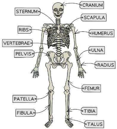
Bones
The musculoskeletal system comprises primarily muscles and a rigid skeleton of bones, complemented by cartilage, tendons, ligaments, and other connective tissues.- An adult human body contains 206 bones.
- Below are some examples:
- The cranium refers to the skull located in the head.
- The vertebrae are a series of bones forming the spinal column.
- The scapula is the technical term for the shoulder blade.
- The humerus is the prominent bone in the upper arm, extending from the shoulder to the elbow.
- The ribs are located in the upper chest, creating a protective cage-like structure.
- The sternum is the breastbone situated at the front of the chest.
- The radius and ulna are found in the forearm, between the elbow and wrist. In diagrams, the radius is shown on the outer side (thumb side), while the ulna is on the inner side (little finger side).
- The pelvis is the anatomical name for the hip bone.
- The femur is a substantial bone in the upper leg, connecting the hip to the knee.
- The tibia and fibula are located in the lower leg, between the knee and ankle. The tibia, or shinbone, is at the front, while the fibula is the smaller bone at the back.
- The patella is positioned in front of the knee joint.
- The talus is located in the ankle, just below the tibia and fibula.
The skeletal system diagram
Structure of the Skeleton
- A primary function of the skeleton is to provide a framework that facilitates movement.
- A joint is the point where two or more bones connect, enabling motion.
- The arrangement and structure of the skeletal bones support this movement.
- The skeleton serves as an anchor for tendons and muscles. Muscle contractions pull on bones to produce movement.
- Different joint types allow varied movements; for instance, the elbow and shoulder have distinct joint structures.
The shape and size of bones are critical to their roles:
- Long bones facilitate large-scale movements, such as running or standing.
- Short bones support precise, controlled movements, like those of the fingers.
- Flat bones, such as the ribs, shield internal organs from injury.
Functions of the Skeleton
- The musculoskeletal system serves multiple purposes.
- Bones provide structural support, maintaining posture and keeping body parts properly aligned.
- Flat bones, such as the cranium and ribs, safeguard vital organs from impact-related damage.
- In high-impact activities like rugby or boxing, flat bones play a critical protective role.
- Bones, along with muscles, cartilage, tendons, ligaments, and connective tissue, enable physical movement.
- Muscles attach to bones, facilitating motion.
- The skeletal structure influences body shape, which can enhance athletic performance in sports like basketball, where longer arm and leg bones provide an advantage.
- Bones store essential minerals like calcium and phosphorus, releasing them when needed by the body.
- Certain bones, such as the femur, contain hollow spaces filled with bone marrow.
- Bone marrow is responsible for producing blood cells, including red blood cells (which transport oxygen for respiration) and white blood cells (which combat infections by targeting pathogens).
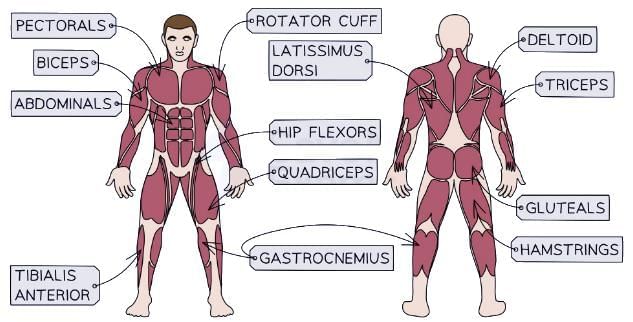
Muscles in the Body
The human body contains approximately 600 muscles, which serve various functions, including:
- Enabling bone movement.
- Cardiac muscles drive the heartbeat.
- Smooth muscles around organs aid processes like digestion.

Muscles of the human body diagram
Lengths of strong connective tissue called tendons, connect muscles to bones
- They are tough and inelastic meaning they do not stretch when a muscle is contracting and pulling on a bone
- There are a few muscles with very long tendons and also a few that are directly attached to the bone
Structure of Synovial Joints
- Synovial joints are the most prevalent type in the human body.
- They feature a joint cavity filled with synovial fluid, which lubricates the joint to minimize friction.
- This fluid is secreted by the synovial membrane, which encases the joint.
- A joint capsule, made of durable fibrous tissue, surrounds the membrane, sealing the joint and enhancing stability.
- Synovial joints support a range of movements, depending on their structure, including the joint type and ligaments.
The Human Hip Joint
- The hip joint is a ball-and-socket synovial joint.
- It involves articulation between the femur (the ball) and the pelvis (the socket).
- Cartilage covers both bones, creating a smooth surface to prevent friction.
- Synovial fluid, enclosed by a membrane within the ball-and-socket structure, lubricates the joint for smooth motion.
- Bursae, small fluid-filled sacs, further reduce friction between bones during movement.
- Ligaments, composed of strong connective tissue, encircle the joint to secure the bones in place.
- Skeletal muscles, attached to the bones via tendons, enable the femur to move within the pelvic socket.
Hip ball and socket diagram
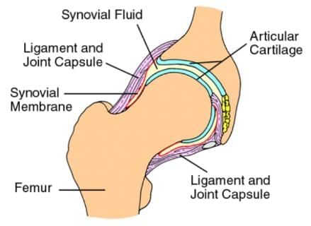 The hip is an example of a ball and socket joint
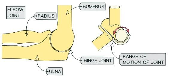
Types of Freely Movable Joints
- The extent of movement at a joint depends on its specific type.
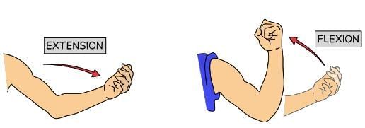
- A hinge joint permits motion primarily in one direction, resembling the opening and closing of a hinge.
- This movement involves flexion (bending) and extension (straightening).
- Some examples of hinge joints are: elbows, knees and ankles
Hinge joint diagram
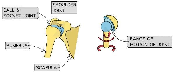
 The shoulder is an example of a ball and socket joint
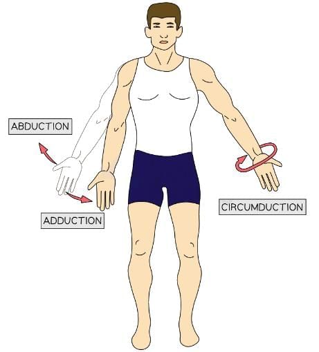
- A ball-and-socket joint allows multidirectional movement, including rotation backward, forward, and sideways. These motions are described as abduction (moving away from the body’s midline), adduction (moving toward the midline), and rotation.
- Examples include the hip and shoulder joints.
Ball and socket joint diagram
 The shoulder is an example of a ball and socket joint
Joint Design
Different joint types, such as hinge or ball-and-socket, result in distinct patterns of bone movement. The specific way bones articulate at a joint, along with their connections to muscles via tendons and ligaments, determines the type of movement possible. This enables a variety of motions essential for activities like sports.
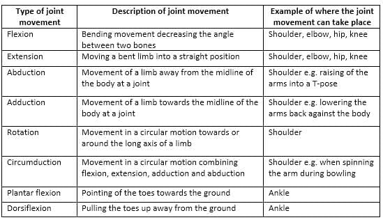
Types of joint movement table
Movement at the elbow joint diagram
Movement at the shoulder joint diagram
How Muscles Move the Skeleton
Multiple muscle groups collaborate at each joint to enable movement in various directions. Key muscles, known as prime movers, are primarily responsible for initiating joint action.Muscles often operate in pairs to produce movement in opposing directions:
- The agonist (or prime mover) contracts and shortens, pulling the bone to create movement.
- The antagonist relaxes and lengthens during the same motion.
Muscle contractions can lead to different outcomes:
- Isometric contractions occur when a muscle tenses without changing length, resulting in no movement.
- Examples, include holding a weight steady or maintaining a plank position.
- Isotonic contractions involve muscle lengthening or shortening, causing joint movement.
- Concentric contraction: The muscle shortens during contraction.
- Eccentric contraction: The muscle lengthens during contraction.
FAQs on The Structure & Functions of the Musculoskeletal System - Physical Education for GCSE/IGCSE - Year 11
| 1. What are the main functions of the skeleton in the human body? |  |
| 2. How are synovial joints structured, and what role do they play in movement? | |
| 3. What are the different types of freely movable joints, and how do they differ from each other? | |
| 4. What is the structure of the human hip joint, and why is it important for mobility? | |
| 5. How do muscles work together with the skeleton to facilitate movement? | |


shortcuts and tricks
,Viva Questions
,video lectures
,ppt
,Extra Questions
,The Structure & Functions of the Musculoskeletal System | Physical Education for GCSE/IGCSE - Year 11
,The Structure & Functions of the Musculoskeletal System | Physical Education for GCSE/IGCSE - Year 11
,Sample Paper
,study material
,MCQs
,mock tests for examination
,Important questions
,past year papers
,Free
,The Structure & Functions of the Musculoskeletal System | Physical Education for GCSE/IGCSE - Year 11
,Summary
,Semester Notes
,practice quizzes
,Exam
,Previous Year Questions with Solutions
,Objective type Questions
;


The Structure & Functions of the Musculoskeletal System Free PDF Download
Importance of The Structure & Functions of the Musculoskeletal System
The Structure & Functions of the Musculoskeletal System Notes
The Structure & Functions of the Musculoskeletal System Year 11 Questions
Study The Structure & Functions of the Musculoskeletal System on the App
|
© EduRev
|
Education Revolution
|


|


within 7 days!