NCERT Summary: Summary of Biology- 2 | Science & Technology for UPSC CSE PDF Download

Muscular and Skeletal System
➢ Skeletal Systems of Various Animals
- Movement is a major characteristic of animals. This movement is a result of the contraction of muscles. The skeleton helps transmit that movement. Skeletons are either a fluid-filled body cavity, exoskeletons, or internal skeletons.
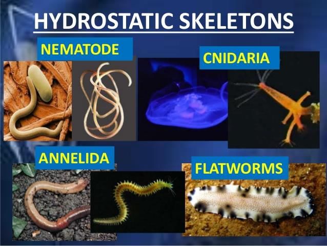
- Hydrostatic skeletons consist of fluid-filled closed chambers. Internal pressure generated by muscle contractions cause movement as well as maintain the shape of the animals, such as the sea anemone and worms. The sea anemone has one set of longitudinal muscles in the outer layer of the body and a layer of circular muscles in the inner layer of the body. The anemone can elongate or contract its body by contracting one or the other set of muscles.
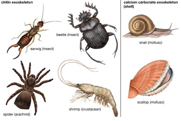
- Exoskeleton are characteristic of the Phylum Arthropoda. Exoskeleton are hard segments that cover the muscles and visceral organs. Muscles for movement attach to the inner surface of the exoskeleton.
 Exoskeletons
 - Exoskeletons restrict the growth of the animal, thus it must shed its exoskeleton (or molt) to form a new one that has room for growth. The bulk and weight of the exoskeleton and associated mechanical problems limit the size animals can attain.
Note:
» Spiders use a combination of an exoskeleton for protection and fluid pressure for movement.
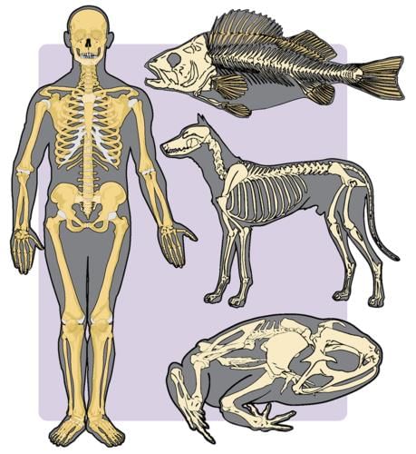
» Vertebrates have developed an internal mineralized (in most cases) endoskeleton composed of bone and/ or cartilage. Muscles are on the outside of the endoskeleton.
» Cartilage and bone are types of connective tissue.
 Endoskeletons
- Sharks and rays have skeletons composed entirely of cartilage, other vertebrates have an embryonic cartilage skeleton progressively replaced by bone as they mature and develop.
- Some areas of the human body, however, retain cartilage in the adult, in joints and flexible structures such as the ribs, trachea, nose, and ears.
➢ The Skeleton and Muscles
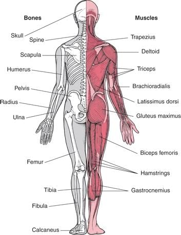
- The skeleton and muscles function together as the musculoskeletal system. This system (often treated as two separate systems, the muscular, and skeletal) plays an important homeostatic role: Allowing the animal to move to more favorable external conditions.
 Musculoskeletal system
 - Certain cells in the bones produce immune cells as well as important cellular components of the blood.
- Bone also helps regulate blood calcium levels, serving as a calcium sink. Rapid muscular contraction is important in generating internal heat, another homeostatic function.
➢ Types of Skeletons
- The axial skeleton consists of the skull, vertebral column, and rib cage.
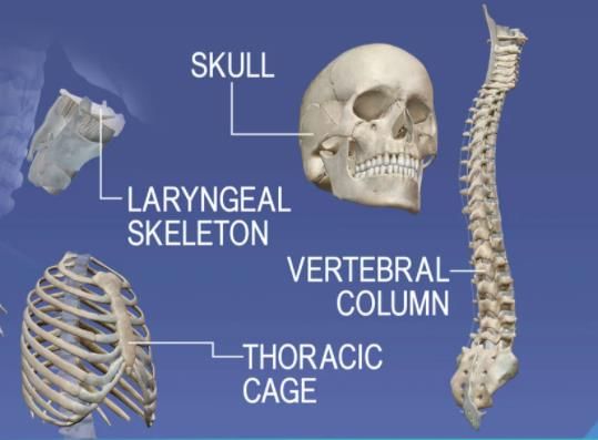 Axial skeleton
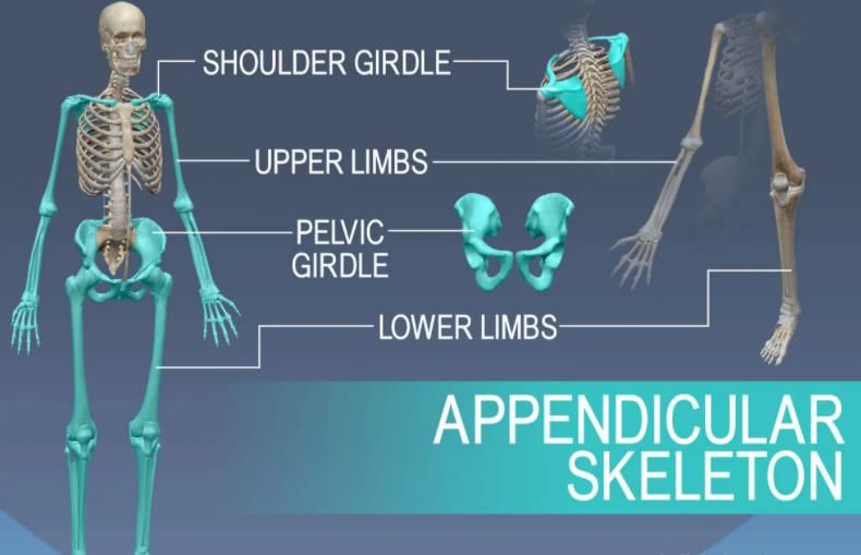
 - The appendicular skeleton contains the bones of the appendages (limbs, wings, or flippers/fins), and the pectoral and pelvic girdles.
- The human skull, or cranium, has a number of individual bones tightly fitted together at immovable joints.
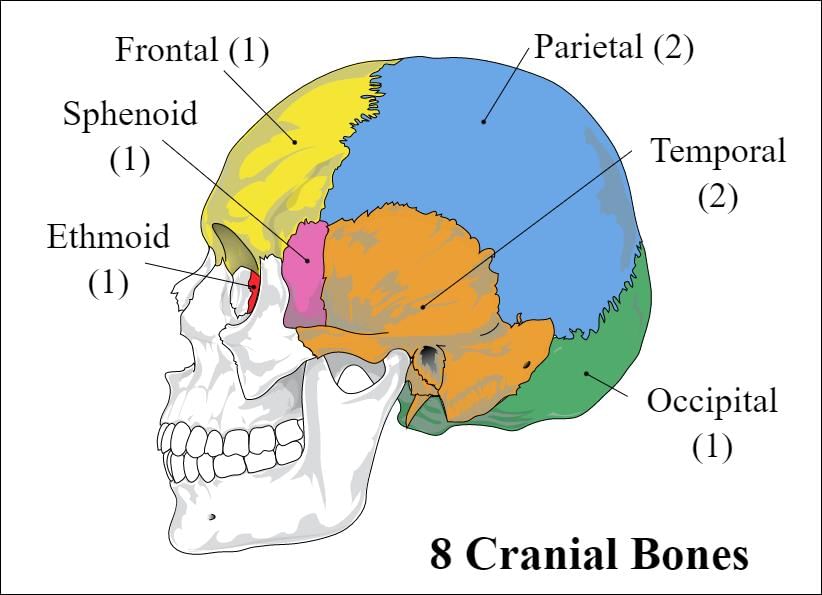 Skull bones
 - At birth many of these joints are not completely structured together as bone, leading to a number of “soft spots” or fontanels, which do not completely join until the age of 14-18 months.
- The vertebral column has 33 individual vertebrae separated from each other by a cartilage disk. These disks allow certain flexibility to the spinal column, although the disks deteriorate with age, producing back pain. The sternum is connected to all the ribs except the lower pair. Cartilage allows for the flexibility of the rib cage during breathing.
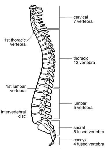 Vertebral column
 - The arms and legs are part of the appendicular skeleton.
- The upper bones of the limbs are single: Humerus (arm) and femur (leg).
- Below a joint (elbow or knee), both limbs have a pair of bones (radius and ulna in the arms, tibia, and fibula in legs) that connect to another joint (wrist or ankle).
- The carpals make up the wrist joint, the tarsals are in the ankle joint.
- Each hand or foot ends in 5 digits (fingers or toes) composed of metacarpals (hands) or metatarsals (feet).
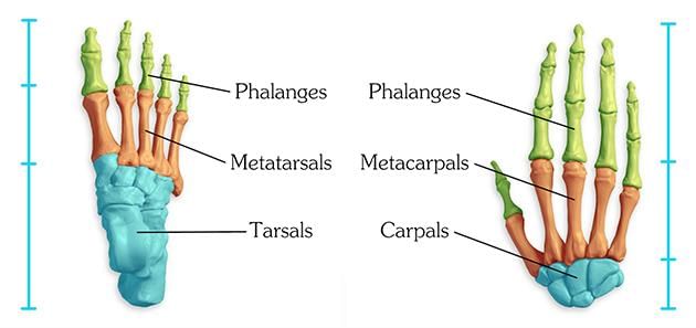 Foot and Hand Bones
 - Limbs are connected to the rest of the skeleton by collections of bones known as girdles. The pectoral girdle consists of the clavicle (collar bone) and scapula (shoulder blade).
- The humerus is joined to the pectoral girdle at a joint and is held in place by muscles and ligaments. A dislocated shoulder occurs when the end of the humerus slips out of the socket of the scapula, stretching ligaments and muscles. The pelvic girdle consists of two hip bones that form a hollow cavity, the pelvis.
- The vertebral column attaches to the top of the pelvis, the femur of each leg attaches to the bottom. The pelvic girdle inland animals transfer the weight of the body to the legs and feet. Pelvic girdles in fish, which have their weight supported by water, are primitive, land animals have more developed pelvic girdles.
- Pelvic girdles in bipeds are recognizable different from those or quadrupeds.
➢ Bone
- Although bones vary greatly in size and shape, they have certain structural similarities. Bones have cells embedded in a mineralized (calcium) matrix and collagen fibers. Compact bone forms the shafts of long bones, it also occurs on the outer side of the bone. Spongy bone forms the inner layer.
- Compact bone has a series of Haversian canals around which concentric layers of bone cells (osteocytes) and minerals occur. New bone is formed by the osteocytes. The Haversian canals form a network of blood vessels and nerves that nourish and monitor the osteocytes.
- Spongy bone occurs at the ends of long bones and is less dense than compact bone. The spongy bone of the femur, humerus, and sternum contains red marrow, in which stem cells reproduce and form the cellular components of the blood and immune system. Yellow marrow, at the center of these bones, is used to store fats. The outer layer of the bones is known as the periosteum.
- The inner layer of the periosteum forms new bone or modifies existing bone to meet new conditions. It is rich in nerve endings and blood and lymphatic vessels. When fractures occur, the pain is carried to the brain by nerves running through the periosteum.
➢ Skeletal Muscle Systems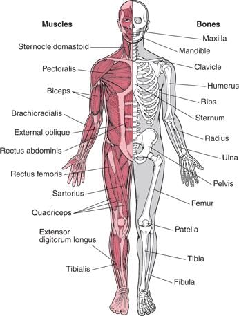 Musculoskeletal System
- Vertebrates move by the actions of muscles on bones. Tendons attach many skeletal muscles across joints, allowing muscle contraction to move the bones across the joint.
- Muscles generally work in pairs to produce movement: When one muscle flexes (or contracts) the other relaxes, a process known as antagonism.
- Muscles have both electrical and chemical activity. There is an electrical gradient across the muscle cell membrane: The outside is more positive than the inside. The stimulus causes an instantaneous reversal of this polarity, causing the muscle to contract (the mechanical characteristic) producing a twitch or movement.
➢ Skeletal Muscle Structure
- Muscle fibers are multinucleated, with the nuclei located just under the plasma membrane. Most of the cell is occupied by striated, thread-like myofibrils. Within each myofibril there are dense Z lines. A sarcomere (or muscle functional unit) extends from Z line to Z line. Each sarcomere has thick and thin filaments. The thick filaments are made of myosin and occupy the center of each sarcomere. Thin filaments are made of action and anchor to the Z line.
- Muscles contract by shortening each sarcomere. The sliding filament model of muscle contraction has thin filaments on each side of the sarcomere sliding past each other until they meet in the middle. Myosin filaments have club-shaped heads that project toward the actin filaments.
- Myosin heads attach to binding sites on the actin filaments. The myosin heads swivel toward the center of the sarcomere, detach and then reattach to the nearest active site of the actin filament. Each cycle of attachment, swiveling, and detachment shortens the sarcomere 1%. Hundreds of such cycles occur each second during muscle contraction.
- Energy for this comes from ATP, the energy coin of the cell. ATP binds to the cross-bridges between myosin heads and actin filaments. The release of energy powers the swiveling of the myosin head. Muscles store little ATP and so must recycle the ADP into ATP rapidly. Creatine phosphate is a muscle storage product involved in the rapid regeneration of ADP into ATP.
- Calcium ions are required for each cycle of myosin-actin interaction. Calcium is released into the sarcomere when a muscle is stimulated to contract. This calcium uncovers the actin-binding sites. When the muscle no longer needs to contract, the calcium ions are pumped from the sarcomere and back into storage.
➢ Contraction of Non-Muscular Cells
- Actin and myosin, whose interaction causes muscle contraction, occur in many other cells. Actin is attached to the inner surface of the plasma membrane. The interaction of cytoplasmic myosin and this actin causes contraction of the cell, such a the coordinated contractions of intestinal cells to absorb nutrients.
- Some fish have modified muscles that discharge electricity. These fish have electric organs consisting of modified muscles known as electroplates. The South American electric eel has more than 6000 plates arranged into 70 columns. The maximum discharge is 100 watts.
➢ Interaction of the Two Systems
- Vertebrates move by application of the principles of the lever. Levers amplify or increase the force or velocity of motion.
- The amount of amplification depends on the length of the lever. There are three types of the skeletal system, all interact with muscles using the lever.
The Nervous System
➢ Divisions of the Nervous System
- The nervous system monitors and controls almost every organ system through a series of positive and negative feedback loops.
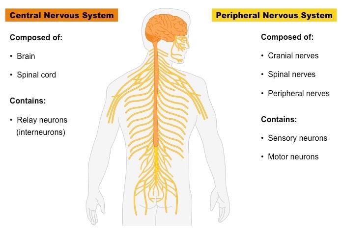
- The Central Nervous System (CNS) includes the brain and spinal cord.
- The Peripheral Nervous System (PNS) connects the CNS to other parts of the body and is composed of nerves (bundles of neurons).
- Not all animals have highly specialized nervous systems.
- Those with simple systems tend to be either small and very mobile or large and immobile.
- Large, mobile animals have highly developed nervous systems: The evolution of nervous systems must have been an important adaptation in the evolution of body size and mobility.
➢ Nervous System in Various Organisms
- Coelenterates, cnidarians, and echinoderms have their neurons organized into a nerve net. These creatures have radial symmetry and lack a head. Although lacking a brain or either nervous system (CNS or PNS) nerve nets are capable of some complex behavior.
- Bilaterally symmetrical animals have a body plan that includes a defined head and a tail region. The development of bilateral symmetry is associated with cephalization, the development of a head with the accumulation of sensory organs at the front end of the organism.
- Flatworms have neurons associated into clusters known as ganglia, which in turn form a small brain. Vertebrates have a spinal cord in addition to a more developed brain. Chordates have a dorsal rather than ventral nervous system. Several evolutionary trends occur in chordates: Spinal cord, continuation of cephalization in the form of larger and more complex brains, and development of a more elaborate nervous system.
➢ The Neuron
- Nervous tissue is composed of two main cell types:
(i) Neurons
(ii) Glial cells - Neurons transmit nerve messages. Glial cells are in direct contact with neurons and often surround them.
- The neuron is the functional unit of the nervous system. Humans have about 100 billion neurons in their brain alone! While variable in size and shape.
1. Parts of Neuron
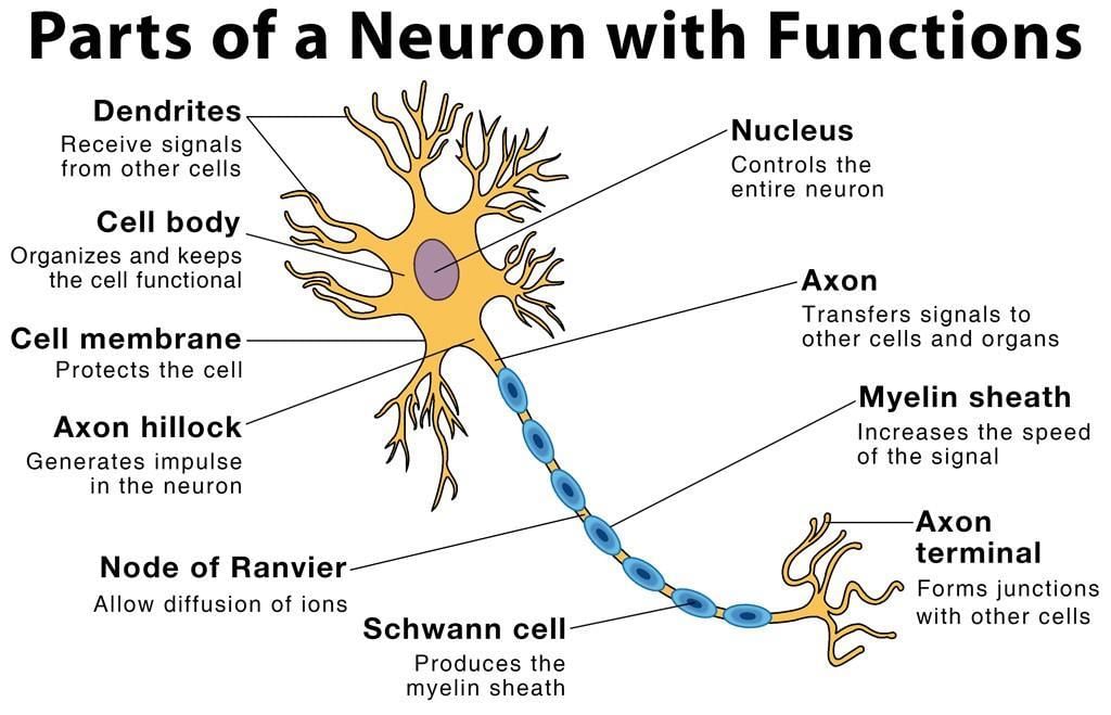
All neurons have three parts:
- Dendrites receive information from another cell and transmit the message to the cell body.
- The cell body contains the nucleus, mitochondria and other organelles typical of eukaryotic cells.
- The axon conducts messages away from the cell body.
2. Types of Neuron
Three types of neurons occur:
(i) Sensory neurons typically have a long dendrite and short axon and carry messages from sensory receptors to the central nervous system.
(ii) Motor neurons have a long axon and short dendrites and transmit messages from the central nervous system to the muscles (or to glands).
(iii) Interneurons are found only in the central nervous system where they connect neuron to neuron. Some axons are wrapped in a myelin sheath formed from the plasma membranes of specialized glial cells known as Schwann cells. Schwann cells serve as supportive, nutritive, and service facilities for neurons. The gap between Schwann cells is known as the Node of Ranvier (refer to above figure) and serves as points along the neuron for generating a signal. Signals jumping from node to node travel hundreds of times faster than signals traveling along the surface of the axon. This allows our brain to communicate with our toes in a few thousandths of a second.
➢ The Nerve Message
- The plasma membrane of neurons, like all other cells, has an unequal distribution of ions and electrical charges between the two sides of the membrane. The outside of the membrane has a positive charge, inside has a negative charge.
- Resting potential results from differences between sodium and potassium positively charged ions and negatively charged ions in the cytoplasm.
- Sodium ions are more concentrated outside the membrane, while potassium ions are more concentrated inside the membrane. This imbalance is maintained by the active transport of ions to reset the membrane known as the sodium-potassium pump.
- The sodium-potassium pump maintains this unequal concentration by actively transporting ions against their concentration gradients. The action potential begins at one spot on the membrane, but spreads to adjacent areas of the membrane, propagating the message along the length of the cell membrane.
- After the passage of the action potential, there is a brief period, the refractory period, during which the membrane cannot be stimulated. This prevents the message from being transmitted backward along the membrane.
➢ Steps in an Action Potential
- At rest, the outside of the membrane is more positive than the inside.
- Sodium moves inside the cell causing an action potential, the influx of positive sodium ions makes the inside of the membrane more positive than the outside.
- Potassium ions flow out of the cell, restoring the resting potential net charges.
- Sodium ions are pumped out of the cell and potassium ions are pumped into the cell, restoring the original distribution of ions.
➢ Synapses
- The junction between a nerve cell and another cell is called a synapse.
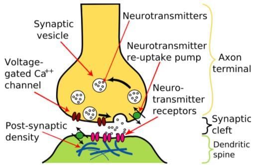 CNS Synapse
 - Messages travel within the neuron as an electrical action potential. The space between the two cells is known as the synaptic cleft. To cross the synaptic cleft requires the actions of neurotransmitters. Neurotransmitters are stored in small synaptic vesicles clustered at the tip of the axon. Neurotransmitters tend to be small molecules, some are even hormones.
- The neurotransmitters cross the cleft, binding to receptor molecules on the next cell, prompting transmission of the message along that cell’s membrane. Diseases that affect the function of signal transmission can have serious consequences.
- Parkinson’s disease has a deficiency of the neurotransmitter dopamine. Progressive death of brain cells increases this deficit, causing tremors, rigidity and unstable posture.
|
90 videos|490 docs|209 tests
|
FAQs on NCERT Summary: Summary of Biology- 2 - Science & Technology for UPSC CSE
| 1. What is the function of the muscular system? |  |
| 2. How does the skeletal system support the body? | |
| 3. What are the types of muscles in the human body? | |
| 4. How do muscles and bones work together? | |
| 5. What is the role of the nervous system in muscle and skeletal function? | |


video lectures
,Semester Notes
,Free
,study material
,Previous Year Questions with Solutions
,mock tests for examination
,Exam
,shortcuts and tricks
,NCERT Summary: Summary of Biology- 2 | Science & Technology for UPSC CSE
,Sample Paper
,practice quizzes
,ppt
,Viva Questions
,past year papers
,NCERT Summary: Summary of Biology- 2 | Science & Technology for UPSC CSE
,Summary
,Extra Questions
,Important questions
,NCERT Summary: Summary of Biology- 2 | Science & Technology for UPSC CSE
,MCQs
,Objective type Questions
;


NCERT Summary: Summary of Biology- 2 Free PDF Download
Importance of NCERT Summary: Summary of Biology- 2
NCERT Summary: Summary of Biology- 2 Notes
NCERT Summary: Summary of Biology- 2 UPSC Questions
Study NCERT Summary: Summary of Biology- 2 on the App
|
© EduRev
|
Education Revolution
|


|


