Human physique and somatotypes | Anthropology Optional for UPSC PDF Download
Human Physique
Body physique refers to an individual’s body form, the configuration of the entire body rather than its specific features. Physique has been related to a variety of behavioral, occupational, disease, and performance variables, primarily in adults. The history of classification and analysis of human physique can be traced back to the very ancient times when people with strong bodies who had the ability to fight, hunt and organize achieved distinction and got noticed by the society.
- In 5th century BC, Hippocrates (a great Greek Philosopher and Physician) described two different types of people as follows:
(i) Habitus phthisicus: People with thin and lean body with long extremities. These individuals had a greater susceptibility to tuberculosis.
(ii) Habitus apoplecticus: People with short, thick and massive bodies who were very much prone to the diseases of the cardiovascular system. - After Hippocrates, not much advances took place in the field of analyzing and categorizing human body physique till 17th century when a luminary, Elsholz, started studying the body morphology with the help of anthropometric measurements, at the University of Padua, Italy.
Viola’s Classification (1921)
An Italian physician Viola, in the beginning of the 20th century, devised a method of human physique analysis with the help of body measurements. These measurements were combined with each other to derive certain indices and values which were used for classifying humans.
The measurements used were: upper extremity length, lower extremity length, thoracic length, thoracic breadth, thoracic depth, upper abdominal breadth, upper abdominal length, lower abdominal breadth, lower abdominal length and abdominal depth.
The following indices were later calculated from different body measurements:
Thoracic index = Thoracic length + Thoracic breadth + Thoracic depth
Upper abdominal index = Upper abdominal breadth + Upper abdominal length + Abdominal depth
Lower abdominal index = Lower abdominal breadth + Lower abdominal length + Abdominal depth
Then, Total abdominal index was calculated as
Total abdominal index = Upper abdominal index + Lower abdominal index
These indexes were further combined together to get the values of trunk and extremities as follows:
Trunk value = Thoracic index + Total abdominal index
Limb value = Upper extremity length + Lower extremity length
On the basis of these measurements, indices and values, four different types of human physiques were identified which were longitype, brachitype, normotype and mixed type:
- Longitype physique: The longitype had long limbs relative to their trunk volume, large thorax relative to their abdomen, a large transverse diameter relative to anterior posterior diameter.
- Brachitype physique: The brachitype was characterized by massiveness and robustness of body, the reverse of longitype. They had short limb relative to trunk, short transverse diameter relative to the antero-posterior diameter, short thorax relative to the abdomen.
- Normotype physique: The normatype physique falls between longitype and brachytype physique characterized by normally proportional limbs versus trunk, thorax versus abdomen, transverse versus antero-posterior widths.
- Mixed type physique: Mixed type shows disproportion in human body. It lacks uniformity in the physique. It is longitype by way of certain characteristic, brachitype by the other and mixed type by other characteristic. In the present day terminology, this may be referred to as dysplasia.
Kretschmer’s Classification (1925)
A German psychiatrist E. Kretschmer, in the beginning of the 20th century, gave a detailed account of the characteristics of three categories of humans which were named as pyknic or fatty, athletic or muscular and leptosome or lean. His method was based on making anthroscopic observations on the individuals. He also correlated the physique with the characteristics including the temperament of the person. This method is still very much popular with psychologists who aimed at studying the behavior and body composition. But other scientists who tried to use his method found it very difficult to apply because majority of the people did not confirm to the characteristics of any of these groups.
Different types of human physiques identified by him were as follows:
- Pyknic: These are thick and short. Mainly, the massiveness of the human body is the characteristic of this type. The people of this type possess head, which is large and heavy, thorax and abdomen are massive or more developed with respect to the extremities.
- Athletic: This type of human physique has the characteristics of typical athletes. They have strong and heavily muscled bodies. They have very less fat, but have considerable amounts of muscles.
- Asthenic (leptosome): These people were long, thin and linear. Their body lacks not only fat but muscles as well. These people seem to lack body strength, and thus, can be considered fragile.
- Dysplastic: This type of physique does not show uniformity, and hence, is disproportional.
Sheldon’s Method of Somatotyping (1940)
William Herbert Sheldon (1898-1977) was an American psychologist and physician. He introduced the concept and word ‘somatotype’ in ‘The Varieties of Human Physique’ (1940). He defined somatotype as ‘quantification of three primary components determining the morphological structure of an individual expressed as a series of three numerals, the first referring to endomorphy, the second to mesomorphy, and the third to ectomorphy’. The conceptual approach is based on the premise that continuous variation occurs in the distribution of physique and thus the variation is related to differential contributions of three specific components, named on the basis of three embryonic germ layers.
A brief description of the three components of physique is given as follows:
- Endomorphy: It is characterized by the predominance of the digestive organs and softness and roundness of contours throughout the body. In other words, with increased fat storage, a wide waist and a large bone structure. Endomorphs are referred to as fat.
- Mesomorphy: It is characterized by the predominance of muscle and bone, skin is made thick by heavy connective tissue. The physique is normally heavy, hard and rectangular in outline. In other words, with medium bones and solid torso, low fat levels, wide shoulders with a narrow waist. Mesomorphs are referred to as muscular.
- Ectomorphy: It is characterized by linearity and fragility of build; with limited muscular development and predominance of surface area over body mass in other words, with long and thin muscles or limbs or low fat storage. Ectomorphs are referred to as slim.
The contribution of the three components defines an individual’s somatotype.Sheldon’s method of estimating somatotype utilizes height and weight and three standardized photograph of front, side and rear views of the naked participants i.e., 4000 college men standing before a calibrated grid.
He summarized his photoscopic (he called it anthroposcopic) somatotype method as follows: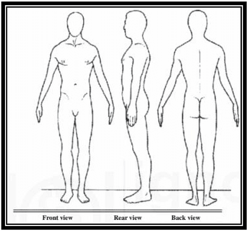 Figure 1: Different views for taking Sheldon’s photoscopic somatotype
(a) Calculation of height/³ √weight ratio (HWR) or reciprocal ponderal index
(b) Calculation of ratios of 17 transverse measurements/diameters (taken from photographic negatives) to stature.
(1) Four on head and neck
(2) Three on the thoracic trunk
(3) Three on the arms
(4) Three on the abdominal trunk
(5) Four on the legs
(c) Inspection of the somatotype photograph, referring to a table of known somatotypes distributed against the criterion of HWR, comparing the photograph with a file of correctly somatotyped photographs, and recording the estimated somatotype.
(d) Comparison of the 17 transverse measurements ratios with the range of scores for each ratio, to give final score.
Each component of physique is assessed individually. Rating are based on a 7- point scale, with 1 representing the least expression, 4 representing moderate expression and 7 representing the fullest expression of that particular component being assessed. The rating of each component determines the somatotype which is expressed by three numerals to sum of no less than 9 and no more than 12. The first number refers to endomorphy, second to mesomorphy and third to ectomorphy. Sheldon identified 76 different somatotype and most common are 3-4-4, 4-3-3 and 3-5-2.
Parnell’s Method of Somatotyping (1954)
R.W. Parnell (1954), a British physician, described a method to objectively somatotype human subjects by physical anthropometry instead of the visual inspection of photographs of the subjects. According to Parnell, Sheldon’s photometric method takes a long time to somatotype a person, may be more than an hour. Secondly, if the choice of dominance of the components in the beginning is wrong, the whole process may result in wrong assessment of the somatotype.
Parnell’s effort was to describe a short physical anthropometric method for obtaining somatotype with the following purposes:
- To provide objective guidance on the dominance of somatotype components in a healthy person.
- To estimate the Sheldonian somatotype objectively and as accurately at least as the agreement achieved between experts in photoscopic somatotyping.
- To make an estimate of women’s somatotype possible although in the absence of a published reference somatotype data, the estimate cannot be compared.
- To reduce on cost, labour and time while doing somatotypes.
He described a method known as Parnell’s M.4 deviation chart method to objectively somatotype human subjects by physical anthropometry. Therefore, Parnell's method of somatotyping was considered to be more objective as compared to Sheldon's method. Parnell (1958) remarked that the phenotype is the body as it appears at a particular point in time. Because of this, Parnell indicated that his method for somatotyping was not a good variable for prediction purposes. Parnell labeled his physical types on the chart as Fat, Muscularity, and Linearity, which correspond to the classification of Sheldon’s viz.; endomorph, mesomorph, and ectomorph respectively.
Parnell developed the scoring method to use anthropometric measurement and recorded the results in M.4 deviation charts to use them along with the pictures. Parnell’s method of somatotyping requires height, weight and measures of muscle girth (biceps and calf), femoral and humeral epicondylar diameters and skinfold thickness in three areas viz.; triceps, subscapular and suprailiac. The M.4 deviation chart included tables to obtain anthropometric somatotypes (Figure 2). Assessments of Fat, Muscularity and Linearity were obtained to the nearest quarter point on a seven scale, giving phenotypes similar to Sheldon’s somatotypes. Parnell replaced Sheldon' component name with fat, muscular (muscularity), and thin type (linearity), abbreviated as F, M and L, respectively. The fat (endomorphy type) type decision is based on skinfold measurement while the muscular type (mesomorphy type) works based on height, bone diameter, and limb thickness. The thin type (ectomorphy type) works based on HWRs. As shown in the M.4 Deviation Charts for Adults, the three element scales were collected from various age brackets (Parnell, 1954; Parnell, 1958). This method of somatotyping emphasized the phenotype, not the somatotype.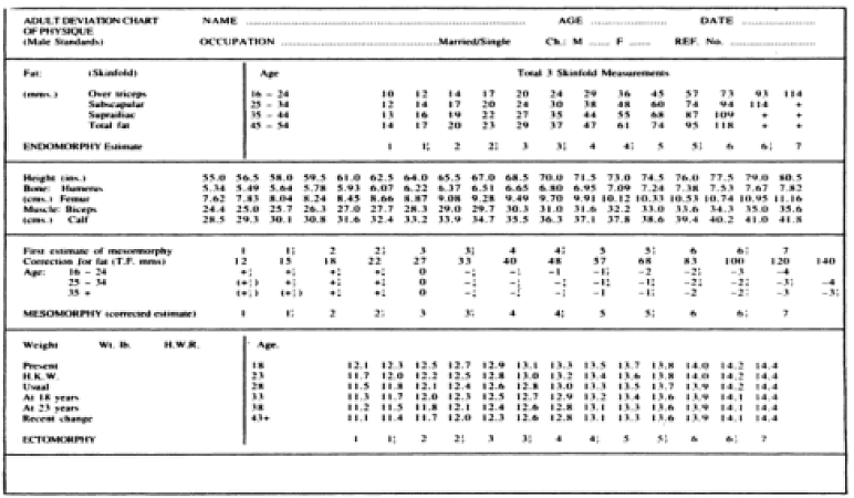 Figure 2: The Parnell M.4 adult Deviation chart with the values for the Harpenden chart for determining the somatotype.
 |
Download the notes
Human physique and somatotypes
|
Download as PDF |
Heath and Carter Method of Somatotyping (1967)
The Heath-Carter method of somatotyping is the most commonly used today. There are three ways of obtaining the somatotype.
- The anthropometric method, in which anthropometry is used to estimate the criterion somatotype.
- The photoscopic method, in which ratings are made from a standardized photograph.
- The anthropometric plus photoscopic method, which combines anthropometry and ratings from a photograph - it is the criterion method.
Because most people do not get the opportunity to become criterion raters using photographs, the anthropometric method has proven to be the most useful for a wide variety of applications.
The Anthropometric Somatotype Method
- Equipment for anthropometry: Anthropometric equipment includes a stadiometer or height scale and headboard, weighing scale, small sliding caliper, a flexible steel or fiberglass tape measure, and a skinfold caliper. The small sliding caliper is a modification of a standard anthropometric caliper or engineer’s vernier type caliper. For accurate measuring of biepicondylar breadths the caliper branches must extend to 10 cm and the tips should be 1.5 cm in diameter (Carter, 1980). Skinfold calipers should have upscale interjaw pressures of 10 gm/mm2 over the full range of openings. The Harpenden and Holtain calipers are highly recommended. The Slim Guide caliper produces almost identical results and is less expensive. Lange and Lafayette calipers also may be used but tend to produce higher readings than the other calipers (Schmidt & Carter, 1990).
- Measurement techniques: Ten anthropometric dimensions are needed to calculate the anthropometric somatotype: stretch stature, body mass, four skinfolds (triceps, subscapular, supraspinale, medial calf), two bone breadths (biepicondylar humerus and femur), and two limb girths (arm flexed and tensed, calf).
The following descriptions are adapted from Carter and Heath (1990):
(i) Stature (height): Taken against a height scale or stadiometer. Take height with the subject standing straight, against an upright wall or stadiometer, touching the wall with heels, buttocks and back. Orient the head in the Frankfort plane (the upper border of the ear opening and the lower border of the eye socket on a horizontal line), and the heels together. Instruct the subject to stretch upward and to take and hold a full breath. Lower the headboard until it firmly touches the vertex.
(ii) Body mass (weight): The subject, wearing minimal clothing, stands in the center of the scale platform. Record weight to the nearest tenth of a kilogram. A correction is made for clothing so that nude weight is used in subsequent calculations.
(iii) Skinfolds: Raise a fold of skin and subcutaneous tissue firmly between thumb and forefinger of the left hand and away from the underlying muscle at the marked site.
Apply the edge of the plates on the caliper branches 1 cm below the fingers of the left hand and allow them to exert their full pressure before reading at 2 sec the thickness of the fold. Take all skinfolds on the right side of the body. The subject stands relaxed, except for the calf skinfold, which is taken with the subject seated.
(iv) Triceps skinfold: With the subject's arm hanging loosely in the anatomical position, raise a fold at the back of the arm at a level halfway on a line connecting the acromion and the olecranon processes.
(v) Subscapular skinfold: Raise the subscapular skinfold on a line from the inferior angle of the scapula in a direction that is obliquely downwards and laterally at 45 degrees.
(vi) Supraspinale skinfold: Raise the fold 5-7 cm (depending on the size of the subject) above the anterior superior iliac spine on a line to the anterior axillary border and on a diagonal line going downwards and medially at 45 degrees. (This skinfold was formerly called suprailiac, or anterior suprailiac. The name has been changed to distinguish it from other skinfolds called "suprailiac", but taken at different locations.)
(vii) Medial calf skinfold: Raise a vertical skinfold on the medial side of the leg, at the level of the maximum girth of the calf.
(viii) Biepicondylar breadth of the humerus: The width between the medial and lateral epicondyles of the humerus, with the shoulder and elbow flexed to 90 degrees. Apply the caliper at an angle approximately bisecting the angle of the elbow. Place firm pressure on the crossbars in order to compress the subcutaneous tissue.
(ix) Biepicondylar breadth of the femur: Seat the subject with knee bent at a right angle. Measure the greatest distance between the lateral and medial epicondyles of the femur with firm pressure on the crossbars in order to compress the subcutaneous tissue.
(x) Upper arm girth, elbow flexed and tensed: The subject flexes the shoulder to 90 degrees and the elbow to 45 degrees, clenches the hand, and maximally contracts the elbow flexors and extensors. Take the measurement at the greatest girth of the arm.
(xi) Calf girth: The subject stands with feet slightly apart. Place the tape around the calf and measure the maximum circumference.
The following equations were used for calculating somatotype:
Endomorphy = – 0.7182 + 0.1451 (X) – 0.00068 (X2) + 0.0000014 (X3)
Where, X = (sum of triceps, subscapular and supraspinale skinfolds) multiplied by (170.18/height in cm). This is called height-corrected endomorphy and is the preferred method for calculating endomorphy.
Mesomorphy = 0.858 × biepicondylar humerus + 0.601 × biepicondylar femur + 0.188 × corrected arm girth + 0.161 × corrected calf girth – height × 0.131 + 4.50
Where, Corrected arm girth = Upper arm circumference (in cm) – Triceps skinfold (in mm)/10 and Corrected calf girth = Calf circumference (in cm) – Calf skinfold (in mm)/10
Ectomorphy was calculated using three different equations according to the heightweight ratio (HWR) which can be calculated as height divided by cube root of weight.
If HWR was greater than or equal to 40.75 then,
Ectomorphy = 0.732 × HWR - 28.58
If HWR was less than 40.75 and greater than 38.25 then,
Ectomorphy = 0.463 × HWR - 17.63
If HWR was equal to or less than 38.25 then,
Ectomorphy = 0.1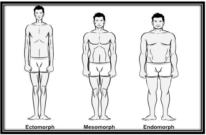 Figure 3: Typical somatotype of ectomorph, mesomorph and endomorph
 - Plotting somatotypes on the 2-D somatochart: X and Y were calculated to plot individual somatotypes on a somatochart using the formula given by Carter (1980). Value of X is obtained by subtracting the value of Endomorphy from the value of Ectomorphy.
X-coordinate = Ectomorphy – Endomorphy
Value of Y was calculated as follows:
Y-coordinate =2 × Mesomorphy – (Endomorphy + Ectomorphy) - Somatochart and Somatoplot: A somatochart is a schematic, triangular shaped, two-dimensional representation of the theoretical range of known somatotypes. The somatotype triangle has all the three sides of equal length, and is arc-shaped. The corners of the triangle represent the extremes in each component. The left corner at the base of the triangle represents extreme in endomorphy, the right corner at the base represents extreme in ectomorphy and the top corner represents the extreme in mesomorphy. The somatotypes can be plotted on the somatotype triangle as a dots or individual somatotypes whose visual inspection can be very useful in interpreting the somatotypes. A typical somatochart has been displayed in Figure 4. Where the individual somatotypes can be plotted which are called somatoplots.
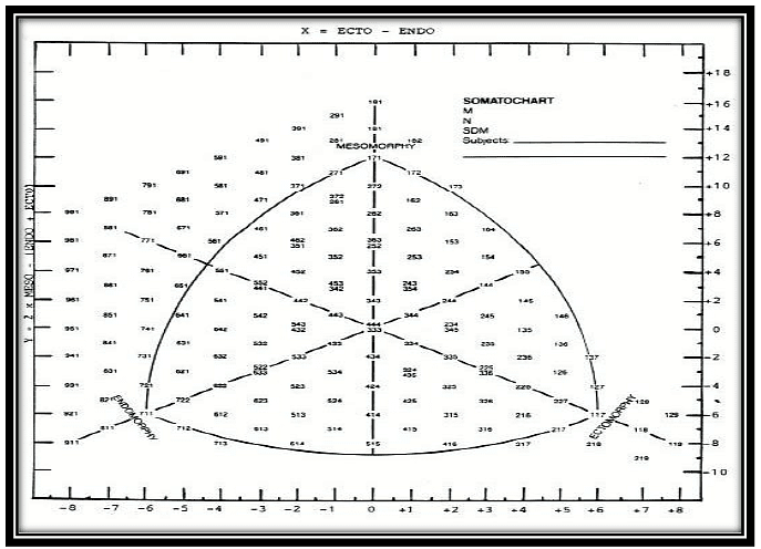 Figure 4: A typical somatochart
 - Somatotype Categories: Somatotypes with similar relationships between the dominance of the components are grouped into categories named to reflect these relationships. Figure 5 shows somatotype categories as represented on the somatochart. The definitions are given below.
The definitions of 13 categories are based on the areas of the 2-D somatochart (Carter and Heath, 1990):
1. Central: no component differs by more than one unit from the other two.
2. Balanced endomorph: endomorphy is dominant and mesomorphy is greater than ectomorphy are equal (or do not differ by more than one-half unit).
3. Mesomorphic endomorph: endomorphy is dominant and mesomorphy is geater than ectomorphy.
4. Mesomorph-endomorph: endomorphy and mesomorphy are equal (or do not differ by more than one-half unit).
5. Endomorphic mesomorph: mesomorphy is dominant and endomorphy is greater than ectomorphy.
6. Balanced mesomorph: mesomorphy is dominant and endomorphy and ectomorphy are equal (or do not differ by more than one-half unit).
7. Ectomorphic mesomorph: mesomorphy is dominant and ectomorphy is greater than endomorphy.
8. Mesomorph-ectomorph: mesomorph and ectomorphy are equal (or do not differ by more than one-half unit), and endomorphy is smaller.
9. Mesomorphic ectomorph: ectomorphy is dominant and mesomorphy is greater than endomorphy.
10. Balanced ectomorph: ectomorph is dominant and endomorphy and mesomorphy are equal (or do not differ by more than one-half unit).
11. Endomorphic ectomorph: ectomorphy is dominant and endomorphy is greater than mesomorphy.
12. Endomorph- ectomorph: endomorphy and ectomorphy are equal (or do not differ by more than one-half unit), and mesomorphy is lower.
13. Ectomorphic endomorph: endomorphy is dominant and ectomorphy is greater than mesomorphy.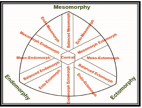 Figure 5: Somatotype categories on two-dimensional Somatochart
 - Somatotype Distributions: Somatotype Dispersion Distance (S.D.D): It refers to the distance between mean Somatotype and each individual somatotype. It is ideal for comparing two mean somatotypes.
It was calculated by using the given formula by Ross and Wilson (1973):
SDD = [3× (X1 – X2)2 + (Y1 –Y2)2]½
Where, X1 and Y1 are the scalar coordinates of mean somatoplot and X2 and Y2 are the coordinates of individual somatoplot. The S.D.D is represented in Y distance units, i.e., in terms of distances at Y- axis of a somatoplot.
Somatotype Dispersion Mean (S.D.M): It can be calculated as the mean of somatotype dispersion distance (SDD). It is useful in knowing about the distribution of somatotypes in a group from its mean somatotypes.
Where, SDD = Somatotype Dispersion Distance
N = Total Number of observations
Dispersion of Somatotype Distance (D.S.D): It can be calculated as the standard deviation of the somatotype dispersion distance.
|
108 videos|243 docs
|


