Human Blood | Famous Books for UPSC Exam (Summary & Tests) PDF Download
| Table of contents |

|
| Introduction |

|
| Blood Vessels |

|
| Body Fluids and Circulation |

|
| Formed Elements |

|
| Coagulation of Blood |

|
| Lymph (Tissue Fluid) |

|
| Blood Groups |

|
| Rh grouping |

|
Introduction
Blood serves as a means of transportation, carrying substances such as digested food from the small intestine to various body parts and delivering oxygen from the lungs to the body's cells. Additionally, it facilitates the removal of waste materials from the body. It consists of a liquid component known as plasma, within which a variety of cells are suspended.
- One particular type of cell found in blood is the red blood cell (RBC), which contains a red pigment called hemoglobin. Hemoglobin binds with oxygen, enabling its transport throughout the body and ultimately to all cells. The presence of hemoglobin gives blood its characteristic red color.
- White blood cells (WBC) are also present in the blood and play a crucial role in combating germs that may enter the body. Another type of cell called platelets contributes to the formation of blood clots.
Blood Vessels
There are two types of blood vessels: arteries and veins. Veins are responsible for carrying carbon dioxide-rich blood (impure blood) from all parts of the body back to the heart. However, the pulmonary vein is an exception as it carries oxygen-rich blood (pure blood) from the lungs to the heart. Veins have thin walls. On the other hand, arteries carry oxygen-rich blood from the heart to all parts of the body. The pulmonary artery is an exception as it carries carbon dioxide-rich blood from the heart to the lungs. Arteries have thick walls due to the high pressure they experience. Blood flows from the heart to arteries and from arteries to various parts of the body. Arteries branch out into smaller vessels, which eventually divide into extremely thin tubes called capillaries when they reach the tissues. Capillaries then connect to form veins, which ultimately empty into the heart.
Body Fluids and Circulation
Blood is a specialized connective tissue that consists of plasma, a fluid matrix, and formed elements.
Plasma
Plasma is a straw-colored, thick fluid that makes up nearly 55 percent of the blood. Approximately 90-92 percent of plasma is composed of water, while proteins contribute 6-8 percent. The major proteins found in plasma are fibrinogen, globulins, and albumins. Fibrinogen is necessary for the clotting or coagulation of blood. Globulins primarily play a role in the body's defense mechanisms, while albumins help maintain osmotic balance.
- Plasma also contains small amounts of minerals such as sodium (Na+), calcium (Ca++), magnesium (Mg++), bicarbonate (HCO3-), chloride (Cl-), etc. Additionally, glucose, amino acids, lipids, and other substances are present in the plasma as they are in constant transit throughout the body.
- The inactive forms of factors responsible for blood coagulation are also present in the plasma. Plasma without these clotting factors is known as serum.
Formed Elements
Erythrocytes, leukocytes, and platelets are collectively referred to as formed elements, constituting approximately 45 percent of the blood.
Red Blood Cells (RBC)
Red blood cells, or erythrocytes, are the most abundant cells in the blood. An average healthy adult male has around 5 million to 5.5 million RBCs per cubic millimeter of blood. RBCs are produced in the red bone marrow in adults. They lack a nucleus in most mammals and have a biconcave shape. These cells derive their red color and name from the presence of a complex protein called hemoglobin, which contains iron. RBCs have an average lifespan of 120 days, after which they are broken down in the spleen (where old RBCs are cleared).
White Blood Cells (WBC)
White blood cells, or leukocytes, are colorless due to the absence of hemoglobin. They are nucleated and are relatively fewer in number, averaging 6000-8000 per cubic millimeter of blood. Leukocytes are generally short-lived. There are two main categories of WBCs: granulocytes and agranulocytes. Granulocytes include neutrophils, eosinophils, and basophils, while agranulocytes consist of lymphocytes and monocytes. Neutrophils are the most abundant cells (60-65 percent) among the total WBC count, while basophils are the least abundant (0.5-1 percent). Neutrophils and monocytes (6-8 percent) are phagocytic cells that destroy foreign organisms entering the body. Basophils secrete substances like histamine, serotonin, and heparin, and play a role in inflammatory reactions. Eosinophils (2-3 percent) are involved in resisting infections and are also associated with allergic reactions. Lymphocytes (20-25 percent) can be classified into two major types: "B" and "T" cells. Both B and T lymphocytes contribute to the body's immune responses.
Coagulation of Blood
- When the body experiences an injury or trauma, blood undergoes coagulation or clotting as a protective mechanism to prevent excessive blood loss. Over time, a dark reddish-brown scum forms at the site of the cut or injury. This scum is a clot or coagulum composed mainly of a network of threads called fibrins, which trap dead and damaged components of blood.
- Fibrins are created through the conversion of inactive fibrinogens present in the plasma, facilitated by the enzyme thrombin. Thrombin, in turn, is formed from another inactive substance called prothrombin, with the help of an enzyme complex known as thrombokinase. This complex is generated through a series of linked enzymatic reactions (cascade process) involving several inactive factors present in the plasma.
- Platelets in the blood are stimulated by an injury or trauma to release specific factors that activate the coagulation process. Additionally, factors released by the tissues at the injury site can also initiate coagulation. Calcium ions play a crucial role in the clotting process.
Lymph (Tissue Fluid)
- As blood flows through the capillaries in tissues, some water and small water-soluble substances move out into the spaces between the cells, while larger proteins and most formed elements remain within the blood vessels. This fluid that is released is known as interstitial fluid or tissue fluid.
- Interstitial fluid, or tissue fluid, has a mineral distribution similar to that of plasma. Nutrient and gas exchange between the blood and cells always occurs through this fluid. The lymphatic system, an extensive network of vessels, collects this fluid and drains it back into the major veins. The fluid present in the lymphatic system is called lymph.
- Lymph is a colorless fluid that contains specialized lymphocytes responsible for the body's immune responses. It also serves as an important carrier for nutrients, hormones, and other substances. Fats are absorbed through the lymphatic system via lacteals located in the intestinal villi.
Blood Groups
As you may be aware, there are certain differences in the blood of human beings, despite its apparent similarity. Various blood grouping systems have been established, and two widely recognized ones are the ABO and Rh systems used worldwide.
ABO Grouping
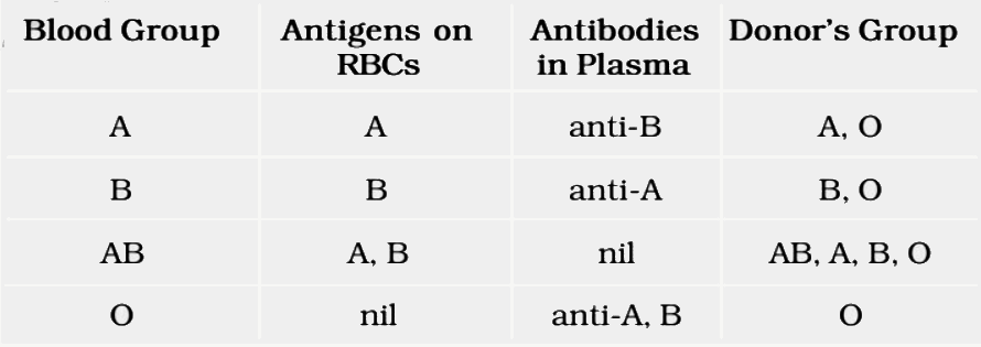
- ABO grouping is determined by the presence or absence of two surface antigens, namely A and B, on the red blood cells (RBCs). Additionally, the plasma of different individuals contains two natural antibodies, which are proteins produced in response to specific antigens.
- The distribution of these antigens and antibodies in the four blood groups—A, B, AB, and O—is provided in the table below.

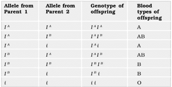
- ABO blood groups are controlled by the gene I. The plasma membrane of the red blood cells has sugar polymers that protrude from its surface and the kind of sugar is controlled by the gene. The gene (I) has three alleles IA, IB and i.
- The alleles IA and IB produce a slightly different form of the sugar while allele i does not produce any sugar.
- Because humans are diploid organisms, each person possesses any two of the three I gene alleles.
- IA and IB are completely dominant over i, in other words when IA and i are present only IA expresses (because i does not produce any sugar), and when IB and i are present IB expresses.
- But when IA and IB are present together they both express their own types of sugars: this is because of co-dominance. Hence red blood cells have both A and B types of sugars.
- Since there are three different alleles, there are six different combinations of these three alleles that are possible, and therefore, a total of six different genotypes of the human ABO blood types. How many phenotypes are possible?

- You probably know that during blood transfusion, any blood cannot be used; the blood of a donor has to be carefully matched with the blood of a recipient before any blood transfusion to avoid severe problems of clumping (destruction of RBC).
- From the above mentioned table it is evident that group ‘O’ blood can be donated to persons with any other blood group and hence ‘O’ group individuals are called ‘universal donors’.
- Persons with ‘AB’ group can accept blood from persons with AB as well as the other groups of blood. Therefore, such persons are called ‘universal recipients’.
Rh grouping
- In addition to the ABO system, there is another important blood grouping based on the presence of the Rh antigen, which is similar to an antigen found in Rhesus monkeys (hence the name Rh). The majority of humans (approximately 80 percent) have this Rh antigen on the surface of their red blood cells (RBCs) and are classified as Rh positive (Rh+). Those individuals in whom the Rh antigen is absent are categorized as Rh negative (Rh-).
- If an Rh-negative person is exposed to Rh-positive blood, specific antibodies against the Rh antigens will be formed. Therefore, Rh compatibility must also be considered before blood transfusions.
- A particular case of Rh incompatibility can arise when an Rh-negative pregnant mother carries an Rh-positive fetus. During the first pregnancy, the Rh antigens of the fetus are not exposed to the Rh-negative blood of the mother due to the separation provided by the placenta. However, during the delivery of the first child, there is a chance of the mother's blood being exposed to small amounts of Rh-positive blood from the fetus.
- In response, the mother's blood starts producing antibodies against the Rh antigen. In subsequent pregnancies, these Rh antibodies from the Rh-negative mother can cross into the bloodstream of the Rh-positive fetus and destroy the fetal red blood cells. This condition, known as erythroblastosis fetalis, can be life-threatening for the fetus or result in severe anemia and jaundice.
- To prevent this, it is crucial to administer anti-Rh antibodies to the Rh-negative mother immediately after the delivery of the first child.
|
1243 videos|2222 docs|849 tests
|


