Cnidaria: Defensive structures and their mechanism | Zoology Optional Notes for UPSC PDF Download
| Table of contents |
|
| Defense Structure in Cnidaria |
|
| Distribution of Nematocyst |
|
| Mechanism of Defense |
|
| Types of nematocysts |
|

Defense Structure in Cnidaria
- Coelenterates possess specialized defensive structures known as stinging cells or nematocysts within their body walls.
- The presence of these cnidocytes gives rise to the name "Cnidaria" for the phylum.
- Cnida, a fluid-filled membranous capsule, resides within each cnidocyte.
- Cnidocytes serve various functions, including defense, locomotion, adhesion, and prey capture.
- The presence of these defensive structures is a key characteristic of coelenterates.
- Cnidocytes are specialized cells found in cnidarians that contain stinging organelles called cnidae, such as nematocysts.
- The term "cnidocyte" originates from the Greek words "knide," meaning nettle, and "blast," meaning germ.
- In many cnidarians such as Hydra, cnidocytes originate from modified interstitial cells in the epidermis, though in some groups, they may also occur in the gastrodermis.
- When fully developed, cnidocytes migrate to the tentacles via amoeboid movement through the mesoglea.
Structure of cnidocyte
- cnidocyte is an oval or rounded cell with a basal nucleus on one side. Inside the cnidocyte an oval or pyriform bladder called as stinging capsule is present. This stinging capsule is also called as nematocyst. The nematocyst consists of a tiny bulb made of chitin.
- The bulb contains a toxic fluid, often termed hypnotoxin, which is primarily composed of proteins, including neurotoxins and enzymes. On end of this bulb is extended as a narrow, long, hollow tube like filament which is coiled round the poisonous sac.
- This filament is called as thread tube. The base of the thread tube is swollen to form a shaft. Inside the shaft there are three large spines called as barbs and three spiral rows of minute spines called as barbules. The shaft is externally covered by a lid-like structure called as operculum.
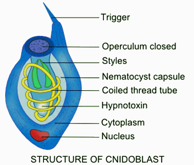
- The outer end of the cnidocyte projects freely beyond the epidermal surface as a tiny, pointed hair-like process called cnidocil or trigger. Groups of supporting rods surround the central core of cnidocil.
- The central core is structurally similar to the cilium with fibers in 9+2 pattern. The cytoplasm of the cnidocyte contains contractile muscle fibrils.
- The cell organelles present in the cytoplasm of the cnidocyte include endoplasmic reticulum, free ribosomes, Golgi bodies, mitochondria and multi-vesicular bodies as revealed by electron microscopic studies.
Distribution of Nematocyst
- Nematocysts are distributed individually or, very rarely, in groups throughout the outer layer of a cnidarian's body. They are notably absent from the base of the organism, but are abundant in the mouth region and on the tentacles, where they assemble into clusters known as "batteries."
- A "battery" of nematocysts consists of two prominent central nematocysts encircled by 10-12 smaller ones, all enclosed within a single large epithelial-muscle cell.
- Notably, these specialized cells, known as cnidocytes, do not originate in the tentacles; instead, they are initially formed in the outer epidermal layer and later migrate to the tentacles.
- During this migration, a significant number of cnidocytes congregate and encyst in clusters within the gastrovascular cavity, ultimately forming a battery of nematocysts.
Mechanism of Defense
- The discharge or explosion of nematocysts takes place when cnidocil is stimulated by food, prey or enemy. Both the presence of food and touch together initiates the process of explosion and not any one alone.
- Hence both the mechanical stimulations like contact of food and chemical stimulations like approaching enemy are involved in the mechanism of action of nematocysts.
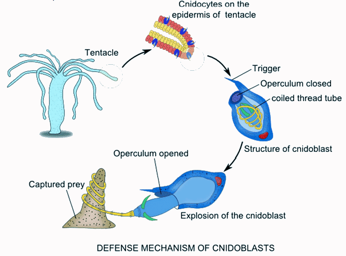
- Though the exact mechanism of discharge and enzymes involved are not known, but is very much evident that the discharge of nematocysts is a local response triggered by mechanical and chemical stimuli and does not require direct involvement of the nervous system, though neural coordination may influence cnidocyte deployment.The wall of nematocyst remains impermeable to water except during discharge.
- On stimulation, the wall of the capsule suddenly increases its permeability causing rapid intake of water and consequently the osmotic pressure inside the capsule increases. Now as a result the operculum is forced to open up, then the coiled thread tube turns inside out and finally the whole nematocyst explodes to the outside.
- As the thread tube everts, the barbs and barbules present inside the shaft unfold to the outside. The thread tube once discharged cannot be withdrawn in other words; the nematocyst once exploded cannot be used again. After the explosion the cnidocytes migrate to the gastro vascular cavity and are digested. The exploded nematocysts are replaced within 48 hours.
Types of nematocysts
- There are about 30 different types of nematocysts found in phylum Coelenterata. Their type is constant for particular species. As far as Hydra is concerned, there are four basic types of nematocysts which serve various functions.
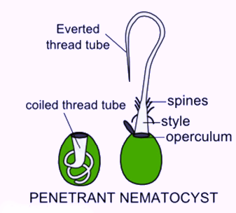
- The following is the description of all of them. Penetrant nematocyst: These are also known as stenotele.
- These kinds of nematocysts are very large and complex compared to other types. These nematocysts are pear-shaped almost occupying the entire space of cnidocyte in which it lies. Its thread is also long and hollow, coiled transversely and bearing three large barbs and three rows of small spines.
- When the thread tube is discharged, it shoots out with great explosive force to pierce the victim body and injects the poisonous fluid which paralyses or kills it outright. The hydra then seizes its prey with tentacles and draws it into its mouth.
Volvent nematocyst: These are also known as desmoneme. These kinds of nematocysts are small and pear-shaped. They contain a short, thick, spineless elastic thread tube forming single loop. When discharged, it tightly coiled round the small projections like hair or bristles of the prey and thus stopping the movement of the prey.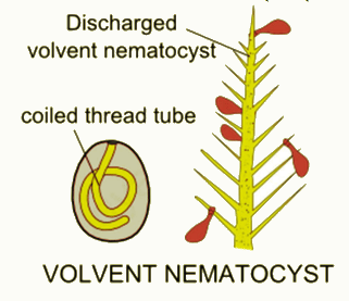
Stereoline glutinant nematocyst: These are also known as small glutinant atrichous isorhizas. These kinds of nematocysts are oval or elongated in shape. They do not have shaft. They discharge a straight unarmed thread tube open at the tip. This kind of nematocysts is useful in attachment and anchorage.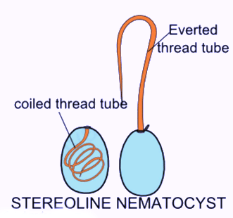
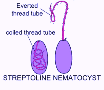
Streptoline glutinant nematocyst: These are also known as large glutinant or holotrichous isorhizas. These kinds of nematocysts are oval or cylindrical. Their thread tube is long with a narrow shaft which forms three or four coils. It bears a spiral row of small spines. These are mainly useful in attachment and to impede the movement of small animals.
|
198 videos|351 docs
|
FAQs on Cnidaria: Defensive structures and their mechanism - Zoology Optional Notes for UPSC
| 1. What are the primary defensive structures found in Cnidaria? |  |
| 2. How do nematocysts function in the defense mechanism of Cnidaria? | |
| 3. In which parts of the Cnidarian body are nematocysts typically distributed? | |
| 4. What are the different types of nematocysts found in Cnidaria, and what are their specific functions? | |
| 5. How do the defensive mechanisms of Cnidaria compare to those of other marine organisms? | |


mock tests for examination
,Important questions
,Cnidaria: Defensive structures and their mechanism | Zoology Optional Notes for UPSC
,Previous Year Questions with Solutions
,Cnidaria: Defensive structures and their mechanism | Zoology Optional Notes for UPSC
,shortcuts and tricks
,Semester Notes
,video lectures
,Cnidaria: Defensive structures and their mechanism | Zoology Optional Notes for UPSC
,Extra Questions
,past year papers
,practice quizzes
,Exam
,Summary
,Objective type Questions
,Free
,Viva Questions
,Sample Paper
,study material
,ppt
,MCQs
;


Cnidaria: Defensive structures and their mechanism Free PDF Download
Importance of Cnidaria: Defensive structures and their mechanism
Cnidaria: Defensive structures and their mechanism Notes
Cnidaria: Defensive structures and their mechanism UPSC Questions
Study Cnidaria: Defensive structures and their mechanism on the App
|
© EduRev
|
Education Revolution
|


|


within 7 days!