Fate Map | Zoology Optional Notes for UPSC PDF Download
| Table of contents |

|
| Introduction |

|
| Construction of Fate Map |

|
| Natural markings |

|
| Fate map of typical chordate blastula |

|
Introduction
The destiny of a cell refers to its eventual role in the normal developmental process. To discern the fate of a specific cell, researchers label the cell and observe the structures it integrates into. Once the destinies of all cells in an embryo are determined, a fate map is constructed. This map, presented as a diagram, illustrates the organism at an early developmental stage, outlining the future roles of each cell or region in the later stages of development. Essentially, a fate map provides insights into which portions of the egg or early embryo contribute to specific tissues or structures in the more advanced stages of development.
In certain animals, a precise and rigid fate map exists, where specific sections of the egg or blastomeres in the cleavage stage consistently contribute to particular regions in the larva or adult organism. Examples of such animals include the nematode C. elegans and urochordate tunicates, like sea squirts. Conversely, some animals lack a highly predictable fate map, as observed in mammals, while others possess distinct fate maps.
Construction of Fate Map
Several techniques have been developed for constructing a fate map, with the most crucial ones involving the tracking of natural colors and artificial markings. In practical terms, a "mark" is introduced onto or into the egg or embryo using various agents, including traditional substances like charcoal, dye, or soot, as well as modern agents such as fluorescent molecules like rhodamine-conjugated dextran, proteins like green fluorescent protein (GFP), or enzymes produced by injected genes or mRNAs. Regardless of the method chosen, the underlying principle remains consistent: a mark is made at a specific time "0," and its location is observed at subsequent times "x." Additionally, it's essential to have a reference for orienting the marks on an embryo in relation to an asymmetric feature, such as a pigment disparity or a distinctive structure.
Natural markings
Certain eggs, such as those of ascidians, naturally contain pigments in their cytoplasm. In Styela eggs, four distinct colored regions have been identified: a light upper hemisphere, a postero-ventral yellow crescent, an antero-dorsal grey crescent, and a vegetal area with dark grey yolky substance. The destiny of these areas can be easily tracked, revealing that the upper clear cytoplasm contributes to epidermal ectoderm, the grey crescent area differentiates into prospective neurectodem and notochord, the yellow crescent becomes prospective mesoderm, and the dark grey yolky area forms prospective endoderm.
Artificial markings involve three methods to label early blastomeres for tracing their fate:
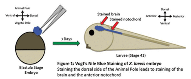
- Vital Staining: Early embryologists utilized "vital dyes" that could stain cells without causing harm. Walter Vogt, in 1929, pioneered this technique, using agar chips impregnated with vital dyes like Nile Blue or Nile Red. These chips were placed on specific cells, allowing the dye to be absorbed into the yolk platelets within those cells. This method facilitated the tracking of cell movements and the identification of the tissues they contributed to. Examples of vital stains include mild blue, sulfate, neutral fed, Jenus green, etc.

- Carbon Particle Marking: Introduced by Spratt in 1946, this technique involves applying tiny carbon particles to the surface of blastomeres. These particles adhere to the cell surface, enabling the tracking of cell movements and determination of their fate, particularly in processes like primitive streak formation in chick embryos.
- Radioactive Isotope Labeling: Radioactive isotopes such as C14 and P are employed to label early blastomeres. By carefully monitoring the path of these radioactive isotopes, the fate of blastomeres can be ascertained.
Fate map of typical chordate blastula
The chordate blastula exhibits distinct presumptive areas, including:
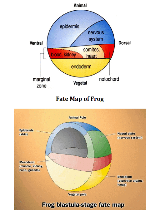
- An extensive ectodermal region in the animal hemisphere, forming the epidermal layer of the skin, known as epidermal ectoderm.
- A relatively smaller ectodermal region below the epidermal ectoderm, referred to as neurectodem, contributing to the development of the neural tube and nervous system.
- A crescent-shaped area beneath the neurectoderm, identified as the notochordal area, giving rise to the embryo's notochord.
- Two lateral areas on either side of the notochordal area, constituting the potential mesoderm.
- The majority of blastomeres in the yolky vegetal hemisphere collectively forming the potential endoderm.
- At the caudal margin of the notochordal area, a small strip of blastomeres known as the prechordal plate region. This region primarily gives rise to some of the head mesoderm.

|
181 videos|346 docs
|


