Morphogenesis and Morphogen | Zoology Optional Notes for UPSC PDF Download
Introduction
In the preceding unit, you delved into the diverse patterns of cleavage, the cleavage mechanism, and the transformation of a single-layered blastula into a gastrula with three germ layers. Unit 15 focuses on postgastrulation changes, encompassing the rearrangement of embryo cells to establish a definitive body form and the differentiation of organs and organ systems from germ layers, collectively termed morphogenesis.
Objectives
Upon completion of this unit, you should be able to:
- List various types of morphogenetic processes and cell movements.
- Describe morphogenetic processes leading to the formation of the neural tube in amphibians and chicks.
- Explain the formation of the heart as a mesodermal derivative.
- Discuss the origin of germ cells from primordial germ cells in the endoderm.
Morphogenetic Processes
In Unit 14, the transition from a fertilized egg to a blastula was explored. Now, the focus shifts to the rearrangement of cells within the gastrula, leading to the development of adult organs from three primary layers: ectoderm, mesoderm, and endoderm.
Cellular Diversity and Differentiation
A single fertilized egg generates diverse structures, a phenomenon known as differentiation. This process involves cytodifferentiation, histodifferentiation, and the development of organ shapes, all facilitated by morphogenetic processes. These processes include cell division direction and amount, changes in cell shape, cell migration, cell growth, cell death, and alterations in cell membrane and extracellular matrix.
Types of Morphogenetic Processes
Epithelial Transformations
Early embryonic changes and organ rudiment formation involve folding and spreading movements in epithelial cells. These movements are vital for shaping the embryo. Several morphogenetic processes contribute to these transformations:
Palisading: Thickening of epithelium through cell elongation precedes any change, visible in the formation of neural plates and ectodermal structures like the lens, ear, and nasal rudiments.
Evagination and Invagination: Outward folding is termed evagination, while inward folding is invagination. Examples include the formation of neural and laryngo-tracheal tubes.
Groove Formation: Folding along a line results in groove formation, exemplified by the formation of the neural tube and laryngo-tracheal tube.
Inpocketing or Infolding: The formation of lens vesicles or otic vesicles demonstrates inpocketing or infolding, creating pouches from epithelial thickening.
Branched Structures: Folds and pouches undergo modification to form branched structures, influencing the development of various glands.
Cell Shape Changes: Folding or bending of a cell sheet may change the shape of individual cells, influencing the overall shape of the epithelium. This is observed in processes like epiboly during amphibian gastrulation.
Cell Spreading: Cells spread to cover specific areas during development, accompanied by changes in cell shape such as thinning and flattening.
Cell Migration: Mesenchymal cells and primordial germ cells detach from major layers, migrating to new locations and developing into programmed structures.
Role of Cell Death
Selective cell death plays a crucial role in shaping various structures during embryo development. Examples include regions of cell death between developing digits in a chick, contributing to the formation of separate digits.
In summary, Unit 15 explores the intricate morphogenetic processes that shape embryonic development, leading to the formation of diverse organs and tissues.
Modes of Cell Movement
In the previous subsection, various types of morphogenetic processes were explored, highlighting the mobile nature of embryonic cells. This section delves into the changes in cell shape during morphogenetic processes and the factors guiding cells to their designated locations.

Fibroblasts and Cell Mobility
Fibroblasts, the precursors to connective tissue, serve as a model for understanding cell mobility mechanisms. The movement of fibroblasts occurs in two phases:
Adhesion Phase: Cells stretch to the limits of their plasma membrane.
Detachment Phase: The hind part of the cell is pulled forward, propelling the cell by generating thin, fan-shaped regions called lamellae.
- Lamellae have a flat extension called lamellipodium.
- Ruffled lamellipodia are observed at the leading edges of moving cells.
Mechanisms Directing Cell Movement
To understand how cells move to specific positions, different mechanisms are proposed:
Chemotaxis (a): Directed movement in response to a concentration gradient of a chemical factor. Example: migration of embryonic lymphocytes.
Haptotaxis (b): Directed movement in response to a concentration gradient of an adhesive molecule present in the extracellular matrix.
Galvanotaxis (c): Movement in response to a potential difference between cells. Voltage differences between embryonic regions may influence morphogenesis.
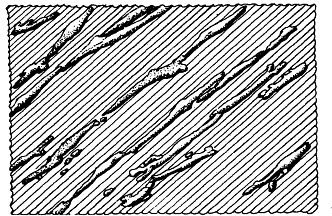
Contact Guidance (d): Physical factors influence cell movement. Cells detect discontinuities in their substratum and migrate accordingly.
 The mammalian cells shown alignhg themselves on the grooved surfam
The Role of Cytoskeletal Structures in Cell Movement
The cytoskeleton, comprising microtubules, microfilaments, and intermediate filaments, plays a crucial role in mediating changes in cell shape and movement during embryonic development.
Microtubules: Hollow cylindrical rods formed by protofilaments. They contribute to the elongation or palisading of epithelial cells.
Microfilaments: Meshwork of actin polymers, involved in the contraction that narrows apical surfaces during cell folding.
Intermediate Filaments: Filaments of intermediate size, with various classes serving functions in different tissues.
Adhesion of Cells to Extracellular Cell Matrix
Cells adhere to each other or substratum surfaces in their environment. Adhesion is mediated by receptor proteins in the plasma membrane. Extracellular matrix (ECM) molecules, including proteoglycans, collagens, and glycoproteins, form a meshwork in the intercellular space.
Basal Lamina: Dense sheet on the basal surface of epithelial cells formed by certain ECM molecules.
Receptor Proteins: Located in the plasma membrane, mediating adhesion to various ECM molecules.
Morphogenesis of an Ectodermal Derivative
During vertebrate body development, distinct regions in the three germ layers of the gastrula undergo segregation to form the rudiments of future organs and tissues. This section focuses on the partitioning of the ectoderm and the intricate process of neurulation in frog and chick embryos.
Neurulation in Amphibians
Neural Plate Formation
- Initiation: Dorsal ectoderm flattens and thickens, forming the neural plate.
- Cell Shape Changes: Neural plate cells transition from cuboidal to columnar, becoming distinct from surrounding ectoderm cells.
- Neural Folds: Edges of the neural plate rise to create neural folds along the embryo's flanks.
- Neural Groove: Central depression forms between neural folds, extending along the middorsal line.
- Neural Tube Formation: Fusion of neural folds results in the formation of the neural tube.
- Regionalization: Neural tube regionalizes, with the cephalic end developing into the brain, characterized by swellings and constrictions.
Neural Crest Cells
- Separation: Neural crest cells separate from the neural ectoderm.
- Migration: These cells migrate to various body parts, giving rise to diverse tissues.
Neurulation in Chick

- Similarities with Amphibians: Neurulation in chick shares similarities with amphibians but exhibits differences.
- Sequential Process: In birds, reptiles, and mammals, neurulation proceeds sequentially, with the anterior part ahead of the posterior region.
- Cell Shape Changes: Neural ectoderm cells undergo changes similar to amphibians, transitioning from cuboidal to columnar shapes during neural plate formation.
- Neural Tube Formation: Begins in the anterior region before progressing posteriorly.
Mechanisms of Neural Plate Formation
- Elongation: Neural plate shaping involves elongation of neural epithelial cells and accompanying apical shrinkage.
- Microtubule Role: Microtubules stabilize the elongated state of cells rather than being directly involved in elongation.
- Bending of Neural Plate: The bending of the neural plate to form the neural tube is likely due to the contraction of actin microfilaments during the rolling process.
Morphogenesis of Mesodermal Derivatives
In this section, the early development of organs derived from the mesoderm, positioned between ectoderm and endoderm tissues, will be explored. The mesoderm cells in the neurula stage organize into five distinct regions, each giving rise to specific organs.
Mesodermal Regions and Organ Derivatives
Chordamesoderm:
- Separates as a middorsal strip from the rest of the mesoderm.
- Establishes the body axis, with the anterior part forming head mesoderm and the remaining part contributing to the notochord.
Dorsal Mesoderm (Paraxial Mesoderm):
- Located on either side of the spinal cord in the back of the embryo.
- Segments into somites, which give rise to connective tissues, muscles, cartilage, and dermis.
Intermediate Mesoderm:
- Thin stalk connecting paraxial mesoderm with the rest of the mesodermal sheet.
- Gives rise to the urogenital system.
Lateral Plate Mesoderm:
- A sheet of loosely connected cells on either side of the gut.
- Splits into somatic mesoderm (associated with ectoderm) and splanchnic mesoderm (associated with endoderm).
- The space between these regions becomes the future coelomic cavity.
- Gives rise to the heart, blood vessels, blood cells, and the lining of body cavities.
- Limb components, excluding muscles, are derived from lateral plate mesoderm.
Head Mesoderm:
- Located in the head region, contributes to the development of head muscles.
Development of Limb (Overview):
- Limb development exemplifies organ differentiation from lateral plate mesoderm.
- Detailed limb development is discussed in Unit 17.
Development of Heart in Amphibians
- The heart and pericardial cavity develop from lateral mesoderm.
- After gastrulation, mesodermal mantles grow anteriorly and ventrally.
- Proliferation of cells below the gut forms a cord, which hollows out to become the endocardium.
- Splanchnic mesodermal layer surrounds the endocardium, forming the epimyocardium.
- Mesodermal mantles fuse ventrally, and the pericardial cavity forms, lined by the pericardium.
Development of Heart in Chick
- In amniotes like chicks, the heart develops initially as a pair of tubes.
- Tubes fuse to form a single tube, and subsequent development leads to a tubular heart.
- Migration of splanchnic mesoderm cells below the foregut forms two tube groups.
- Tubes fuse below the gut to create the endocardium, surrounded by myocardium.
- Development proceeds anteriorly and posteriorly, forming the tubular heart.
- The tubular heart transforms into a four-chambered structure through looping and bending, involving changes in myocardial epithelium.
Development of Blood Cells
In this subsection, we delve into the intricate process of blood cell development, primarily focusing on erythrocytes or red blood cells (RBCs). The understanding of this process is primarily derived from studies on birds and mammals.
Overview of Blood Cell Development
- Blood comprises various cell types, including erythrocytes, leukocytes (granulocytes, monocytes), platelets, plasma cells, and lymphocytes.
- All blood cells, including erythrocytes, have a limited lifespan, necessitating continuous replacement from hematopoietic stem cells in the bone marrow.
- Hematopoietic stem cells are undifferentiated and pluripotent, capable of extensive proliferation and generating differentiated cells as well as embryonic cells of their type.
Hierarchy of Blood Cell Development
CFU - M, L (Myeloid and Lymphoid Colony Forming Unit):
- Pluripotent stem cell giving rise to both red and white blood cells, as well as itself.
- Derives CFU - S and CFU - L.
CFU - S (Somatic Stem Cell) and CFU - L (Lymphoid Stem Cell):
- Pluripotent stem cells with lesser potentiality than CFU - M, L.
- CFU - S can generate erythrocytes, granulocytes, monocytes, and platelets.
- CFU - L can give rise to lymphocytes and plasma cells.
Committed Stem Cells:
- BFU - E (Blood Forming Unit - Erythroid) is committed to the erythroid pathway, specifically forming erythrocytes.
Erythrocyte Development Process
BFU - E Cell:
- Differentiates into proerythroblasts through repeated divisions.
Proerythroblast Stage:
- Active RNA synthesis and proliferation.
Erythroblast Stage:
- Chromosomal condensation and initiation of hemoglobin synthesis.
Polychromatophilic Stage:
- Increased hemoglobin synthesis; decreased RNA synthesis.
Orthochromatic Stage:
- Nucleus inactivated; cell division no longer possible.
Reticulocyte Stage:
- Nucleus extruded; some hemoglobin synthesis persists.
Erythrocyte:
- No more synthetic activity; fully functional cell in the blood stream.
Regulation and Hormonal Influence
- Erythropoietin:
- Hormone secreted in the kidneys.
- Influences the transformation of BFU - E progeny into proerythroblasts.
- Deficient O2 supply enhances erythropoietin production, leading to increased erythrocyte production.
Site of Blood Cell Formation
Bone Marrow:
- Major site for blood cell formation in adult mammals.
- In embryos, hematopoietic stem cells originate in the mesodermal blood islands of the yolk sac and subsequently colonize the liver, spleen, and bone marrow.
- Differentiation begins in the yolk sac, progressing to the fetal liver, and ultimately in the bone marrow.
Embryonic Blood Cell Differentiation:
- Initiates in the yolk sac, then occurs in the fetal liver, and finally in the bone marrow.
- Mouse embryo differentiation timeline: yolk sac (8th day), fetal liver (12th day), bone marrow (16th day onward).
- Human fetus differentiation: yolk sac (9th day), bone marrow (after the first trimester).
Morphogenesis of Endodermal Derivatives
In this section, we shift our focus to the endoderm and its derivatives, specifically examining the development of the gut tube, respiratory apparatus, and primordial germ cells (PGC).
Endodermal Organ Development
- The endoderm, the innermost germ layer, primarily contributes to the gut tube and its accessory organs, respiratory apparatus, and primordial germ cells.
Origin of Endodermal Organs
Pharyngeal Region:
- Paired pouches in the pharyngeal region form gill slits, giving rise to structures such as the eustachian tube, thymus, and parathyroid gland in higher vertebrates.
Thyroid Gland:
- Cords of endodermal cells from the pharynx penetrate ventrally into the underlying mesoderm, forming the thyroid gland.
Trachea and Lungs:
- A midventral groove (laryngo-tracheal groove) in the pharynx leads to the development of the trachea and lungs.
Liver, Pancreas, and Gall Bladder:
- Posterior evaginations at the future duodenum level initiate the development of the liver, pancreas, and gall bladder.
Mesodermal Involvement:
- Organs that bud off from the pharynx and gut are not purely endodermal; mesoderm invests them, supplying blood vessels and shaping their structure.
Primordial Germ Cells (PGCs):
- Migrate from the endoderm and deserve special attention.
Origin and Migration of Primordial Germ Cells in Frog
Frog Embryos:
- Germplasm near the vegetal pole contains germinal granules.
- Primordial germ cells (PGCs) migrate dorsally around the posterior gut region, entering the mesodermal region (genital ridges).
- Gonads develop from these genital ridges, with PGCs dividing and forming gonial cells that undergo meiosis to produce gametes.
Migration Mechanism:
- PGCs align with the dorsal mesentery, and fibronectin mediates their adhesion and alignment during migration to the gonads.
Origin and Migration PGCs in Amniotes
Birds and Reptiles:
- PGCs originate in the epiblast, moving into the germinal crescent region, appearing larger than other embryonic cells.
- PGCs invade blood vessels, passively carried through the bloodstream to reach the genital ridges.
Mammals:
- PGCs, with a high concentration of alkaline phosphatase, emerge in the yolk sac endoderm.
- Migrate through the developing gut into the dorsal mesentery and finally to the genital ridge.
- Movement involves filopodia, chemotaxis, and contact guidance.
Summary
- Understanding morphogenesis involves exploring various processes and mechanisms mediating cell movements in vertebrate embryos.
- Cytoskeletal components like microtubules, microfilaments, and intermediate filaments play crucial roles in cell movement.
- Organ rudiments from the ectoderm, mesoderm, and endoderm undergo morphogenesis, including neurulation, heart development, and blood cell differentiation.
- The gonads, though mesodermal, have primordial germ cells originating from endodermal cells, showcasing intricate migration processes.
|
181 videos|351 docs
|


