General Anatomy Chapter Notes | Anatomy - NEET PG PDF Download
| Table of contents |
|
| Skeleton |
|
| Ossification |
|
| Growing End |
|
| Blood Supply |
|
| Sesamoid Bones |
|
| Pneumatic Bones |
|
| Joints |
|
| Synovial Joints |
|
| Muscles |
|
| Portal Venous Circulation |
|

Skeleton
The human skeleton is made up of 206 bones at adulthood. Skeletons are of two types:- Axial Skeleton: It consists of 80 bones of skull, vertebral column and rib cage.
- Appendicular Skeleton: It consists of 126 bones of limbs, pectoral girdle and pelvic girdle.
Structure of a Long Bone
A long bone has a shaft (diaphysis) and two ends (epiphyses). The metaphysis is a part of the diaphysis adjacent to the epiphyses.1. Diaphysis
- Forms the shaft (central region) of the bone.
- Composed of a thick tube of compact bone that encloses the marrow cavity.
2. Metaphysis
- A part of the diaphysis.
- Serves as the growth zone between the diaphysis and epiphysis during bone development.
3. Epiphyses
- Expanded articular ends of the bone.
- Separated from the shaft by the epiphyseal plate during bone growth.
- Composed of spongy bone surrounded by a thin layer of compact bone.
Ossification
Ossification is the process by which new bone material is laid down by cells called osteoblasts. There are two types of ossification:1. Membranous Ossification: This involves the direct formation of bone into the mesenchyme, which is embryonic connective tissue.
2. Endochondral Ossification: This type involves the formation of bone in a precursor model of cartilage.
Types of Bones and Their Ossification:
1. Membrane (Dermal) Bones: These bones ossify in membrane through intramembranous ossification. They are derived from mesenchymal condensations. Examples include the flat bones of the skull and face, the mandible, and the clavicle.
2. Cartilaginous Bones: These bones ossify in cartilage through endochondral ossification. They are derived from preformed cartilaginous models. Examples include the bones of the extremities (limbs) and weight-bearing parts of the axial skeleton such as the vertebral column and thoracic cage.
3. Membrano-Cartilaginous Bones: These bones are initially formed in membrane but later ossify partly in cartilage. Examples include the clavicle, mandible, occipital bone, temporal bone, and sphenoid bone.
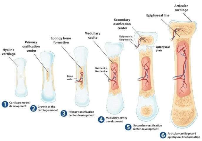
Cartilaginous Ossification
Cartilaginous ossification involves the formation of bone in cartilage and includes primary and secondary centers of ossification.Primary Center of Ossification:
- In long bones, bone tissue first appears in the diaphysis (middle of the shaft).
- Primary centers begin to appear at around 6 weeks of intrauterine life.
- Chondrocytes multiply, forming trabeculae, and cartilage is progressively eroded and replaced by bone, extending towards the epiphysis.
- A layer of perichondrium surrounding the cartilage forms the periosteum, which generates osteogenic cells that create a collar encircling the exterior of the bone and remodels the medullary cavity on the inside.
- The nutrient artery enters through the nutrient foramen, a small opening in the diaphysis, invading the primary center of ossification and bringing osteogenic cells (osteoblasts on the outside, osteoclasts on the inside).
- The canal of the nutrient foramen is directed away from the more active end of the bone when one end grows faster than the other. If bone grows at the same rate at both ends, the nutrient artery is perpendicular to the bone.
Secondary Center of Ossification:
- Secondary centers generally appear at the ends (epiphysis) of long bones.
- Secondary ossification mostly occurs after birth, except for secondary centers around the knee joint (distal femur and proximal tibia), which appear during the last weeks of fetal life or immediately after birth.
- Epiphyseal arteries and osteogenic cells invade the epiphysis, depositing osteoblasts and osteoclasts that erode cartilage and build bone. This process occurs at both ends of long bones but only at one end of digits and ribs.
Ossification Centers Present at Birth:
- Diaphysis of long bones
- Skull bones
- Vertebral column
- Ribs and sternum
- Some foot bones (talus, calcaneus, cuboid)
Primary Centers of Ossification for Carpal and Tarsal Bones:
- Primary centers of ossification for all carpal and tarsal bones, except for the talus, calcaneus, and cuboid, appear after birth.
 Ossification of Long Bone
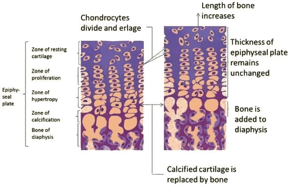 Figs. A and B: Growth of bone—lengthwise: (A) Four zones of epiphyseal cartilage; (B) Conversion of calcified cartilage into bone.
Growing End
- The growing ends of bones in upper limb are upper end of humerus and lower ends of radius and ulna.
- In lower limb, the lower end of femur and upper end of tibia are the growing ends.
- The nutrient foramen is directed away from the growing end of the bone; their directions are indicated by a memory aid: ‘Towards the elbow I go, from the knee I flee’.
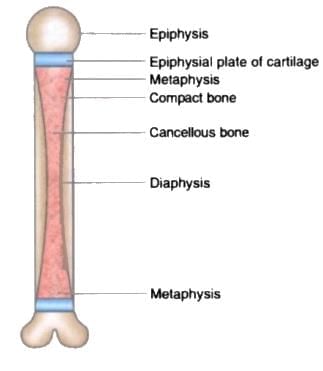 Parts of a young long bone.
Blood Supply
- Nutrient artery enters the diaphysis (shaft) through the nutrient foramen, runs obliquely through the cortex, and divides into ascending and descending branches in the medullary cavity.
- Each branch divides into a number of small parallel channels which terminate in the adult metaphysis by anastomosing with the epiphysial, metaphysial and periosteal arteries.
- It supplies medullary cavity, inner 2/3 of cortex and metaphysis.
- The nutrient foramen is directed away from the growing end of the bone. Memory aid: Towards the elbow I go, from the knee I flee.
- Bony skeleton is divided into two parts: (1) The axial skeleton and (2) The appendicular skeleton.
- Axial skeleton is the central core unit, consisting of the skull, vertebrae, ribs, and sternum.
- Appendicular skeleton comprises the bones of the extremities.
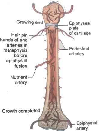 Arterial supply of a long bone. The upper epiphysis (growing end) has not yet fused with the diaphysis.
Sesamoid Bones
- Development and Function: Sesamoid bones form within certain tendons to reduce friction, thereby protecting the tendon from excessive wear.
- Common Locations: These bones are typically found where tendons cross the ends of long bones in the limbs.
Sites of Sesamoid Bones
- In the Ear: The lenticular process of the incus is a sesamoid bone, making it the fourth ossicle of the middle ear.
- In the Hand: Two sesamoid bones are located in the distal portions of the first metacarpal bone, within the tendons of the adductor pollicis and flexor pollicis brevis.
- In the Wrist: The pisiform bone is a sesamoid bone located within the tendon of the flexor carpi ulnaris. It typically develops between ages 9 and 12.
- In the Knee: The patella is a sesamoid bone located within the quadriceps tendon.
- Other Locations:
- Fabella: Found in the lateral head of the gastrocnemius muscle behind the knee joint.
- Sesamoid Bone in the Tendon of Peroneus Longus: Located where the tendon binds around the cuboid bone.
- In the Foot: Two sesamoid bones in the distal portions of the first metatarsal bone, within the tendons of the flexor hallucis brevis.
Pneumatic Bones
Note: Pneumatic bones are the irregular bones which contain air-filled cavities within them.
- They are generally produced during development by excavation of bone by pneumatic diverticula (air sacs) from an air-filled space such as the nasal cavity.
- E.g., maxilla, frontal, sphenoid, and ethmoid bones and a part of the mastoid part of the temporal bone.
Note: At birth, the mastoid is not pneumatized, but becomes aerated over the first year of life.
Types of Epiphysis
- Pressure Epiphysis: These are the parts of the bone involved in weight transmission and are located within the joint capsule (intracapsular). Examples include the heads of the humerus and femur, as well as the condyles of the humerus, femur, and tibia.
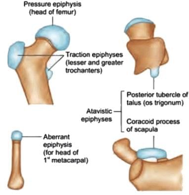 Types of epiphyses: Pressure and traction epiphyses; Atavistic epiphyses; Aberrant epiphysis.
 - Traction Epiphysis: These epiphyses are found at the ends of bones and develop due to the traction exerted by attached muscles. They are located outside the joint capsule (extracapsular). Examples include the greater and lesser tubercles of the humerus and the greater and lesser trochanters of the femur. Traction epiphyses ossify later than pressure epiphyses. The mastoid process is also considered a traction epiphysis.
- Atavistic Epiphysis: Atavistic epiphyses are fused bones that are separate in lower animals but fused in humans. Examples include the coracoid process of the scapula and the posterior tubercle of the talus (known as the trigonum).
- Aberrant Epiphysis: Aberrant epiphyses are unusual and not always present. They represent deviations from the norm. Examples include the epiphysis at the head of the first metacarpal and the epiphyses at the base of other metacarpals.
Joints
Joints between bones are classified structurally into three types based on the connecting material and presence of a cavity: fibrous, cartilaginous, and synovial. Functionally, joints are divided into synarthroses (immovable), amphiarthroses (slightly mobile), and diarthroses (freely mobile).
Structural Classification:
- Fibrous Joints: Bones are connected by fibrous tissue, allowing minimal movement. Examples include:
- Sutures: Joints like the spheno-vomerine suture in the skull.
- Gomphosis: Tooth and socket joints.
- Syndesmosis: Joints like the middle and inferior radioulnar joints or the tibia-fibula connection.
- Cartilaginous Joints: These are divided into primary and secondary types:
- Primary Cartilaginous Joints (Synchondroses): Bones are joined by hyaline cartilage, permitting no movement but allowing growth in length. They are strong, with the bone-cartilage interface resisting separation. Examples include epiphyseal plates (between epiphysis and diaphysis in growing bones), spheno-occipital, and manubrio-sternal synchondroses.
- Secondary Cartilaginous Joints (Symphyses): Bones are connected by hyaline cartilage and fibrocartilage, typically in the midline, allowing slight movement. Examples include the pubic symphysis, midline intervertebral joints, and the sternal angle joint. Intervertebral discs, containing a gel-filled cavity in fibrocartilage, are also secondary cartilaginous joints with limited mobility based on fibrous tissue content.
- Synovial Joints: These are freely mobile joints with a cavity, corresponding to diarthroses in functional classification.
Functional Classification and Examples:
- Synarthrosis (Immovable): Fibrous joints, e.g., skull sutures.
- Amphiarthrosis (Slightly Mobile): Cartilaginous joints, e.g., pubic symphysis.
- Diarthrosis (Freely Mobile): Synovial joints.
Types of Cartilaginous Joints and Examples:
- Synchondrosis: Spheno-occipital joint, epiphysio-diaphyseal joint.
- Symphysis: Midline intervertebral joints, sacrococcygeal joint.
Synovial Joints
Synovial joints are freely mobile and classified by their axes of movement: uniaxial (plane, hinge, pivot), biaxial (condylar, ellipsoid), and multiaxial (saddle, ball and socket).
Types of Synovial Joints and Examples:
- Uniaxial Joints:
- Plane: Allow gliding movements. Examples: acromioclavicular, intercarpal, intertarsal joints.
- Hinge: Permit flexion and extension. Examples: elbow, interphalangeal joints.
- Pivot (Trochoid): Allow rotation. Examples: atlanto-axial, superior and inferior radioulnar joints.
- Biaxial Joints:
- Condylar: Permit movement in two planes. Examples: temporomandibular, knee joint. (Note: Some authors classify these as modified hinge joints.)
- Ellipsoid: Allow flexion, extension, abduction, and adduction. Examples: atlanto-occipital, wrist (radiocarpal), metacarpophalangeal (knuckle) joints. (Note: Some authors classify these as condylar joints.)
- Multiaxial Joints:
- Saddle: Permit movement in multiple planes. Examples: malleus-incus, sternoclavicular, first carpometacarpal, calcaneocuboid joints.
- Ball and Socket: Allow movement in all directions. Examples: incus-stapes, shoulder, hip, talocalcaneonavicular joints.
Few authors consider these joints condylar: Atlanto-occipital, wrist (radio-carpal), metacarpo-phalangeal (knuckle). Few authors consider these joints as modified hinge: Temporo-mandibular, knee joint.
The knee joint is a complex synovial joint involving more than two bones. Structurally, the femorotibial joint resembles a hinge joint but is classified as a condylar synovial joint, formed between the two condyles of the femur and tibia. Additionally, it includes a saddle joint between the femur and patella, allowing for varied movements.
Muscles
Skeletal muscle fibers are oriented either parallel or oblique to the muscle's line of pull. Parallel fibers allow a longer range of contraction, while oblique fibers provide greater force but reduced range. The sartorius muscle exemplifies parallel fiber arrangement.
Muscle Fiber Arrangements:
Parallel Fasciculi: Fibers align with the line of pull, enabling a greater range of movement. Types include:
- Quadrilateral: e.g., thyrohyoid, pronator quadratus.
- Strap-like: e.g., sternohyoid, sartorius.
- Strap-like with tendinous intersections: e.g., rectus abdominis.
- Fusiform: e.g., biceps brachii, digastric.
Oblique Fasciculi: Fibers are angled to the line of pull, increasing power but reducing movement range. Types include:
- Convergent Fasciculi: Fibers converge at the insertion point for maximized contraction. These may be:
- Triangular: e.g., temporalis.
- Fan-shaped: e.g., temporalis.
- Spiral or Twisted Fasciculi: Fibers are twisted or spiraled, enhancing strength and flexibility. Examples: trapezius, latissimus dorsi, pectoralis major, supinator.
- Cruciate Muscles: Fibers or fasciculi are arranged in superficial and deep planes, crossing in an ‘X’ pattern. Examples: sternocleidomastoid, masseter, adductor magnus.
- Sphincteric Fasciculi: Fibers surround an opening, constricting it upon contraction. Examples: orbicularis oculi (around the eye), orbicularis oris (around the mouth).
- Pennate Fasciculi: Fibers resemble a feather, with fleshy fibers (like barbs) inserting into a tendon (like the shaft), increasing force but reducing movement range. Types include:
- Unipennate: e.g., flexor pollicis longus, extensor digitorum longus.
- Bipennate: e.g., rectus femoris, flexor hallucis longus.
- Multipennate: e.g., tibialis anterior, subscapularis, deltoid (acromial fibers).
Common Intramuscular Injection Sites:
- Upper Arm (Deltoid): Injection at 5 cm distal to the acromion or 4 cm proximal to deltoid insertion to avoid the circumflex humeral nerve.
- Gluteal Region: Upper outer (superolateral) quadrant to avoid superior/inferior gluteal vessels and sciatic nerve; injected into gluteus medius.
- Thigh (Vastus Lateralis):
- Infants: Upper lateral quadrant below the greater trochanter.
- Adults: Middle third of the lateral thigh.
Hybrid (Composite) Muscles: These muscles have multiple fiber sets and nerve supplies, often located at boundaries between muscle groups, incorporating fibers from adjacent groups with dual innervation. Examples and their nerve supplies:
- Trapezius: Spinal accessory nerve (motor), ventral rami of C3, C4 (proprioception).
- Digastric: Trigeminal nerve (anterior belly), facial nerve (posterior belly).
- Pectoralis Major: Medial pectoral nerve, lateral pectoral nerve.
- Subscapularis: Upper subscapular nerve, lower subscapular nerve.
- Brachialis: Musculocutaneous nerve (motor), radial nerve (proprioceptive).
- Flexor Digitorum Profundus: Median nerve (lateral half), ulnar nerve (medial half).
- Opponens Pollicis: Median nerve (superficial part), ulnar nerve (deep part).
- Ilio-psoas: Direct branches of L1–L3 anterior rami (psoas major), femoral nerve (iliacus).
- Pectineus: Femoral nerve (anterior fibers), obturator nerve (posterior fibers).
- Biceps Femoris: Tibial part of sciatic nerve (long head), common peroneal nerve (short head).
- Adductor Magnus: Tibial part of sciatic nerve (ischial part), obturator nerve (adductor part).
Portal Venous Circulation
- Definition: Portal circulation involves a capillary network situated between two veins, where blood passes through two sets of capillaries before returning to the heart.
- Hepatic Portal System: Blood from abdominal organs flows through two capillary networks (one in the organ, one in the liver) before returning to the heart.
- Hypophyseal Portal System: A portal circulation connects the median eminence and infundibulum of the hypothalamus to the adenohypophysis (anterior pituitary).
- Renal Glomeruli: The glomerular capillary bed, located between afferent and efferent arterioles, may function similarly to a portal circulation, though most sources, including Gray’s Anatomy, do not classify it as such.
|
42 docs|7 tests
|
FAQs on General Anatomy Chapter Notes - Anatomy - NEET PG
| 1. What is the process of ossification in bones? |  |
| 2. What are sesamoid bones and their significance? | |
| 3. How does blood supply affect bone growth and healing? | |
| 4. What are the characteristics of synovial joints? | |
| 5. What is the role of the growing end in bone development? | |


video lectures
,Semester Notes
,Important questions
,Viva Questions
,ppt
,General Anatomy Chapter Notes | Anatomy - NEET PG
,Free
,General Anatomy Chapter Notes | Anatomy - NEET PG
,past year papers
,Objective type Questions
,practice quizzes
,General Anatomy Chapter Notes | Anatomy - NEET PG
,Summary
,Extra Questions
,Previous Year Questions with Solutions
,shortcuts and tricks
,MCQs
,mock tests for examination
,Sample Paper
,Exam
,study material
;


Chapter Notes: General Anatomy Free PDF Download
Importance of Chapter Notes: General Anatomy
Chapter Notes: General Anatomy
Chapter Notes: General Anatomy NEET PG Questions
Study Chapter Notes: General Anatomy on the App
|
© EduRev
|
Education Revolution
|


|


