Lower Limb - 2 Chapter Notes | Anatomy - NEET PG PDF Download

Muscles (Medial Thigh)
Muscles of medial thigh
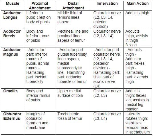
Together, the five listed muscles function as thigh adductors, but their roles extend beyond simple adduction. For instance, they contribute to hip joint flexion when the knee is flexed and are engaged during walking. The spinal cord segmental innervation is noted (e.g., “L2, L3, L4” indicates that the adductor longus is innervated by nerves from the second to fourth lumbar spinal segments). The primary segmental innervation is highlighted in bold (e.g., L3). Injury to these spinal cord segments or their associated motor nerve roots can lead to paralysis of the affected muscles.
- All muscles in the thigh's adductor compartment attach to the femur, except for the gracilis, which attaches to the tibia.
- Rider’s bone is a calcified tendon or sesamoid bone within the tendon of the adductor longus.
Muscles of the Posterior Thigh
The muscles in the back of the thigh are responsible for several important movements. They are mainly involved in extending the hip and flexing the knee, along with some rotational movements. Here's a detailed look at these muscles:
1. Semimembranosus
Location of Attachment: The semimembranosus muscle attaches to the posterior part of the medial condyle of the tibia. A part of its tendon forms the oblique popliteal ligament, which connects to the lateral femoral condyle.
Nerve Supply: This muscle is innervated by the tibial division of the sciatic nerve, specifically from the spinal segments L5, S1, and S2.
Functions: The semimembranosus performs several key actions:
- Extends the thigh at the hip joint.
- Flexes the leg at the knee and rotates it medially when the knee is bent.
- When both the thigh and leg are flexed, it can help extend the trunk.
2. Semitendinosus
Location of Attachment: The semitendinosus muscle has its origin at the ischial tuberosity, similar to the semimembranosus.
Nerve Supply: Like the semimembranosus, it is innervated by the tibial division of the sciatic nerve (L5, S1, S2).
Functions: The semitendinosus muscle is responsible for:
- Thigh Extension: It extends the thigh at the hip joint.
- Leg Flexion: Flexes the leg at the knee and rotates it medially when the knee is flexed.
- Trunk Extension: Assists in extending the trunk when both the thigh and leg are flexed.
3. Biceps Femoris
Location of Attachment: The biceps femoris has two heads:
- Long Head: Originates from the ischial tuberosity.
- Short Head: Originates from the linea aspera and lateral supracondylar line of the femur.
Nerve Supply: The long head is innervated by the tibial division of the sciatic nerve (L5, S1, S2), while the short head is innervated by the common fibular division of the sciatic nerve (L5).
Functions: The biceps femoris performs the following actions:
- Leg Flexion: Flexes the leg at the knee and rotates it laterally when the knee is bent.
- Thigh Extension: Extends the thigh at the hip, such as during the initial phase of walking.
Overview of Hamstrings: Collectively, the semimembranosus, semitendinosus, and biceps femoris (along with the posterior part of the adductor magnus) are known as the hamstrings. These muscles are crucial for:
- Thigh Extension: Extending the thigh at the hip.
- Leg Flexion: Flexing the leg at the knee.
- Opposing Quadriceps: Working against the quadriceps femoris, which is responsible for extending the leg at the knee.
Role in Walking: The hamstring muscles are the primary extensors of the hip during walking. They play a vital role in propelling the body forward with each step.
Sacrotuberous Ligament: Some researchers suggest that the sacrotuberous ligament is a remnant of the tendon of the long head of the biceps femoris, which has degenerated over time. This ligament contributes to the stability of the pelvis and sacrum.
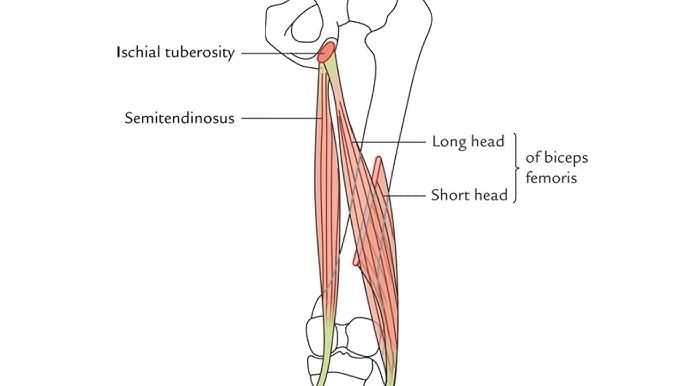 Attachments of semitendinosus and biceps femoris
Muscles of the Gluteal Region (Abductors and Rotators of the Thigh)
Muscles of gluteal region (abductors and rotators of thigh)
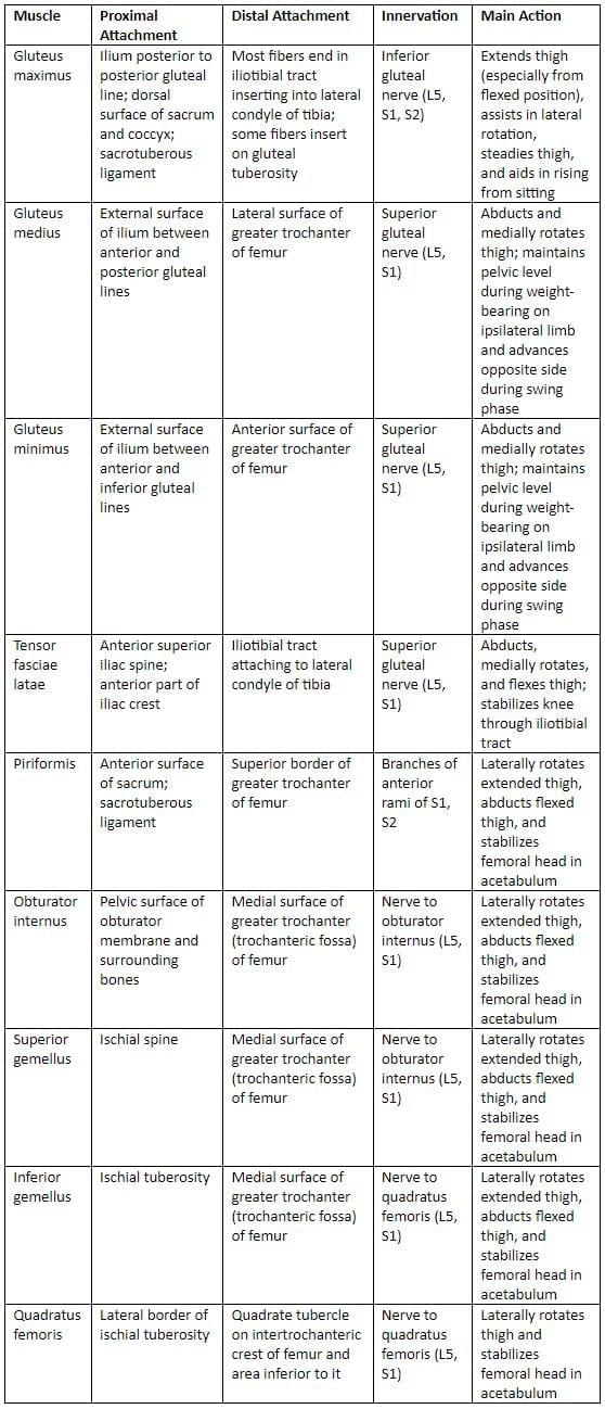
The spinal cord segments innervating specific muscles are denoted, for example, “S1-S2” indicates the piriformis muscle is supplied by nerves from the first two sacral segments. Bolded segments (e.g., S1) represent the primary innervation. Damage to these spinal cord segments or their associated motor nerve roots can lead to paralysis of the affected muscles. The gemelli muscles merge with the obturator internus tendon, attaching to the femur’s greater trochanter, together forming the triceps coxae. The six lateral rotators of the thigh—piriformis, obturator internus, superior and inferior gemelli, quadratus femoris, and obturator externus—also stabilize the hip joint.
- The gluteus maximus aids in transitioning from sitting to standing by extending the trunk relative to the thigh. Acting from the pelvis, it extends a flexed thigh to align with the trunk.
- The superior gemellus and obturator internus are innervated by the nerve to the obturator internus (L5, S1-S2).
- The inferior gemellus and quadratus femoris are innervated by the nerve to the quadratus femoris (L4-L5, S1).
- The piriformis muscle, a key structure in the gluteal region, originates from the anterior surface of the sacrum, the gluteal surface of the ilium, and the sacrotuberous ligament. Its rounded tendon passes through the greater sciatic foramen and inserts at the tip of the greater trochanter.
- The gluteus maximus is the largest muscle in the human body.
Iliotibial Tract
The iliotibial tract is a specialized extension of the deep fascia (known as fascia lata) situated on the outer aspect of the thigh.
This tract acts as a crucial attachment site for the lateral portions of the gluteus maximus and tensor fasciae latae muscles. It extends down to Gerdy’s tubercle, which is located on the anterolateral aspect of the tibia, specifically on the lateral tibial condyle.
Notably, the iliotibial tract does not connect to the tibial tuberosity. This structure plays a vital role in stabilizing the knee joint in both semi-flexed and fully extended positions, making it essential for activities such as walking and running.
However, individuals engaged in activities like long-distance running or cycling may develop Iliotibial Band Syndrome, a condition characterized by pain along the iliotibial tract.
Trendelenburg Test
- Left Superior Gluteal Nerve Injury: When there is an injury to the left superior gluteal nerve, which causes paralysis of the left gluteus medius, the patient's right lower limb drops during the swing phase of walking. This makes it difficult to lift the foot off the ground, leading to a lurch to clear it.
- Motor Loss: Injury to the superior gluteal nerve results in specific motor loss, weakening hip abduction. This leads to a disabling limp because the gluteus medius and minimus are ineffective.
- Standing on One Foot: When asked to stand on one foot, the gluteus medius and minimus should contract as the opposite foot leaves the ground. This contraction prevents the pelvis from tilting.
- Gluteus Medius and Minimus Dysfunction: In the case of a superior gluteal nerve lesion, when the patient stands on one leg, the pelvis on the unsupported side drops. This indicates that the gluteus medius and minimus on the supported side are not working, resulting in a positive Trendelenburg test.
- Pelvic Drop and Gait: When the pelvis drops on the unsupported side, the lower limb appears too long and fails to clear the ground during the swing phase. To compensate, the person leans away from the unsupported side, lifting the pelvis to create enough space for the foot during the swing phase. This results in a lurching gait. If both sides are affected, it becomes a waddling gait.
- Conditions Associated with Trendelenburg Test: The Trendelenburg test may also be positive in cases of fracture of the greater trochanter (where the gluteus medius attaches) or a dislocated hip joint.
Hybrid Muscles
- Pectineus: This muscle is innervated by both the femoral and obturator nerves.
- Adductor Magnus: This muscle is innervated by the obturator nerve and the tibial part of the sciatic nerve.
- Biceps Femoris: The biceps femoris is innervated by the tibial part of the sciatic nerve and the common peroneal nerve.
- The sartorius muscle is the longest muscle in the human body, running from the hip to the knee.
- The gluteus maximus is the strongest extensor of the thigh at the hip joint, particularly active when walking uphill, climbing stairs, or rising from a seated position.
- The iliopsoas muscle is a major flexor of the thigh, attaching to the lesser trochanter of the femur.
- The tensor fascia lata and rectus femoris muscles can flex the thigh at the hip joint and extend the leg at the knee joint effectively.
- In the lower limb, the gracilis muscle is the most commonly used for surgical grafting procedures.
Anterior and Lateral Leg Muscles
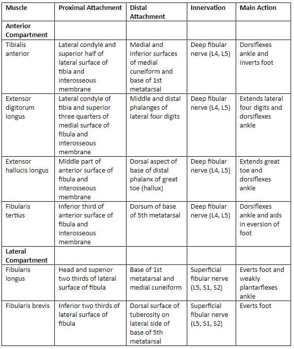
The spinal cord segmental innervation for these muscles is indicated, with specific nerve roots supplying each muscle. For example, "L4, L5" means the tibialis anterior is supplied by nerves from the fourth and fifth lumbar segments. Damage to these spinal cord segments or the motor nerve roots can lead to paralysis of the affected muscles.
Muscles of Anterior and Lateral Leg Region
- Extensor TDH(Tom, Dick and Harry):
- T. Tibialis anterior
- D. Extensor digitorum longus
- H. Extensor hallucis longus
- Peroneus (fibularis) tertius is found in the anterior leg compartment with the extensors.
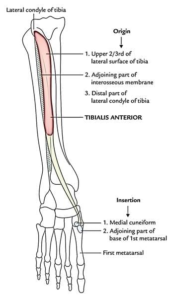 Attachments of tibialis anterior
Posterior Leg Muscles
Superficial muscles of the posterior leg region.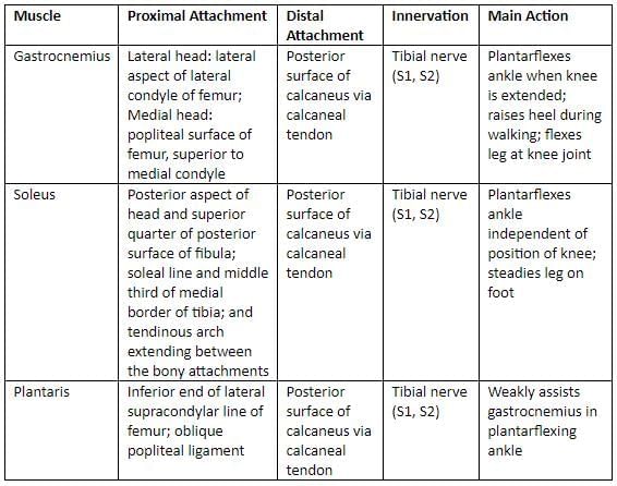
The innervation by spinal cord segments is indicated (e.g., “S1, S2” means the nerves for these muscles come from the first and second sacral segments of the spinal cord). Damage to these segments or the motor nerve roots can result in paralysis of the affected muscles.
Deep muscles of the posterior leg region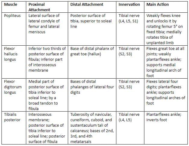
The innervation by spinal cord segments is indicated (e.g., ‘S2, S3’ means the nerves supplying the flexor hallucis longus are derived from the second and third sacral segments of the spinal cord). Damage to these segments or the motor nerve roots can lead to paralysis of the affected muscles.
Posterior Leg (Calf) Muscles: Superficial and Deeper Group
- Superficial (GPS). Triceps surae - Gastrocnemius, plantaris, and soleus.
- Deep. Flexor TDH (Tom, Dick, and Harry): Tibialis posterior, flexor digitorum longus, flexor hallucis longus → Cause flexion at ankle and toe joints.
The popliteus muscle starts inside the knee joint from the outer side of the femur and connects with the outer part of the knee joint. It attaches to the back of the tibia. This muscle is supplied by the tibial nerve and helps to unlock the knee by rotating the tibia in a foot that is not planted. It also works with the hamstring muscles to assist in bending the knee.
The tibialis posterior originates from the inner back edges of the tibia and fibula, as well as the interosseous membrane. It has many attachments on the bones of the foot but does not connect to the talus bone.
The plantaris has a long, thin tendon that looks like a nerve, which is why it is sometimes called freeman's nerve. This muscle is vestigial and may be absent in 5–10% of people, and its tendon can be used for grafting.
- The muscle in the leg with the highest concentration of muscle spindles is the soleus.
- The muscle that works as an organ of proprioception in the leg is the plantaris.
- The soleus is referred to as the peripheral heart because it aids in pumping blood in the circulatory system.
In cases of posterior compartment syndrome, the patient often holds the foot in a plantar flexion position to relieve pressure on the fascia and muscles. Pain is felt during dorsiflexion due to the stretching of the posterior leg muscles.
The plantaris has a thin tendon that can stretch and rupture during severe dorsiflexion.
- The muscle called the “workhorse” of foot plantar flexion is the soleus.
Arteries of the Lower Limb
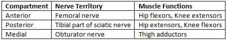
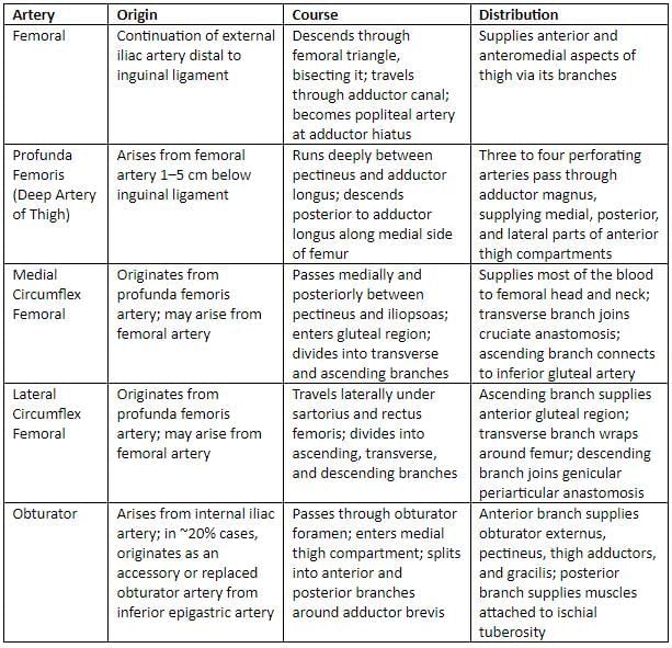
- The profunda femoris artery is the primary blood supply to all three thigh compartments, including the posterior compartment.
- The anterior compartment is also supplied by the femoral artery, while the medial compartment receives additional supply from the obturator artery.
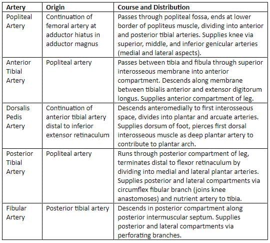
- The popliteal artery gives rise to five genicular arteries (including the middle genicular artery) that supply the knee joint, consisting of two superior (medial and lateral) and two inferior (medial and lateral) arteries.
- The popliteal pulse is the most challenging peripheral pulse to palpate.
- The popliteal artery is the most susceptible artery to aneurysm formation in the body.
Arteries of the Anterior and Medial Thigh
Arteries of the Leg Region
Femoral Artery and Its Branches
- Superficial external pudendal artery is a branch of the femoral artery.
- Superficial epigastric artery is also a branch of the femoral artery.
- The midinguinal point (MIP) is found halfway between the anterior superior iliac spine and the pubic symphysis. At this point, the pulse of the femoral artery can be felt, typically within ± 1 cm on either side.
- It is crucial to refer to this location as the midinguinal point rather than the midpoint of the inguinal ligament.
- The femoral artery is located superficially in the femoral triangle, making it the preferred artery for cannulation and injecting dye for procedures like angiography.
- It is also the preferred vessel for coronary angiography and angioplasty.
- The primary artery supplying the muscles of the thigh is the profunda femoris artery.
- The largest branch of the profunda femoris artery is the lateral circumflex femoral artery.
- A distinctive characteristic of the lateral compartment of the leg is that it does not have its own artery.
- The nutrient artery of the femur, which is a branch of the posterior tibial artery, is the largest nutrient artery in the body.
- The peroneal artery is the largest and most important branch of the posterior tibial artery.
Veins of Lower Limb
Femoral vein is the continuation of the popliteal vein, starting from the adductor opening and ending behind the inguinal ligament as the external iliac vein. It is located posterior and lateral to the femoral artery in the distal adductor canal.
Tributaries
- Veins accompanying the superficial epigastric, superficial circumflex iliac, and external pudendal arteries join the long saphenous vein before entering the saphenous opening.
- Other tributaries include the profunda femoris vein, lateral and medial circumflex femoral veins, and the deep external pudendal vein.
The femoral vein typically contains four or five valves, with the two most consistent valves found just below the entry of the profunda femoris and near the inguinal ligament.
The great saphenous vein originates from the dorsal venous arch and ascends 2.5 cm in front of the medial malleolus. In the thigh, it relates to the medial cutaneous nerve and passes through the saphenous opening to join the femoral vein 2.5 cm below the inguinal ligament.
The saphenous opening (fossa ovalis) is a gap in the fascia lata, sealed by a membrane of areolar tissue called the cribriform fascia, which is penetrated by the great saphenous vein. The middle of the opening is approximately 4 cm inferolateral to the pubic tubercle.
The small (short) saphenous vein forms when the dorsal vein from the fifth digit (smallest toe) merges with the lateral end of the dorsal venous arch. It then runs upwards behind the lateral malleolus, alongside the lateral edge of the tendo calcaneus, and is accompanied by the sural nerve on its lateral side. It continues up the middle of the back of the leg and eventually drains into the popliteal vein.
- This vein contains 7–13 valves, with one situated near its termination.
- The great saphenous vein, located in front of the medial malleolus at the ankle, is the preferred site for venesection (cut-down).
- The femoral vein is the preferred choice for intravenous infusions in infants, children, and patients experiencing peripheral circulatory failure.
- The longest vein in the body is the great saphenous vein.
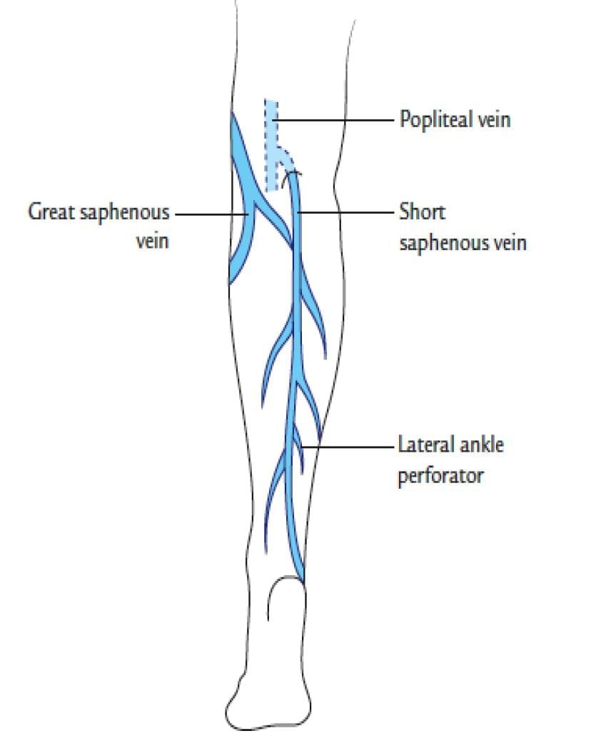 Short saphenous vein
Lymphatics
- Cloquet and Rosenmüller lymph nodes are located in the femoral canal. This is a crucial area where important blood vessels pass through.
- Most of the lymph from the lower limb is drained by the lower vertical group of superficial inguinal lymph nodes.
- The Vein of Leonardo da Vinci, also known as the vein of the posterior arch, is a branch of the great saphenous vein.
Adductor Canal
The Adductor Canal, also referred to as the Sub-sartorial or Hunter's canal, extends from the apex of the femoral triangle to the popliteal fossa.
Foot
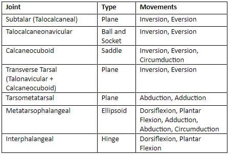
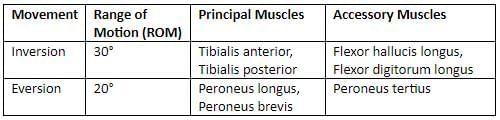
- The primary site for inversion and eversion is the subtalar joint, with significant movements occurring at the talocalcaneonavicular joint.
- Additional joints involved include the transverse tarsal (midtarsal) joints, specifically the calcaneocuboid and talonavicular joints.
Movements of inversion and eversion
- The talocalcaneonavicular joint is a ball and socket synovial joint.
- The tarsometatarsal joint (Lisfranc joint) forms the articulation between the tarsal bones and the metatarsals.
Muscles of the Sole
The dorsal interossei play a crucial role in moving the toes apart from each other, while the plantar interossei are responsible for bringing the toes together. All the interossei, along with the lumbricals, are involved in bending the metatarsophalangeal (MTP) joints and straightening the interphalangeal (IP) joints. When the lumbricals and interossei are paralysed, it can lead to a condition known as claw foot.
Functions of Dorsal and Plantar Interossei
- The dorsal interossei are responsible for moving the toes away from the midline of the second metatarsal.
- The plantar interossei bring the third, fourth, and fifth toes together.
All the interossei in the sole primarily receive their nerve supply from the lateral plantar nerve. However, the first interosseous muscle is supplied by the medial plantar nerve.
Plantar Arches
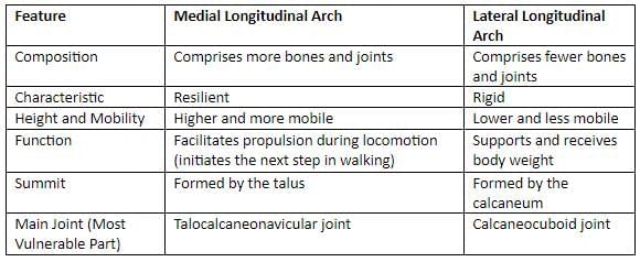
Differences between the medial and lateral longitudinal arches.
- The transverse arch of the foot is formed by the bases of the five metatarsals, the cuboid, and the cuneiforms. The wedge-shaped intermediate and lateral cuneiforms help maintain this arch.
- The transverse arches are supported by the interosseous, plantar, and dorsal ligaments, the short muscles of the first and fifth toes (notably the transverse head of the Adductor hallucis), and the tendon of the Peroneus longus, which spans the arch's piers.
- The tendons of the Peroneus longus and Tibialis posterior are critical for maintaining the transverse arch.
- The talus serves as the keystone for the medial longitudinal arch, while the cuboid is the keystone for the lateral longitudinal arch.
- The talocalcaneonavicular joint is the most vulnerable part of the medial longitudinal arch, and the calcaneocuboid joint is the most vulnerable part of the lateral longitudinal arch.
- The spring ligament is the most crucial ligament for supporting the foot's arches.
- The most common foot deformity is Talipes equinovarus.
- The extensor digitorum brevis is the only intrinsic muscle located on the dorsum of the foot.
- Plantar fasciitis is also known as Policeman’s heel.
Gait Cycle
- During the walking cycle, the body's kinetic energy is lowest in the mid-stance phase, when only one foot is on the ground (single support phase), and highest during the double support phase, when both feet are on the ground.
- The tibialis anterior muscle remains active during both the swing and stance phases of the walking cycle.
|
41 docs|16 tests
|
FAQs on Lower Limb - 2 Chapter Notes - Anatomy - NEET PG
| 1. What are the primary muscles located in the medial thigh region and their functions? |  |
| 2. Which muscles are classified as posterior thigh muscles, and what roles do they play in movement? | |
| 3. What is the function of the iliotibial tract, and which muscles are associated with it? | |
| 4. Can you explain the significance of hybrid muscles in the lower limb? | |
| 5. What are the major arteries and veins of the lower limb, and what are their roles? | |


Lower Limb - 2 Chapter Notes | Anatomy - NEET PG
,Summary
,MCQs
,video lectures
,Viva Questions
,Objective type Questions
,Important questions
,study material
,practice quizzes
,past year papers
,shortcuts and tricks
,Extra Questions
,Lower Limb - 2 Chapter Notes | Anatomy - NEET PG
,Sample Paper
,mock tests for examination
,Previous Year Questions with Solutions
,Semester Notes
,ppt
,Lower Limb - 2 Chapter Notes | Anatomy - NEET PG
,Free
,Exam
;


Chapter Notes: Lower Limb - 2 Free PDF Download
Importance of Chapter Notes: Lower Limb - 2
Chapter Notes: Lower Limb - 2
Chapter Notes: Lower Limb - 2 NEET PG Questions
Study Chapter Notes: Lower Limb - 2 on the App
|
© EduRev
|
Education Revolution
|


|


