Anatomy of Monocot & Dicot Plants: Root, Stem & Leaf | Biology Class 11 - NEET PDF Download
| Table of contents |
|
| Dicotyledonous Root |
|
| Monocotyledonous root |
|
| Dicotyledonous Stem |
|
| Monocotyledonous Stem |
|
| Dorsiventral (Dicotyledonous) Leaf |
|
| Isobilateral (Monocotyledonous) Leaf |
|

For a clearer understanding of how tissues are organized in the roots, stems, and leaves of plants, it is helpful to examine the transverse sections (cross-sections) of the mature regions of these organs.
Plants are broadly classified into two categories based on the number of cotyledons (seed leaves) in their seeds:
- Dicotyledonous Plants (Dicots): These plants have two cotyledons in their seeds. Examples include beans, sunflowers, and oak trees.
- Monocotyledonous Plants (Monocots): These plants have a single cotyledon in their seeds. Examples include rice, wheat, and lilies.
By studying the transverse sections of the mature roots, stems, and leaves of dicots and monocots, we can observe the differences in tissue organization and structure between these two groups of plants.
Dicotyledonous Root
- Structure Overview: The transverse section of a sunflower root shows the internal tissue organization.
- Outermost Layer (Epiblema): Thin layer of cells, some of which develop into unicellular root hairs to assist in water and nutrient absorption.
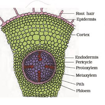 Dicot Root
- Cortex: Made up of several layers of thin-walled parenchyma cells with intercellular spaces. The innermost layer of the cortex is called the endodermis.
- Endodermis: A single layer of barrel-shaped cells without intercellular spaces. The walls of these cells have a water-impermeable waxy material called suberin, forming Casparian strips.
- Pericycle: Located next to the endodermis, consisting of a few layers of thick-walled parenchyma cells. This is where lateral roots and vascular cambium initiate during secondary growth.
- Pith: Usually small or inconspicuous in dicot roots.
- Conjunctive Tissue: Parenchymatous cells located between the xylem and phloem.
- Vascular Bundles: Typically, there are two to four patches of xylem and phloem. A cambium ring may develop between the xylem and phloem later.
- Stele: All tissues inside the endodermis, including the pericycle, vascular bundles, and pith, constitute the stele.
Monocotyledonous root
Monocotyledonous roots have a similar structure to dicot roots, including features like the epidermis, cortex, endodermis, pericycle, vascular bundles, and pith.
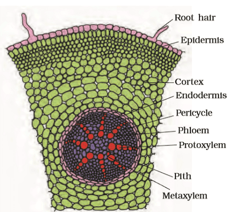 Monocot Root
- However, monocot roots typically have more than six xylem bundles(polyarch), while dicot roots have fewer.
- The pith in monocot roots is large and well-developed.
- Unlike dicot roots, monocotyledonous roots do not undergo secondary growth.
Dicotyledonous Stem
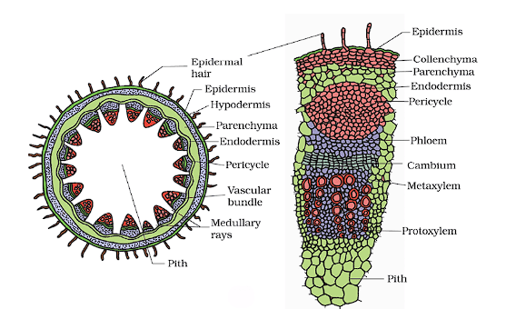
In a typical young dicotyledonous stem, the transverse section reveals various distinct features:
 Dicot Stem
- Epidermis: The outermost protective layer, covered with a thin cuticle. It may have trichomes (hair-like structures) and a few stomata (pores).
- Cortex: Located between the epidermis and pericycle, the cortex consists of multiple layers and has three sub-zones:
- Outer Hypodermis: Just below the epidermis, composed of a few layers of collenchymatous cells that provide mechanical strength to the young stem.
- Cortical Layers: Below the hypodermis, consisting of rounded, thin-walled parenchymatous cells with noticeable intercellular spaces.
- Endodermis: The innermost layer of the cortex, rich in starch grains, also known as the starch sheath.
- Pericycle: Located on the inner side of the endodermis and above the phloem, consisting of semi-lunar patches of sclerenchyma.
- Medullary Rays: Layers of radially placed parenchymatous cells found between vascular bundles.
- Vascular Bundles: Arranged in a characteristic ring formation, each bundle is conjoint, open, and features endarch protoxylem.
- Pith: Occupying the central portion of the stem, composed of rounded parenchymatous cells with large intercellular spaces.
Monocotyledonous Stem
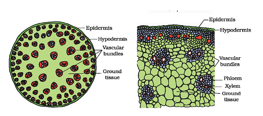
- Sclerenchymatous hypodermis: The outer layer of the monocot stem is made up of sclerenchymatous cells, which provide strength and support.
- Vascular bundles: These are scattered throughout the stem and are surrounded by a bundle sheath of sclerenchyma cells. The vascular bundles are conjoint(containing both xylem and phloem) and closed(without cambium).
 Monocot Root
- Ground tissue: There is a large amount of parenchymatous ground tissue filling the space between vascular bundles.
- Phloem parenchyma: This tissue is absent in the vascular bundles.
- Water-containing cavities: These are present within the vascular bundles, providing storage for water.
- Peripheral vs. central vascular bundles: The vascular bundles located at the periphery of the stem are generally smaller than those found in the center.
Dorsiventral (Dicotyledonous) Leaf
Epidermis: The epidermis covers both the upper (adaxial) and lower (abaxial) surfaces of the leaf and has a noticeable cuticle.
The abaxial epidermis usually has more stomata than the adaxial epidermis, which may even lack stomata.
Mesophyll: The mesophyll, located between the upper and lower epidermis, contains chloroplasts and is responsible for photosynthesis.
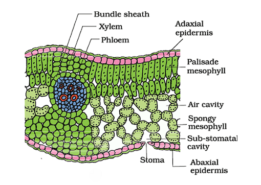 Dicot Leaf
It is made up of parenchyma tissue and consists of two types of cells:
(a) Palisade Parenchyma: These are elongated cells arranged vertically and parallel to each other, located just below the upper epidermis.
(b) Spongy Parenchyma: These oval or round cells are loosely arranged and situated below the palisade cells, extending down to the lower epidermis. There are large air spaces and cavities between these cells.Vascular System: The vascular system includes vascular bundles found in the veins and midrib of the leaf.
(a) The size of the vascular bundles depends on the size of the veins, which vary in thickness in the reticulate venation of dicot leaves.
(b) The vascular bundles are surrounded by a layer of thick-walled bundle sheath cells.
(c) In the vascular bundles, the xylem is positioned in the upper part.
Isobilateral (Monocotyledonous) Leaf
The anatomy of isobilateral leaf is similar to that of the dorsiventral leaf in many ways. It shows the following characteristic differences.
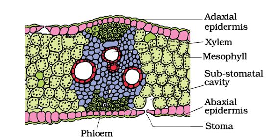 Monocot Leaf
Stomata: In isobilateral leaves, stomata are present on both surfaces of the epidermis.
Mesophyll: The mesophyll in isobilateral leaves is not differentiated into palisade and spongy parenchyma.
Bulliform Cells: In grasses, certain adaxial epidermal cells along the veins modify into large, empty, colorless bulliform cells. These cells play a crucial role in water regulation.
Water Regulation: When bulliform cells are turgid (full of water), they help expose the leaf surface. Conversely, when they are flaccid (due to water stress), they cause the leaves to curl inward to minimize water loss.
Venation: The parallel venation in monocot leaves is reflected in the similar sizes of vascular bundles (except in main veins) observed in vertical sections of the leaves.
|
150 videos|398 docs|136 tests
|
FAQs on Anatomy of Monocot & Dicot Plants: Root, Stem & Leaf - Biology Class 11 - NEET
| 1. What are the main differences between dicotyledonous and monocotyledonous roots? |  |
| 2. How can you differentiate between dicotyledonous and monocotyledonous stems? | |
| 3. What structural features are characteristic of a dorsiventral dicotyledonous leaf? | |
| 4. What are the key characteristics of an isobilateral monocotyledonous leaf? | |
| 5. Why is understanding the anatomy of monocot and dicot plants important? | |


Stem & Leaf | Biology Class 11 - NEET
,shortcuts and tricks
,Anatomy of Monocot & Dicot Plants: Root
,Anatomy of Monocot & Dicot Plants: Root
,Stem & Leaf | Biology Class 11 - NEET
,mock tests for examination
,Extra Questions
,Previous Year Questions with Solutions
,Objective type Questions
,Summary
,video lectures
,Sample Paper
,Exam
,ppt
,Viva Questions
,practice quizzes
,MCQs
,Semester Notes
,study material
,past year papers
,Anatomy of Monocot & Dicot Plants: Root
,Stem & Leaf | Biology Class 11 - NEET
,Free
,Important questions
;


Anatomy of Monocot & Dicot Plants: Root, Stem & Leaf Free PDF Download
Importance of Anatomy of Monocot & Dicot Plants: Root, Stem & Leaf
Anatomy of Monocot & Dicot Plants: Root, Stem & Leaf Notes
Anatomy of Monocot & Dicot Plants: Root, Stem & Leaf NEET Questions
Study Anatomy of Monocot & Dicot Plants: Root, Stem & Leaf on the App
|
© EduRev
|
Education Revolution
|


|


