Digestive Glands: Pancreas, Salivary Glands & Liver | Biology for Grade 11 PDF Download
The human digestive system is a complex network of organs and glands working together to break down food and absorb nutrients. Among the various glands associated with the alimentary canal, the salivary glands, liver, and pancreas play crucial roles in the digestion process. These glands secrete various digestive enzymes and fluids that help in the breakdown of food molecules into simpler substances that can be absorbed by the body. In this context, understanding the functions and mechanisms of these glands is essential to maintain a healthy digestive system.
What is Pancreas?
- The pancreas is an abdominal organ located behind the stomach and surrounded by the spleen, liver and small intestine. It is a vital part of the digestive system and is responsible for regulating blood sugar levels.
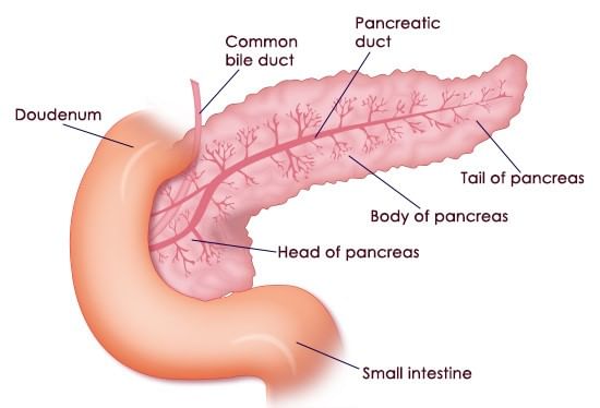 Pancreas
 - The pancreas secretes digestive enzymes such as amylase, proteases and lipase into the duodenum. These enzymes help in digesting sugar, proteins and fat respectively. Islets of Langerhans are embedded in the pancreas that secretes hormones such as insulin and glucagon into the blood.
(a) Pancreas Location
- The pancreas is located in the abdomen. A part of it is placed between the stomach and the spine. The other part finds its place in the curve of the first section of the small intestine, known as the duodenum.
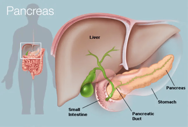 Location of Pancreas
 - The head of the pancreas is on the right side of the abdomen and is connected to the duodenum through the pancreatic duct. The tail of the pancreas extends to the left side of the body.
(b) Pancreatic Diseases
- Due to the inaccessibility of the pancreas, the evaluation of pancreatic diseases could be difficult.
- Disorders that affect the pancreas include precancerous conditions, pancreatitis, and pancreatic cancer. Each disorder exhibits different symptoms and needs different treatments.
(i) Pancreatitis
Pancreatitis is swelling when the pancreatic enzyme is secreted and begins to digest the organ itself. It could exist as painful attacks or a chronic condition that lasts for years.
(ii) Precursors to Pancreatic Cancer
The primary reason for pancreatic cancer is yet to be known, but there are risk factors that increase the danger of developing diseases. Some of the factors include smoking or hereditary cancer syndromes.
(iii) Pancreatic Cancer
Pancreatic adenocarcinoma is one of the most common forms of pancreatic cancer. It is an exocrine tumour that arises from the cells that line the pancreatic duct. A tumour of the endocrine gland accounts for less than 5% of all pancreatic tumours and is referred to as islet or neuroendocrine.
(c) Pancreas Function
The pancreas performs the following functions:
(i) Exocrine Function
The pancreas consists of exocrine glands that produce enzymes trypsin and chymotrypsin that are essential for digestion. These enzymes contain chymotrypsin and trypsin to digest proteins, amylase for the digestion of carbohydrates and lipase to break down fats. These pancreatic juices are liberated into the system of ducts and culminated in the pancreatic duct when the food enters the stomach.
(ii) Endocrine Function
The endocrine part of the pancreas comprises Islets of Langerhans that release insulin and glucagon directly into the bloodstream. They help in regulating the blood sugar levels of the body.
Salivary Glands
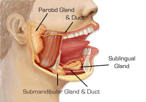 Salivary Glands
In mammals, 4 pair of salivary glands are present. These are:
1. Infra-orbital Glands
- Gland is located just below the eye-orbit.
- The duct of these glands open in the upper-jaw near the 2nd molar teeth.
2. Parotid Glands (Largest Salivary Glands)
- These glands are located just below the external auditory canal. Their duct is called Parotid duct/Stenson's duct which open in the upper jaw i.e. the Buccal-vestibule.
- Whenever in human, these glands are infected by viruses this disease is called as Mumps. Due to this, the gland swells up.
3. Sub Maxillary or Submandibular Glands
- These are located at the junction of the upper and the lower jaw.
- Their duct is called Wharton's duct (largest salivary duct). These ducts open in the lower jaw just behind the Incisor teeth.
4. Sublingual Glands
- These are the smallest salivary glands. These glands are found in the lower jaw. Many ducts arise from these glands called as the Ducts of Rivinus or Bartholin's ducts. These ducts open in the bucco-pharyngeal cavity on the ventral side of the tongue.
- Maximum saliva is secreted by the Sub-lingual glands. (Smallest salivary duct) Salivary glands are Exocrine glands. The secretion of salivary glands is termed as the saliva.
Composition of Saliva
- Water - 99.5 %
- Mucus, starch -digesting Ptyalin enzyme, lysozyme and thiocyanates and few ions like sodium, potassium, chloride, IgA antibody, urea and uric acid etc., are present.
- Ptyalin is secreted only by the parotid gland. Lysozyme and Thiocyanates mainly kill bacteria. They also check the growth of bacteria in bucco-pharyngeal cavity.
- Salivation is stimulated by cranial nerve VII & IX.
- In addition to it 5th pair of molar gland is found in Cat which is situated near to the upper molar teeth and also opens near upper molar teeth.
What is Liver?
The liver is located in the upper right portion of the abdomen. It is the largest gland in the human body that performs several important functions. It is the only organ that has the ability to regenerate efficiently.
(a) Structure of Liver
- Human Liver is made up of four lobes. Left lobe is small right proper lobe is large, two addition lobe quadrate and caudate lobe are also found on posterior side of right proper lobe.
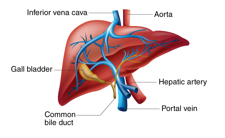 Human Liver
- It develops from endoderm. (Weight 1.5 kg, both exocrine and endocrine)
- In human it is found in right side of abdominal cavity, below the diaphragm.
- The liver is the largest gland of body.
- Right and left liver lobe are separate from each other by the falciform ligament, (Fibrous connective tissue) Which is made up of fold of peritoneum.
- Right and left hepatic duct develop from right and left liver lobe Both these ducts combine to form a Common Hepatic duct.
- Gall bladder is situated below right lobe of liver.
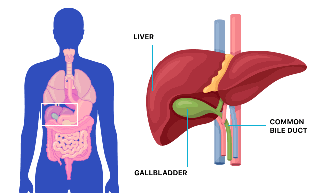 Gall bladder
 - Cystic duct of gall bladder is connected to common hepatic duct and form a common bile duct which also called ductus choledocus or common bite duct.
- Internally liver is made up of numerous polygonal lobules. These lobules are covered by fibrous connective tissue, covering layer is called Glisson's Capsule.
- Each lobule consists of radial rows of hepatic cells, two row of hepatic cells are combindely called as hepatic cord. Each hepatic cord is lined by endothelial layer.
- In between the hepatic cord, a space is present called as hepatic sinusoid. These sinusoids are filled with blood.
- Sinusoids are lined by the endothelial cells mostly but, a few macrophages cells are also present.These are called as kupffer's cells. (Phagocyte cells)
- The bile canaliculi run in between the two layers of hepatic cells in each hepatic cord. Hepatocytes (hepatic cells) pour bile into the canaliculi. Canaliculi open into branch of hepatic duct which is situated at the angular part of lobule in the Glisons capsule.
- All branches of hepatic duct of right and left lobe are combined to form right and left Hepatic duct which come out from the liver and forms a common hepatic duct.
- Hepatic artery and hepatic portal vein enter into liver and divide to form many branches. These branches are also found at the angular part of Glisson's capsule. Its fine branches open in to hepatic sinusoids. Branch of hepatic portal vein, branch of hepatic artery and branch of hepatic duct are collectively called as Portal triad.
- All hepatic sinusoids of one Glisson's capsule are open into central vein or intra lobular vein, all Central veins are combined and form one pair hepatic vein which, comes out from liver and opens into inferior vena cave.
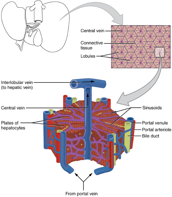 3-D view of Lobule of Liver
(b) Functions of Liver
Liver is known as chemical factory of the body. Most of the biochemical functions of the body are done by the liver.
- Secretion & synthesis of bile: This is the main function of liver. Bile is yellowish-green, alkaline fluid. In bile juice, bile salts, sodium bicarbonate, glycocholate, taurocholate, bile pigments, cholesterol, Lecithin etc. are present. Bile salts help in emulsification of fats. Bile prevents the food from putrification. It kills harmful bacteria.
- Carbohydrate Metabolism: The main centre of carbohydrate metabolism is liver. Following steps are related with carbohydrate metabolism:
(i) Glycogenesis: The conversion and storage of extra amount of glucose into glycogen from the digested food is called glycogenesis. The main stored food in the liver is glycogen.
(ii) Glycogenolysis: The conversion of glycogen into glucose back when glucose level in blood falls down is called glycogenolysis.
(iii) Gluconeogenesis: At the time of need, liver converts non-carbohydrate compounds (Eg. Amino acids, Fatty acids) into glucose. This conversion is called gluconeogenesis. This is the neo-formative process of glucose.
(iv) Glyconeogenesis: Synthesis of glycogen from lactic acid (which comes from muscles) is called glyconeogenesis. - Storage of fats: Liver stores fats in a small amount. Hepatic cell plays an important role in fat metabolism. The storage of fats increases in the liver of alcohol addict persons (Fatty liver). this storage of fats decreases the activity of liver. the damage of liver due to alcohol intake is called Alcoholic Liver cirrhosis.
- Deamination and Urea formation: Deamination is the removal of an amino group from a molecule. Enzymes that catalyse this reaction are called deaminases. Liver converts ammonia (obtained form deamination) into urea through ornithine cycle. So after the spoilage of liver, the ammonia level in the animal body is increased and the animal dies.
- Purification of blood: The spleen and liver separate dead blood cells and bacteria from the blood. Kupffer cells in liver and phagocytes in spleen perform this function.
- Synthesis of plasma proteins: Many types of proteins are present in blood plasma. Except gamma globulins all type of plasma proteins are synthesized in the liver.
- Most of the blood clotting factor are synthesized in the liver.
- Synthesis of heparin: Heparin is an anticoagulant (Mucopolysaccharide). Some heparin is also formed by basophils, that are special type of white blood cells.
- Synthesis of Vitamin A: The liver converts the b-carotene into vitamin A : B- caroteine is a photosynthetic pigment which is obtained from plants. It is abundantly found in carrot.
- Liver stores vitamin A, D, E, K and B12
- Storage of minerals: Liver stores iron in the form of ferritin. Liver also stores the, copper, zinc, cobalt, molybdenum etc Liver is a good source of iron.
- Detoxification: In this process liver converts the toxic substances into non-toxic substances. The toxic substances are formed by metabolic activities of the body. Example: Prussic acid is converted into neutral Potassium sulfocynide (It is a non-toxic salt) by the liver.
- Hematopoiesis: The formation of blood cells is called haemopoesis. In empbryonic stage R.B.C. and WBC are formed by liver.
- Yolk synthesis: Most of the yolk is synthesized in liver.
- Secretion of enzymes: Some enzymes are secreted by liver, participate in metabolism of proteins, fats and carbohydrates. For example, Dehydrogenase, cytochrome oxidase etc.
- Prothrombin and fibrinogen proteins are also formed in hepatic cells. These help in blood clotting.
- Factors I, II, V, VII, IX and X are formed in liver, which are responsible for blood clotting.
(c) Liver Diseases
- Fascioliasis: This is caused by a parasite “liver fluke”. The parasite can lie dormant in the liver for months or even years.
- Cirrhosis: This can be caused due to alcohol consumption, toxins and hepatitis. Here, the scar cells replace liver cells in a process known as fibrosis. The functionality of liver cells is destroyed, which might lead to liver failure.
- Hepatitis: It is the inflammation of the liver caused by viruses such as hepatitis A, B and C. In most cases, it leads to liver failure.
- Alcoholic Liver Disease: Uncontrolled alcohol consumption leads to liver damage. It is the most common cause of cirrhosis.
- Fatty Liver Disease: This is the result of alcohol abuse or obesity. In this disease, the vacuoles of fat build-up in the liver cells.
- Liver Cancer: Alcohol and hepatitis are the major cause of liver cancer. Hepatocellular carcinoma and cholangiocarcinoma are the two types of liver cancer.
|
219 videos|306 docs|270 tests
|
FAQs on Digestive Glands: Pancreas, Salivary Glands & Liver - Biology for Grade 11
| 1. What is the function of the pancreas? |  |
| 2. What are some common diseases of the pancreas? |  |
| 3. What is the composition of saliva? |  |
| 4. What are the functions of the liver? |  |
| 5. What are some common liver diseases? |  |


