


All Exams >
JAMB >
Biology for JAMB >
All Questions
All questions of Circulation in Animals for JAMB Exam
Which of the following engulfs pathogens rapidly?- a)Basophils
- b)Acidophils
- c)Monocytes
- d)Neutrophils
Correct answer is option 'D'. Can you explain this answer?
Which of the following engulfs pathogens rapidly?
a)
Basophils
b)
Acidophils
c)
Monocytes
d)
Neutrophils
|
|
Vijay Bansal answered |
Neutrophils are the type of WBCs which can engulf the pathogen rapidly. Neutrophils are more in number i.e. upto 60% of Total Leukocyte Count. They immediately engulf bacteria and viruses to destroy them by phagocytosis.

How many contraction nodes are found in heart of Human :-- a)One
- b)Two
- c)Many
- d)None
Correct answer is option 'B'. Can you explain this answer?
How many contraction nodes are found in heart of Human :-
a)
One
b)
Two
c)
Many
d)
None
|
|
Sathvika Tadri answered |
Two contraction nodes...SAN and AVN..option B
Which of the following is a non-granulocyte?- a)Neutrophils
- b)Monocytes
- c)Basophils
- d)Eosinophils
Correct answer is option 'B'. Can you explain this answer?
Which of the following is a non-granulocyte?
a)
Neutrophils
b)
Monocytes
c)
Basophils
d)
Eosinophils
|
|
Anjana Chakraborty answered |
Non-Granulocytes in Blood
Blood cells are divided into two groups: granulocytes and agranulocytes. Granulocytes have granules in their cytoplasm and include neutrophils, eosinophils, and basophils. Agranulocytes, on the other hand, lack granules and include lymphocytes and monocytes.
Non-Granulocyte
A non-granulocyte is simply an agranulocyte or a leukocyte that lacks granules in its cytoplasm. Monocytes are non-granulocytes and belong to the agranulocyte group of leukocytes.
Monocytes
Monocytes are the largest leukocytes in the blood, accounting for 2-8% of all leukocytes. They have a large kidney-shaped nucleus and are characterized by their ability to differentiate into macrophages, which are cells that engulf and digest foreign particles, dead cells, and cellular debris in the body.
Functions of Monocytes
Monocytes have several functions in the body, including:
1. Phagocytosis: Monocytes, when differentiated into macrophages, are capable of phagocytosis, which is the process of engulfing and digesting foreign particles, dead cells, and cellular debris in the body.
2. Antigen presentation: Monocytes play a crucial role in the immune response by presenting antigens to T cells, which are responsible for activating the immune response.
3. Cytokine production: Monocytes produce cytokines, which are signaling molecules that regulate the immune response.
Conclusion
In conclusion, monocytes are non-granulocytes and belong to the agranulocyte group of leukocytes. They are characterized by their ability to differentiate into macrophages, which are cells that engulf and digest foreign particles, dead cells, and cellular debris in the body. Monocytes also play a crucial role in the immune response by presenting antigens to T cells and producing cytokines.
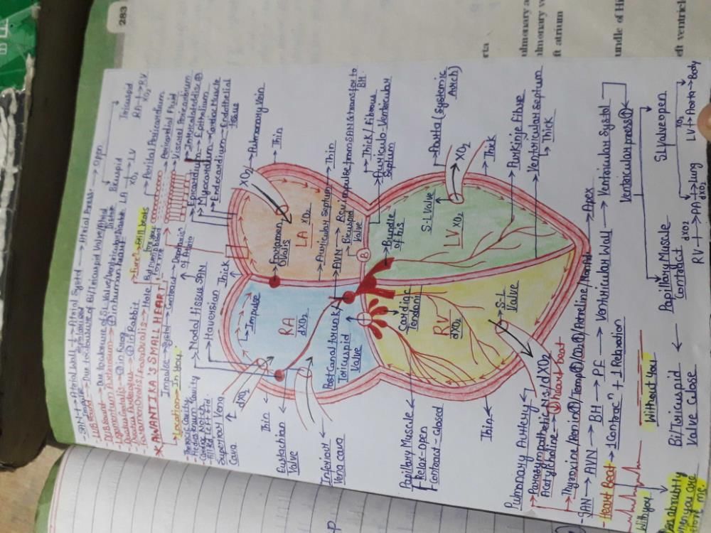
The valve situated between the left atrium and the left ventricle is called
(1) Bicuspid valve
(2) Tricuspid valve
(3) Mitral valve
(4) Eustachian tube- a)2 and 4 are correct.
- b)1 and 2 are correct.
- c)1 and 3 are correct.
- d)1, 2 and 3 are correct.
Correct answer is option 'C'. Can you explain this answer?
The valve situated between the left atrium and the left ventricle is called
(1) Bicuspid valve
(2) Tricuspid valve
(3) Mitral valve
(4) Eustachian tube
(1) Bicuspid valve
(2) Tricuspid valve
(3) Mitral valve
(4) Eustachian tube
a)
2 and 4 are correct.
b)
1 and 2 are correct.
c)
1 and 3 are correct.
d)
1, 2 and 3 are correct.
|
|
Bhaskar Ala answered |
The valve situated between left atria and the left ventricle is BICUSPID VALVE also known as MITRAL VALVE.. closure of this valve produce first heart sound "LUB"..
Can you explain the answer of this question below: Find out the wrong match:
- A:
Eosinophils – Allergic response
- B:
Basophils – Secrete histamine and serotonin
- C:
Monocytes – Secrete heparin
- D:
Neutrophils – Phagocytic and eat foreign organisms
The answer is c.
Find out the wrong match:
Eosinophils – Allergic response
Basophils – Secrete histamine and serotonin
Monocytes – Secrete heparin
Neutrophils – Phagocytic and eat foreign organisms
|
|
Vijay Bansal answered |
Monocytes and macrophages synthesize and secrete thrombospondin.
Mark the odd one:- a)Neutrophil
- b)Erythrocyte
- c)Monocyte
- d)Lymphocyte
Correct answer is option 'B'. Can you explain this answer?
Mark the odd one:
a)
Neutrophil
b)
Erythrocyte
c)
Monocyte
d)
Lymphocyte
|
|
Rajeev Saxena answered |
Erythrocytes bcs its stands for rbc's whereas others are different types of wbc's
Which of these has a closed type of circulatory system :-- a)Cockroach
- b)Fish
- c)Mollusca
- d)Scorpion
Correct answer is option 'B'. Can you explain this answer?
Which of these has a closed type of circulatory system :-
a)
Cockroach
b)
Fish
c)
Mollusca
d)
Scorpion
|
|
Gaurav Kumar answered |
Phylum Arthropoda and Mollusca include open circulatory system . Fish is member of phylum Chordata which contain close circulatory system.
Cockroach and scorpion are members of class inscecta phylum Arthropoda.
Cockroach and scorpion are members of class inscecta phylum Arthropoda.
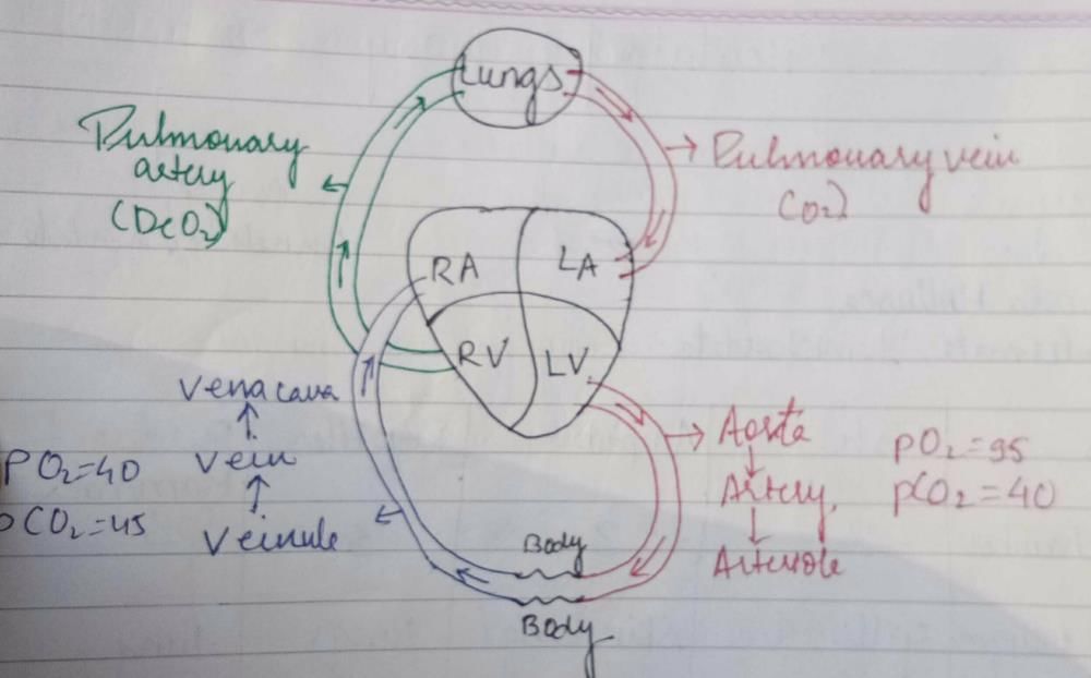
Blood circulation take following course in heart of man :-- a)Left auricle - left ventricle - body - right auricle -right ventricle
- b)Right auricle - left ventricle
- c)Left auricle - left ventricle - lungs-right auricle -right ventricle
- d)None of them
Correct answer is option 'A'. Can you explain this answer?
Blood circulation take following course in heart of man :-
a)
Left auricle - left ventricle - body - right auricle -right ventricle
b)
Right auricle - left ventricle
c)
Left auricle - left ventricle - lungs-right auricle -right ventricle
d)
None of them
|
|
Ananya Das answered |
The blood pumped by the right ventricle enters the pulmonary artery, whereas the left ventricle pumps blood into the aorta. The deoxygenated blood pumped into the pulmonary artery is passed onto the lungs from where the oxygenated blood is carried by the pulmonary veins into the left atrium. This pathway constitutes the pulmonary circulation. The oxygenated blood entering the aorta is carried by a network of arteries, arterioles, and capillaries to the tissues from where the deoxygenated blood is collected by a system of venules, veins and vena cava and emptied into the right atrium. This is the systemic circulation. The systemic circulation provides nutrients, O2 and other essential substances to the tissues and takes CO2 and other harmful substances away for elimination.
So, the correct answer is option A.
So, the correct answer is option A.
The blood during diastole :-- a)Leaves the heart
- b)Enters the heart
- c)Enters lungs
- d)Leaves the ventricles
Correct answer is option 'B'. Can you explain this answer?
The blood during diastole :-
a)
Leaves the heart
b)
Enters the heart
c)
Enters lungs
d)
Leaves the ventricles
|
|
Rajeev Saxena answered |
Diastole This should really be called “ventricular diastole”, but don't worry about this for GCSE. During diastole the thick muscular walls of the ventricles relax. Again, this happens to both sides of the heart. The pressure of the blood in the ventricles falls low enough for the bicuspid valve to open.
Read the following :
i. Blood vessels include arteries and veins. Arteries carry blood from heart to body parts and veins bring back blood from body parts to heart.
ii. The blood vessel which carry blood from heart to lungs is pulmonary artery and it carries oxygenated blood.- a)Statement ii) is correct and i) is wrong.
- b)both are correct
- c)Statement ii) is wrong and i) is correct.
- d)both are wrong
Correct answer is option 'C'. Can you explain this answer?
Read the following :
i. Blood vessels include arteries and veins. Arteries carry blood from heart to body parts and veins bring back blood from body parts to heart.
ii. The blood vessel which carry blood from heart to lungs is pulmonary artery and it carries oxygenated blood.
i. Blood vessels include arteries and veins. Arteries carry blood from heart to body parts and veins bring back blood from body parts to heart.
ii. The blood vessel which carry blood from heart to lungs is pulmonary artery and it carries oxygenated blood.
a)
Statement ii) is correct and i) is wrong.
b)
both are correct
c)
Statement ii) is wrong and i) is correct.
d)
both are wrong
|
|
Geetika Shah answered |
- The arteries carry blood from the heart to all over body parts. Hence, statement (i) is correct.
- The vessels which carry blood from the heart to the lungs is the pulmonary artery but it carries deoxygenated blood. Hence, statement (ii) is correct.
The specialised patch of modified heart muscles from where contraction initiates, is/are :- a)Pacesetter of heart
- b)both SAN and AVN together
- c)Pacemaker of heart
- d)none of these
Correct answer is option 'C'. Can you explain this answer?
The specialised patch of modified heart muscles from where contraction initiates, is/are :
a)
Pacesetter of heart
b)
both SAN and AVN together
c)
Pacemaker of heart
d)
none of these
|
|
Gaurav Kumar answered |
- The nodal musculature has the ability to generate action potentials without any external stimuli.
- The number of action potentials that could be generated in a minute varies at different parts of the nodal system.
- The SAN can generate the maximum number of action potentials and is responsible for initiating and maintaining the rhythmic contractile activity of the heart.
- Therefore, it is called the pacemaker.
Which one of the following is correct?- a)Plasma = Blood − Lymphocytes
- b)Neuron = Cyton + Dendron + Axon + Synapse
- c)Lymph = Plasma + WBCs + RBCs
- d)Blood = Plasma + RBCs + WBCs + Platelets
Correct answer is option 'D'. Can you explain this answer?
Which one of the following is correct?
a)
Plasma = Blood − Lymphocytes
b)
Neuron = Cyton + Dendron + Axon + Synapse
c)
Lymph = Plasma + WBCs + RBCs
d)
Blood = Plasma + RBCs + WBCs + Platelets
|
|
Rajat Kapoor answered |
It has four main components: plasma, red blood cells, white blood cells, and platelets. Blood has many different functions, including: transporting oxygen and nutrients to the lungs and tissues. forming blood clots to prevent excess blood loss.
In human beings, the duration of the cardiac cycle is- a)8.0 second
- b)0.5 second
- c)0.8 second
- d)0.08 second
Correct answer is 'C'. Can you explain this answer?
In human beings, the duration of the cardiac cycle is
a)
8.0 second
b)
0.5 second
c)
0.8 second
d)
0.08 second
|
|
Rohan Singh answered |
Cardiac cycle a complex process which involves a sequence of activities and that too within 0.8 sec. The cardiac cycle consists of a systole and a diastole. The cardiac cycle begins with a joint diastole (relaxation of all four chambers of the heart).
The maximum surface area of the circulatory system is seen in- a)Heart
- b)Capillaries
- c)Arterioles
- d)Veins
Correct answer is option 'B'. Can you explain this answer?
The maximum surface area of the circulatory system is seen in
a)
Heart
b)
Capillaries
c)
Arterioles
d)
Veins
|
|
Raghav Bansal answered |
An adult human has been estimated to have some 60,000 miles (96,560 km) of capillaries with a total surface area of some 8001000 m2. The total volume of this system is roughly 5 liters, the same as the total volume of blood.


The SA node is located in- a)Upper lateral wall of the left atrium
- b)Lower lateral wall of the right atrium
- c)Lower lateral wall of the left atrium
- d)Upper lateral wall of the right atrium
Correct answer is option 'D'. Can you explain this answer?
The SA node is located in
a)
Upper lateral wall of the left atrium
b)
Lower lateral wall of the right atrium
c)
Lower lateral wall of the left atrium
d)
Upper lateral wall of the right atrium
|
|
Sathvika Tadri answered |
SA node (sino-atrial node) is located in right upper corner of right atrium .of heart..option D is the answer
Antigens are present- a)Inside the cytoplasm
- b)Inside the nucleus
- c)On the nuclear membrane
- d)On the cell surface
Correct answer is option 'D'. Can you explain this answer?
Antigens are present
a)
Inside the cytoplasm
b)
Inside the nucleus
c)
On the nuclear membrane
d)
On the cell surface

|
Ayush Joshi answered |
Cell markers, also known as cell surface antigens, serve as monograms to help identify and classify cells. The majority of them are molecules or antigens within cell's plasma membrane. Unique to different cell types, there exist specific combinations of markers or antigens.
Blood enters the heart because muscles of the- a)Ventricles contract
- b)Ventricles relax
- c)Atria contract
- d)Atria relax
Correct answer is option 'D'. Can you explain this answer?
Blood enters the heart because muscles of the
a)
Ventricles contract
b)
Ventricles relax
c)
Atria contract
d)
Atria relax
|
|
Geetika Shah answered |
Atrial diastole is characterized by the relaxation of the atrial muscles. During this stage, the blood enters into the atria, as the volume of the blood in the atria increases the pressure within it decreases.
So, the correct answer is option D.
So, the correct answer is option D.
In the systemic circulation, blood vessel that carries blood from the intestine to liver is named :- a)Hepatic portal arch
- b)none of these
- c)Hepatic portal artery
- d)Hepatic portal vein
Correct answer is option 'D'. Can you explain this answer?
In the systemic circulation, blood vessel that carries blood from the intestine to liver is named :
a)
Hepatic portal arch
b)
none of these
c)
Hepatic portal artery
d)
Hepatic portal vein
|
|
Naina Bansal answered |
Lienal vein is an old term for splenic vein. The portal vein or hepatic portal vein is a blood vessel that carries blood from the gastrointestinal tract, gallbladder, pancreas and spleen to the liver. This blood contains nutrients and toxins extracted from digested contents.
Rh incompatibility develops when :- a)foetus is Rh-ve and father is Rh+ve
- b)foetus is Rh+ve and mother is Rh-ve
- c)foetus is Rh-ve and mother is Rh+ve
- d)None of these
Correct answer is option 'B'. Can you explain this answer?
Rh incompatibility develops when :
a)
foetus is Rh-ve and father is Rh+ve
b)
foetus is Rh+ve and mother is Rh-ve
c)
foetus is Rh-ve and mother is Rh+ve
d)
None of these

|
Bhargavi Choudhury answered |
A special case of Rh incompatibility (mismatching) has been observed between the Rh-veblood of a pregnant mother with Rh+ve blood of the foetus.
Which of the following statements doesn’t hold true for blood clotting cascade?
i. Ruptured platelets release thromboplastin.
ii. Prothrombinis converted into thrombin in the presence of vitamin K and Na+ions.
iii. Fibrinogen is converted into fibrin through enzymatic action of thrombin in the presence of Ca++ions
iv. Fibrin makes clot via process of polymerisation.- a)only statement i) and iv) are wrong
- b)only statement ii) and iii) are wrong
- c)all statements are wrong
- d)only statement iv) is wrong
Correct answer is option 'B'. Can you explain this answer?
Which of the following statements doesn’t hold true for blood clotting cascade?
i. Ruptured platelets release thromboplastin.
ii. Prothrombinis converted into thrombin in the presence of vitamin K and Na+ions.
iii. Fibrinogen is converted into fibrin through enzymatic action of thrombin in the presence of Ca++ions
iv. Fibrin makes clot via process of polymerisation.
i. Ruptured platelets release thromboplastin.
ii. Prothrombinis converted into thrombin in the presence of vitamin K and Na+ions.
iii. Fibrinogen is converted into fibrin through enzymatic action of thrombin in the presence of Ca++ions
iv. Fibrin makes clot via process of polymerisation.
a)
only statement i) and iv) are wrong
b)
only statement ii) and iii) are wrong
c)
all statements are wrong
d)
only statement iv) is wrong

|
Krish Patel answered |
Ruptured platelets release thromboplastin. Fibrinogen present in blood plasma is converted into fibrin through enzymatic action of thrombin. Fibrin makes clot via process of polymerisation.
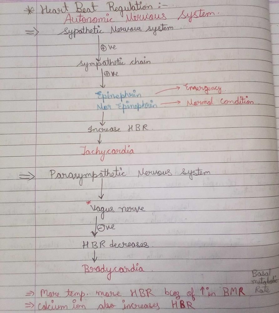
Heart of Man is :-- a)Myogenic
- b)Neurogenic
- c)Cardiogenic
- d)Digenic
Correct answer is option 'A'. Can you explain this answer?
Heart of Man is :-
a)
Myogenic
b)
Neurogenic
c)
Cardiogenic
d)
Digenic
|
|
Rocky Handsome answered |
The muscles of the human heart are stimulated to contract by nerve impulses generated by the Sino Atrial(SA) node. It is a cluster of cells which are part of the heart muscle.
•Hence the human heart is myogenic.
•Hence the human heart is myogenic.
Each cardiac cycle takes 0.8 seconds to occur. Calculate how many cardiac cycles occur in 4 minutes ?- a)250
- b)75
- c)400
- d)300
Correct answer is option 'D'. Can you explain this answer?
Each cardiac cycle takes 0.8 seconds to occur. Calculate how many cardiac cycles occur in 4 minutes ?
a)
250
b)
75
c)
400
d)
300

|
Nilotpal Gupta answered |
4 min = 240 sec. Therefore, 240/0.8 = 300 cardiac cycles.

In a normal man blood pressure is :-- a)120/80mm of Hg
- b)80/100 mm of Hg
- c)80/120 mm of Hg
- d)100/80 mm of Hg
Correct answer is option 'A'. Can you explain this answer?
In a normal man blood pressure is :-
a)
120/80mm of Hg
b)
80/100 mm of Hg
c)
80/120 mm of Hg
d)
100/80 mm of Hg
|
|
Harshitha Reddy answered |
Normal Blood Pressure
The normal blood pressure in a man is 120/80 mm of Hg.
Explanation
Blood pressure is the force exerted by blood against the walls of arteries. Blood pressure is measured in millimeters of mercury (mm Hg). The two numbers in blood pressure readings represent systolic pressure (the top number) and diastolic pressure (the bottom number).
The normal blood pressure reading for an adult is 120/80 mm of Hg. The systolic pressure represents the pressure when the heart beats and pumps blood into the arteries. The diastolic pressure represents the pressure when the heart is at rest between beats.
If the blood pressure reading is consistently higher than normal, it may indicate hypertension or high blood pressure. Hypertension can lead to serious health problems like heart disease, stroke, and kidney disease.
On the other hand, if the blood pressure reading is consistently lower than normal, it may indicate hypotension or low blood pressure. Hypotension can cause dizziness, fainting, and other health problems.
It's important to monitor blood pressure regularly and take necessary steps to maintain normal blood pressure. A healthy lifestyle with a balanced diet, regular exercise, and stress management can help maintain normal blood pressure levels.
Manoj has AB blood group, so he will have the following antibodies in his blood plasma :- a)a antibody
- b)both a and b antibodies
- c)b antibody
- d)No antibodies are present
Correct answer is option 'D'. Can you explain this answer?
Manoj has AB blood group, so he will have the following antibodies in his blood plasma :
a)
a antibody
b)
both a and b antibodies
c)
b antibody
d)
No antibodies are present

|
Kunal Rane answered |
AB blood group does not have any antibodies in plasma.
Blood pressure is expressed as the ratio of systolic over diastolic pressure. The difference between systolic and diastolic pressure is called pulse pressure. What will be its value for a normal healthy adult?- a)40 mm Hg
- b)30 mm Hg
- c)70 mm Hg
- d)50 mm Hg
Correct answer is option 'A'. Can you explain this answer?
Blood pressure is expressed as the ratio of systolic over diastolic pressure. The difference between systolic and diastolic pressure is called pulse pressure. What will be its value for a normal healthy adult?
a)
40 mm Hg
b)
30 mm Hg
c)
70 mm Hg
d)
50 mm Hg

|
Tejas Chavan answered |
Blood pressure is represented as the ratio of systolic over diastolic pressure. The difference between systolic and diastolic pressure is called pulse pressure. The value of normal healthy pulse pressure is 40 mm Hg.
Blood plasma proteins :
i. decrease in their level causes excessive absorption of water from tissues into blood.
ii. they maintain osmotic pressure.
- a)both are correct.
- b)both are wrong
- c)Statement i) is wrong and ii) is correct.
- d)Statement i) is correct and ii) is wrong
Correct answer is option 'C'. Can you explain this answer?
Blood plasma proteins :
i. decrease in their level causes excessive absorption of water from tissues into blood.
ii. they maintain osmotic pressure.
i. decrease in their level causes excessive absorption of water from tissues into blood.
ii. they maintain osmotic pressure.
a)
both are correct.
b)
both are wrong
c)
Statement i) is wrong and ii) is correct.
d)
Statement i) is correct and ii) is wrong

|
Ishaan Menon answered |
Let's analyze the statements about blood plasma proteins:
Statement i: "Decrease in their level causes excessive absorption of water from tissues into blood."
- This statement is incorrect. A decrease in the level of blood plasma proteins would actually result in a decrease in osmotic pressure, leading to less water being drawn from tissues into the blood. Instead, it would cause water to accumulate in the tissues, leading to edema.
Statement ii: "They maintain osmotic pressure."
- This statement is correct. Blood plasma proteins, especially albumin, play a crucial role in maintaining the osmotic pressure of the blood.
Therefore, the correct option is:
3. Statement i) is wrong and ii) is correct.
For the blood to flow from the right ventricle to the left ventricle in the mammalian heart, it must flow through- a)Right ventricle, Pulmonary veins, Lungs, Pulmonary arteries, Left atrium
- b)Right ventricle, Right atrium, Lungs, Pulmonary veins, Left atrium
- c)Right ventricle, Pulmonary arteries, Lungs, Pulmonary veins, Left atrium
- d)Right ventricle, Systemic aorta, Lungs, Pulmonary veins, Left atrium
Correct answer is option 'C'. Can you explain this answer?
For the blood to flow from the right ventricle to the left ventricle in the mammalian heart, it must flow through
a)
Right ventricle, Pulmonary veins, Lungs, Pulmonary arteries, Left atrium
b)
Right ventricle, Right atrium, Lungs, Pulmonary veins, Left atrium
c)
Right ventricle, Pulmonary arteries, Lungs, Pulmonary veins, Left atrium
d)
Right ventricle, Systemic aorta, Lungs, Pulmonary veins, Left atrium

|
Vijayam6309002257 Vijaya answered |
Blood flows from right ventricle -pulmonary artery -lungs -pulmonary vein -left atrium -left ventricle
In ECG, the depolarisation of atria is indicated by- a)S-wave
- b)R-wave
- c)P-wave
- d)Q-wave
Correct answer is option 'C'. Can you explain this answer?
In ECG, the depolarisation of atria is indicated by
a)
S-wave
b)
R-wave
c)
P-wave
d)
Q-wave

|
Savita Soni answered |
By P wave
11 ncert page no 286
11 ncert page no 286
The mass of tissue seen in the left corner of the right atrium close to the atri-ventricular septum is- a)Purkinje fibres
- b)Bundle of His
- c)AVN
- d)SAN
Correct answer is option 'C'. Can you explain this answer?
The mass of tissue seen in the left corner of the right atrium close to the atri-ventricular septum is
a)
Purkinje fibres
b)
Bundle of His
c)
AVN
d)
SAN

|
Krish Chakraborty answered |
The mass of tissue seen in the lower left corner of the right atrium close to the atrio-ventricular septum called the atrio-ventricular node (AVN).
When blood clot starts contracting, a pale yellow fluid starts oozing out. Its name and composition is :- a)Serum = plasma – [thrombin + blood corpuscles]
- b)Serum = plasma – [fibrinogen + blood corpuscles]
- c)Sera = blood corpuscles – [thrombin + fibrin]
- d)Serum = dissolved fibrin – [plasma + blood corpuscles]
Correct answer is option 'B'. Can you explain this answer?
When blood clot starts contracting, a pale yellow fluid starts oozing out. Its name and composition is :
a)
Serum = plasma – [thrombin + blood corpuscles]
b)
Serum = plasma – [fibrinogen + blood corpuscles]
c)
Sera = blood corpuscles – [thrombin + fibrin]
d)
Serum = dissolved fibrin – [plasma + blood corpuscles]

|
Arnav Iyer answered |
Plasma without the clotting factors is called serum. The clotting factors include fibrinogen and blood corpuscles
In the human heart :
i. The valve between right atrium and right ventricle is tricuspid while valve between left atrium and left ventricle is mitral valve.
ii. Openings of the right and the left ventricles into the pulmonary artery and the aorta are guarded by semilunar valves.
iii. ‘Lub’ the first sound which is low pitched is caused by the closure of semilunar valves while ‘Dub’ the second sound which is high pitched is caused by the closure of bicuspid and tricuspid valves.- a)only ii) and iii) are correct
- b)only i) and ii) are correct
- c)only iii) is correct
- d)only ii) is correct
Correct answer is option 'B'. Can you explain this answer?
In the human heart :
i. The valve between right atrium and right ventricle is tricuspid while valve between left atrium and left ventricle is mitral valve.
ii. Openings of the right and the left ventricles into the pulmonary artery and the aorta are guarded by semilunar valves.
iii. ‘Lub’ the first sound which is low pitched is caused by the closure of semilunar valves while ‘Dub’ the second sound which is high pitched is caused by the closure of bicuspid and tricuspid valves.
a)
only ii) and iii) are correct
b)
only i) and ii) are correct
c)
only iii) is correct
d)
only ii) is correct

|
Rajeev Sharma answered |
The first heart sound (lub) is associated with the closure of the tricuspid and bicuspid valves whereas the second heart sound (dub) is associated with the closure of the semilunar valves.
Meghna suffers from allergic asthma. After a blood test her leucocyte count displayed an abnormal increase in the number of :- a)Eosinophils
- b)Monocytes
- c)Neutrophils
- d)Basophils
Correct answer is option 'A'. Can you explain this answer?
Meghna suffers from allergic asthma. After a blood test her leucocyte count displayed an abnormal increase in the number of :
a)
Eosinophils
b)
Monocytes
c)
Neutrophils
d)
Basophils

|
Soumya Ahuja answered |
In case of allergic asthma, leucocyte count of blood test shows abnormal increase in the number of Eosinophils
Lymph will transport :- a)digested carbohydrates
- b)oxyhemoglobin
- c)digested fats
- d)digested proteins
Correct answer is option 'C'. Can you explain this answer?
Lymph will transport :
a)
digested carbohydrates
b)
oxyhemoglobin
c)
digested fats
d)
digested proteins
|
|
Bhargavi Choudhury answered |
Fats are absorbed through lymph in the lacteals present in the intestinal villi.
By counting the number of which of the following waves, the heartbeat of a person can be determined?- a)QRS complex
- b)P-wave
- c)ST-segment
- d)PQ interval
Correct answer is option 'A'. Can you explain this answer?
By counting the number of which of the following waves, the heartbeat of a person can be determined?
a)
QRS complex
b)
P-wave
c)
ST-segment
d)
PQ interval
|
|
Adebisi Ekpe answered |
Understanding Heartbeat Measurement
The heartbeat of a person can be determined by analyzing specific components of an electrocardiogram (ECG or EKG). Among these components, the QRS complex plays a crucial role.
What is the QRS Complex?
- The QRS complex represents the electrical activity associated with the contraction of the ventricles, which are the heart's main pumping chambers.
- It is a critical part of the cardiac cycle, as it indicates that the heart is actively pumping blood.
Counting the QRS Complexes
- By counting the number of QRS complexes in a given time frame, healthcare professionals can calculate the heart rate.
- Each QRS complex corresponds to one heartbeat. Therefore, counting these complexes over a specific period allows for an accurate measurement of heart rate.
Other Components Explained
- P-Wave: Represents atrial depolarization. Its primary role is to indicate the contraction of the atria but does not directly reflect the heart rate.
- ST-Segment: Indicates the period between ventricular depolarization and repolarization. It is important for diagnosing certain heart conditions but is not used for heart rate determination.
- PQ Interval: Represents the time between atrial and ventricular depolarization. While it provides insight into the conduction system, it does not serve as a direct measure of heart rate.
Conclusion
The QRS complex is essential for determining the heartbeat because it directly correlates to ventricular contractions. Therefore, option 'A' is the correct answer for measuring heart rate effectively.

Which structure separates the right and left atria of the heart?- a)Interventricular septum
- b)Interatrial septum
- c)Atrio-ventricular septum
- d)Pericardium
Correct answer is option 'B'. Can you explain this answer?
Which structure separates the right and left atria of the heart?
a)
Interventricular septum
b)
Interatrial septum
c)
Atrio-ventricular septum
d)
Pericardium

|
Ambition Institute answered |
The right and left atria of the heart are separated by a thin, muscular wall called the interatrial septum. This partition ensures that blood from the systemic and pulmonary circulations does not mix within the heart, allowing for efficient oxygenation and circulation of blood throughout the body.
Assertion (A): Coronary artery disease is primarily caused by a buildup of calcium, fat, cholesterol, and fibrous tissues in the arteries.Reason (R): This accumulation leads to the narrowing of the arterial lumen, which reduces blood flow to the heart muscle.- a)If both Assertion and Reason are true and Reason is the correct explanation of Assertion
- b)If both Assertion and Reason are true but Reason is not the correct explanation of Assertion
- c)If Assertion is true but Reason is false
- d)If both Assertion and Reason are false
Correct answer is option 'A'. Can you explain this answer?
Assertion (A): Coronary artery disease is primarily caused by a buildup of calcium, fat, cholesterol, and fibrous tissues in the arteries.
Reason (R): This accumulation leads to the narrowing of the arterial lumen, which reduces blood flow to the heart muscle.
a)
If both Assertion and Reason are true and Reason is the correct explanation of Assertion
b)
If both Assertion and Reason are true but Reason is not the correct explanation of Assertion
c)
If Assertion is true but Reason is false
d)
If both Assertion and Reason are false

|
Bs Academy answered |
- Assertion Analysis: The assertion is true as coronary artery disease (CAD) is indeed caused by the buildup of substances such as calcium, fat, and cholesterol in the arterial walls.
- Reason Analysis: The reason is also true because the described accumulation does lead to the narrowing of the arteries, which is a key characteristic of CAD.
- Explanation Relation: The reason directly explains the assertion as it describes the process through which CAD occurs. Thus, both the assertion and reason are true, and the reason accurately explains the assertion.
Line in NCERT: "Coronary Artery Disease, often referred to as atherosclerosis, affects the vessels that supply blood to the heart muscle. It is caused by deposits of calcium, fat, cholesterol and fibrous tissues, which makes the lumen of arteries narrower."
The heart sound "DUP" is Produced when :-- a)Mitral valve opens
- b)Mitral valve closes
- c)Semilunar valve at the base of aorta closes
- d)Tricuspid valve opens
Correct answer is option 'C'. Can you explain this answer?
The heart sound "DUP" is Produced when :-
a)
Mitral valve opens
b)
Mitral valve closes
c)
Semilunar valve at the base of aorta closes
d)
Tricuspid valve opens
|
|
Pankaj Singh answered |
The heart sound ‘’DUP’’ is produced when a semilunar valve at the base of aorta classes.
Two distinct sounds can be heard during a heart beat with the help of a stethoscope. These are 'lubb' and 'dup'.Lubb is the first sound which has a low pitch and produced by the closure of tricuspid and bicuspid valves (collectively called atrioventricular valves) at the beginning of ventricular systole.
Dup is the second sound which is sharp and has a high pitch. It is produced by closure of semilunar valves by the end of ventricular systole.
Which type of circulatory system is found in arthropods and molluscs?- a) Closed circulatory system
- b) Open circulatory system
- c) Double circulatory system
- d) Incomplete circulatory system
Correct answer is option 'B'. Can you explain this answer?
Which type of circulatory system is found in arthropods and molluscs?
a)
Closed circulatory system
b)
Open circulatory system
c)
Double circulatory system
d)
Incomplete circulatory system

|
Ciel Knowledge answered |
Arthropods and molluscs possess an open circulatory system where blood flows freely in body cavities.
Which of the following represents the repolarisation of atrial muscles and depolarisation of ventricular muscles in an electrocardiograph?- a)P-wave
- b)PQ interval
- c)QT interval
- d)QRS complex
Correct answer is option 'D'. Can you explain this answer?
Which of the following represents the repolarisation of atrial muscles and depolarisation of ventricular muscles in an electrocardiograph?
a)
P-wave
b)
PQ interval
c)
QT interval
d)
QRS complex
|
|
Ciel Knowledge answered |
It begins with a small negative deflection and represents the repolarisation of atrial muscles and depolarisation of ventricular muscles in an electrocardiograph.
What is the primary cause of coronary artery disease (CAD)?- a)Excessive contraction of the heart muscle
- b)Infections in the heart muscle
- c)Deposits of calcium, fat, cholesterol, and fibrous tissues in the arteries
- d)Overproduction of adrenal medullary hormones affecting the heart
Correct answer is option 'C'. Can you explain this answer?
a)
Excessive contraction of the heart muscle
b)
Infections in the heart muscle
c)
Deposits of calcium, fat, cholesterol, and fibrous tissues in the arteries
d)
Overproduction of adrenal medullary hormones affecting the heart
|
|
Ciel Knowledge answered |
Coronary Artery Disease (CAD) is primarily caused by the buildup of deposits such as calcium, fat, cholesterol, and fibrous tissues within the arteries that supply blood to the heart muscle. This buildup leads to the narrowing of the arterial lumen, which can restrict blood flow and increase the risk of heart attack and other cardiac complications. This process is often referred to as atherosclerosis and is the leading cause of CAD.
Which of these occurs during the atrial systole?- a)Action potential is generated by the AVN initially
- b)Both atria contract simultaneously
- c)Tricuspid valve closes
- d)The semilunar valves remain open
Correct answer is option 'B'. Can you explain this answer?
Which of these occurs during the atrial systole?
a)
Action potential is generated by the AVN initially
b)
Both atria contract simultaneously
c)
Tricuspid valve closes
d)
The semilunar valves remain open
|
|
Chirag Unni answered |
Atrial Systole Overview
Atrial systole is a crucial phase in the cardiac cycle where the atria contract, pushing blood into the ventricles. During this phase, several important physiological events occur.
Key Events During Atrial Systole
- Both Atria Contract Simultaneously: This is the defining characteristic of atrial systole. The sinoatrial (SA) node generates an action potential that spreads through the atria, causing them to contract together. This synchronous contraction efficiently fills the ventricles with blood.
- Action Potential Generation by AV Node: While the atrial contraction is initiated by the SA node, the atrioventricular node (AVN) plays a role in conducting the electrical impulse to the ventricles. However, the AVN does not generate the initial action potential during atrial systole.
- Tricuspid Valve Status: The tricuspid valve remains open during atrial systole, allowing blood to flow from the right atrium to the right ventricle. It closes after atrial contraction as the ventricles begin to contract.
- Semilunar Valves Status: The semilunar valves (pulmonary and aortic valves) remain closed during this phase. They only open when the ventricles contract, allowing blood to exit the heart.
Conclusion
In summary, option 'B' is correct because during atrial systole, both atria contract simultaneously, facilitating efficient blood flow into the ventricles. Understanding these dynamics is essential for grasping cardiac physiology, particularly for NEET aspirants.
What potential health issue can arise from a significant reduction in platelet count?- a)Increased risk of infections
- b)Enhanced ability to clot blood
- c)Excessive blood loss due to clotting disorders
- d)Improved oxygen transport to tissues
Correct answer is option 'C'. Can you explain this answer?
What potential health issue can arise from a significant reduction in platelet count?
a)
Increased risk of infections
b)
Enhanced ability to clot blood
c)
Excessive blood loss due to clotting disorders
d)
Improved oxygen transport to tissues

|
Lead Academy answered |
Platelets, or thrombocytes, are crucial for blood clotting. A significant reduction in their number can lead to clotting disorders, making it difficult for the body to stop bleeding. This can result in excessive blood loss from minor injuries or in surgical situations, posing serious health risks.
In which organisms is an open circulatory system typically found?- a) Arthropods and molluscs
- b) Vertebrates
- c) Annelids and chordates
- d) Fishes and amphibians
Correct answer is option 'A'. Can you explain this answer?
In which organisms is an open circulatory system typically found?
a)
Arthropods and molluscs
b)
Vertebrates
c)
Annelids and chordates
d)
Fishes and amphibians
|
|
Sagnik Jain answered |
Open Circulatory System Overview
An open circulatory system is a type of circulatory system where blood is not always contained within blood vessels. Instead, the blood, or hemolymph, bathes the organs directly in a body cavity known as a hemocoel.
Organisms with Open Circulatory Systems
- Arthropods: This group includes insects, arachnids, and crustaceans. In these organisms, the heart pumps hemolymph into the hemocoel, where it circulates freely around the tissues and organs. The low pressure of this system makes it suitable for small-bodied animals that do not require rapid blood circulation.
- Molluscs: Many molluscs, such as snails and clams, also possess an open circulatory system. Their hearts pump hemolymph into sinuses, where it directly contacts the organs. While cephalopods (like squids and octopuses) have a closed circulatory system, most other molluscs utilize the open type.
Comparison with Other Organisms
- Vertebrates: These organisms have a closed circulatory system where blood circulates within vessels, ensuring efficient transport of oxygen and nutrients, which is crucial for their larger and more complex body structures.
- Annelids and Chordates: Annelids (like earthworms) possess a closed circulatory system, while chordates (including mammals, birds, reptiles) have evolved a more complex closed system.
- Fishes and Amphibians: Both groups have a closed circulatory system. Fishes have a two-chambered heart, while amphibians usually possess a three-chambered heart, allowing for more efficient oxygenation of blood.
Conclusion
In summary, open circulatory systems are characteristic of arthropods and many molluscs, making option 'A' the correct answer. This system suits the physiological needs of these organisms by allowing sufficient nutrient and gas exchange while conserving energy.
What is the role of the lymphatic system in relation to interstitial fluid?- a)It increases the mineral distribution within the interstitial fluid.
- b)It collects and drains the interstitial fluid back to the major veins.
- c)It converts interstitial fluid into blood plasma.
- d)It absorbs and redistributes red blood cells.
Correct answer is option 'B'. Can you explain this answer?
a)
It increases the mineral distribution within the interstitial fluid.
b)
It collects and drains the interstitial fluid back to the major veins.
c)
It converts interstitial fluid into blood plasma.
d)
It absorbs and redistributes red blood cells.
|
|
Harsh Mukherjee answered |
Role of the Lymphatic System
The lymphatic system plays a crucial role in maintaining fluid balance within the body, particularly concerning interstitial fluid. Here's how it functions:
Collection of Interstitial Fluid
- The lymphatic system is responsible for collecting excess interstitial fluid, which is the fluid that exists in the spaces between cells.
- Interstitial fluid is formed as blood plasma leaks out of capillaries to nourish cells and remove waste.
Draining to Major Veins
- Once collected, the lymphatic system drains this excess fluid, now referred to as lymph, back into the circulatory system.
- The lymphatic vessels transport lymph to larger lymphatic ducts, which eventually empty into major veins, such as the subclavian vein, near the heart.
Role in Immune Function
- Besides fluid drainage, the lymphatic system plays a vital role in the immune response by transporting white blood cells and filtering out pathogens through lymph nodes.
Fluid Homeostasis
- By draining interstitial fluid, the lymphatic system helps maintain homeostasis, preventing swelling (edema) and ensuring that tissues receive adequate nutrients and oxygen while efficiently removing waste.
In summary, the lymphatic system's primary role concerning interstitial fluid is to collect and drain it back to major veins, thus maintaining the balance of body fluids and supporting immune function. This function is essential for overall health and well-being.
Where does the atrioventricular bundle (AV bundle) emerge from after passing through the atrioventricular septa?- a) Left atrium
- b) Interventricular septum
- c) Right ventricle
- d) Pulmonary artery
Correct answer is option 'B'. Can you explain this answer?
Where does the atrioventricular bundle (AV bundle) emerge from after passing through the atrioventricular septa?
a)
Left atrium
b)
Interventricular septum
c)
Right ventricle
d)
Pulmonary artery

|
Top Rankers answered |
The atrioventricular bundle (AV bundle) is a bundle of nodal fibers that continues from the atrioventricular node (AVN) and passes through the atrioventricular septa. It emerges on the top of the interventricular septum, where it immediately divides into a right and left bundle. These branches further propagate into minute fibers throughout the ventricular musculature of the respective sides, playing a crucial role in coordinating the electrical impulses that regulate the contraction of the ventricles.
Chapter doubts & questions for Circulation in Animals - Biology for JAMB 2025 is part of JAMB exam preparation. The chapters have been prepared according to the JAMB exam syllabus. The Chapter doubts & questions, notes, tests & MCQs are made for JAMB 2025 Exam. Find important definitions, questions, notes, meanings, examples, exercises, MCQs and online tests here.
Chapter doubts & questions of Circulation in Animals - Biology for JAMB in English & Hindi are available as part of JAMB exam.
Download more important topics, notes, lectures and mock test series for JAMB Exam by signing up for free.
Biology for JAMB
221 videos|172 docs|126 tests
|
|
© EduRev
|
Education Revolution
|


|


Signup to see your scores
go up within 7 days!
Access 1000+ FREE Docs, Videos and Tests
Takes less than 10 seconds to signup