


IIT JAM Exam > IIT JAM Questions > Reducing SDS-PAGE of IgM will produce _______...
Start Learning for Free
Reducing SDS-PAGE of IgM will produce ____________ bands in autoradiogram.
Correct answer is '3'. Can you explain this answer?

Verified Answer
Reducing SDS-PAGE of IgM will produce ____________ bands in autoradiog...
Most Upvoted Answer
Reducing SDS-PAGE of IgM will produce ____________ bands in autoradiog...
Reducing SDS-PAGE of IgM will produce 3 bands in autoradiogram.
Explanation:
IgM, also known as immunoglobulin M, is a type of antibody that is produced by B cells in response to an infection. It is the largest antibody in the human body and is composed of five basic subunits called monomers. Each monomer consists of two heavy chains and two light chains.
When IgM is subjected to reducing SDS-PAGE (sodium dodecyl sulfate polyacrylamide gel electrophoresis), the denaturing conditions of the gel system cause the antibody to dissociate into its subunits. This allows for the separation and identification of the individual subunits based on their molecular weights.
Process of SDS-PAGE:
1. Denaturation: The sample containing IgM is treated with a reducing agent, such as beta-mercaptoethanol, to break the disulfide bonds that hold the subunits together. This results in the dissociation of IgM into its monomers.
2. Electrophoresis: The denatured IgM is loaded onto an SDS-PAGE gel and subjected to electrophoresis. The gel matrix provides a sieving effect, allowing the separation of the monomers based on their size.
3. Staining: After electrophoresis, the gel is stained with a dye, such as Coomassie Brilliant Blue, to visualize the separated proteins. However, in the case of autoradiogram, the separated proteins are transferred onto a nitrocellulose or PVDF membrane.
4. Immunoblotting: The separated proteins on the membrane are then probed with specific antibodies that recognize IgM. This step allows for the detection and visualization of the individual subunits.
Result:
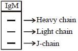
In the case of reducing SDS-PAGE of IgM, three distinct bands will be observed in the autoradiogram. These bands represent the three different subunits of IgM:
1. Heavy chain: The largest band corresponds to the heavy chains of IgM. As IgM is composed of five monomers, each containing two heavy chains, this band represents the presence of 10 heavy chains.
2. Light chain: The two smaller bands represent the light chains of IgM. As IgM is composed of five monomers, each containing two light chains, these bands represent the presence of 10 light chains.
Thus, the reducing SDS-PAGE of IgM will produce three bands in the autoradiogram, corresponding to the heavy chains and light chains of IgM.
Explanation:
IgM, also known as immunoglobulin M, is a type of antibody that is produced by B cells in response to an infection. It is the largest antibody in the human body and is composed of five basic subunits called monomers. Each monomer consists of two heavy chains and two light chains.
When IgM is subjected to reducing SDS-PAGE (sodium dodecyl sulfate polyacrylamide gel electrophoresis), the denaturing conditions of the gel system cause the antibody to dissociate into its subunits. This allows for the separation and identification of the individual subunits based on their molecular weights.
Process of SDS-PAGE:
1. Denaturation: The sample containing IgM is treated with a reducing agent, such as beta-mercaptoethanol, to break the disulfide bonds that hold the subunits together. This results in the dissociation of IgM into its monomers.
2. Electrophoresis: The denatured IgM is loaded onto an SDS-PAGE gel and subjected to electrophoresis. The gel matrix provides a sieving effect, allowing the separation of the monomers based on their size.
3. Staining: After electrophoresis, the gel is stained with a dye, such as Coomassie Brilliant Blue, to visualize the separated proteins. However, in the case of autoradiogram, the separated proteins are transferred onto a nitrocellulose or PVDF membrane.
4. Immunoblotting: The separated proteins on the membrane are then probed with specific antibodies that recognize IgM. This step allows for the detection and visualization of the individual subunits.
Result:
In the case of reducing SDS-PAGE of IgM, three distinct bands will be observed in the autoradiogram. These bands represent the three different subunits of IgM:
1. Heavy chain: The largest band corresponds to the heavy chains of IgM. As IgM is composed of five monomers, each containing two heavy chains, this band represents the presence of 10 heavy chains.
2. Light chain: The two smaller bands represent the light chains of IgM. As IgM is composed of five monomers, each containing two light chains, these bands represent the presence of 10 light chains.
Thus, the reducing SDS-PAGE of IgM will produce three bands in the autoradiogram, corresponding to the heavy chains and light chains of IgM.

|
Explore Courses for IIT JAM exam
|
|
Top Courses for IIT JAMView all


Question Description
Reducing SDS-PAGE of IgM will produce ____________ bands in autoradiogram.Correct answer is '3'. Can you explain this answer? for IIT JAM 2025 is part of IIT JAM preparation. The Question and answers have been prepared according to the IIT JAM exam syllabus. Information about Reducing SDS-PAGE of IgM will produce ____________ bands in autoradiogram.Correct answer is '3'. Can you explain this answer? covers all topics & solutions for IIT JAM 2025 Exam. Find important definitions, questions, meanings, examples, exercises and tests below for Reducing SDS-PAGE of IgM will produce ____________ bands in autoradiogram.Correct answer is '3'. Can you explain this answer?.
Reducing SDS-PAGE of IgM will produce ____________ bands in autoradiogram.Correct answer is '3'. Can you explain this answer? for IIT JAM 2025 is part of IIT JAM preparation. The Question and answers have been prepared according to the IIT JAM exam syllabus. Information about Reducing SDS-PAGE of IgM will produce ____________ bands in autoradiogram.Correct answer is '3'. Can you explain this answer? covers all topics & solutions for IIT JAM 2025 Exam. Find important definitions, questions, meanings, examples, exercises and tests below for Reducing SDS-PAGE of IgM will produce ____________ bands in autoradiogram.Correct answer is '3'. Can you explain this answer?.
Solutions for Reducing SDS-PAGE of IgM will produce ____________ bands in autoradiogram.Correct answer is '3'. Can you explain this answer? in English & in Hindi are available as part of our courses for IIT JAM.
Download more important topics, notes, lectures and mock test series for IIT JAM Exam by signing up for free.
Here you can find the meaning of Reducing SDS-PAGE of IgM will produce ____________ bands in autoradiogram.Correct answer is '3'. Can you explain this answer? defined & explained in the simplest way possible. Besides giving the explanation of
Reducing SDS-PAGE of IgM will produce ____________ bands in autoradiogram.Correct answer is '3'. Can you explain this answer?, a detailed solution for Reducing SDS-PAGE of IgM will produce ____________ bands in autoradiogram.Correct answer is '3'. Can you explain this answer? has been provided alongside types of Reducing SDS-PAGE of IgM will produce ____________ bands in autoradiogram.Correct answer is '3'. Can you explain this answer? theory, EduRev gives you an
ample number of questions to practice Reducing SDS-PAGE of IgM will produce ____________ bands in autoradiogram.Correct answer is '3'. Can you explain this answer? tests, examples and also practice IIT JAM tests.
|
|
Explore Courses for IIT JAM exam
|
|

Signup for Free!
Signup to see your scores go up within 7 days! Learn & Practice with 1000+ FREE Notes, Videos & Tests.
|
© EduRev
|
Education Revolution
|


|


Signup on EduRev and stay on top of your study goals
10M+ students crushing their study goals daily