Class 1 Exam > Class 1 Questions > diagram of stethoscope Related: Learn how a ...
Start Learning for Free
diagram of stethoscope
?Verified Answer
diagram of stethoscope Related: Learn how a stethoscope can help dete...

 This question is part of UPSC exam. View all Class 1 courses
Most Upvoted Answer
diagram of stethoscope Related: Learn how a stethoscope can help dete...
Diagram of a Stethoscope:
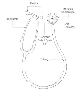
A stethoscope is a medical device used for auscultation, or listening to internal sounds of the body. It consists of several components that work together to amplify and transmit sound waves from the body to the healthcare professional's ears. Here is a diagram outlining the main parts of a stethoscope:
1. Earpieces: The stethoscope has two earpieces, one for each ear. These are designed to fit comfortably into the healthcare professional's ears and help block out external noise.
2. Tubing: The earpieces are connected to the tubing, which is usually made of rubber or plastic. The tubing serves as a pathway for sound waves to travel from the chest piece to the healthcare professional's ears.
3. Chest Piece: The chest piece is the part of the stethoscope that is placed on the patient's body. It consists of two sides: the diaphragm and the bell.
- Diaphragm: The diaphragm is the larger side of the chest piece and is used to detect high-frequency sounds. It is typically used to listen to lung sounds, heart sounds, and bowel sounds.
- Bell: The bell is the smaller side of the chest piece and is used to detect low-frequency sounds. It is usually used to listen to heart murmurs or bruits.
4. Tube Length: The length of the tubing can vary, but it is typically around 22 inches long. This allows the healthcare professional to comfortably position themselves while using the stethoscope.
5. Adjustable Headset: The headset is the part of the stethoscope that connects the earpieces to the tubing. It is usually adjustable, allowing the healthcare professional to customize the fit to their comfort.
6. Additional Features: Some stethoscopes may have additional features such as a tunable diaphragm, which allows the healthcare professional to switch between high-frequency and low-frequency sounds without rotating the chest piece.
How a Stethoscope Helps Determine Blood Pressure:
A stethoscope is an essential tool for measuring blood pressure using the auscultatory method. Here's how it works:
1. Preparation: The patient should be seated comfortably, with their arm supported at heart level. The healthcare professional should ensure the stethoscope is in good working condition, with a clear chest piece and functioning earpieces.
2. Placement: The healthcare professional wraps a cuff around the patient's upper arm and positions the stethoscope's diaphragm over the brachial artery, just below the cuff. The cuff is then inflated to a pressure higher than the patient's systolic blood pressure.
3. Listening for Korotkoff Sounds: The healthcare professional slowly releases the pressure in the cuff while listening for Korotkoff sounds using the stethoscope. These sounds are created by the turbulent blood flow in the artery as the cuff pressure decreases.
4. Identifying Systolic and Diastolic Pressure: The healthcare professional notes the pressure at which the first Korotkoff sound is heard. This corresponds to the systolic blood pressure. As the cuff pressure continues to decrease

|
Explore Courses for Class 1 exam
|

|
Similar Class 1 Doubts
diagram of stethoscope Related: Learn how a stethoscope can help determine blood pressure?
Question Description
diagram of stethoscope Related: Learn how a stethoscope can help determine blood pressure? for Class 1 2025 is part of Class 1 preparation. The Question and answers have been prepared according to the Class 1 exam syllabus. Information about diagram of stethoscope Related: Learn how a stethoscope can help determine blood pressure? covers all topics & solutions for Class 1 2025 Exam. Find important definitions, questions, meanings, examples, exercises and tests below for diagram of stethoscope Related: Learn how a stethoscope can help determine blood pressure?.
diagram of stethoscope Related: Learn how a stethoscope can help determine blood pressure? for Class 1 2025 is part of Class 1 preparation. The Question and answers have been prepared according to the Class 1 exam syllabus. Information about diagram of stethoscope Related: Learn how a stethoscope can help determine blood pressure? covers all topics & solutions for Class 1 2025 Exam. Find important definitions, questions, meanings, examples, exercises and tests below for diagram of stethoscope Related: Learn how a stethoscope can help determine blood pressure?.
Solutions for diagram of stethoscope Related: Learn how a stethoscope can help determine blood pressure? in English & in Hindi are available as part of our courses for Class 1.
Download more important topics, notes, lectures and mock test series for Class 1 Exam by signing up for free.
Here you can find the meaning of diagram of stethoscope Related: Learn how a stethoscope can help determine blood pressure? defined & explained in the simplest way possible. Besides giving the explanation of
diagram of stethoscope Related: Learn how a stethoscope can help determine blood pressure?, a detailed solution for diagram of stethoscope Related: Learn how a stethoscope can help determine blood pressure? has been provided alongside types of diagram of stethoscope Related: Learn how a stethoscope can help determine blood pressure? theory, EduRev gives you an
ample number of questions to practice diagram of stethoscope Related: Learn how a stethoscope can help determine blood pressure? tests, examples and also practice Class 1 tests.

|
Explore Courses for Class 1 exam
|

|
Signup for Free!
Signup to see your scores go up within 7 days! Learn & Practice with 1000+ FREE Notes, Videos & Tests.


