UPSC Exam > UPSC Notes > Medical Science Optional Notes for UPSC > Applied Anatomy
Applied Anatomy | Medical Science Optional Notes for UPSC PDF Download
Inguinal Canal
Boundaries

Openings


Inguinal Region - Applied anatomy


Ischiorectal Fossa - Boundaries
- An anatomical region with a wedge-shaped configuration situated on both sides of the anal canal beneath the pelvic diaphragm.
- Measuring 5 cm in length, 2.5 cm in width, and 5 cm in depth.
- The base, directed downward, is formed by the skin, while the apex, located upwards, marks the point where the obturator fascia intersects with the inferior fascia of the pelvic diaphragm—the origin of the levator ani.
- The medial wall inclines upwards and laterally, with its lower part comprising the external anal sphincter and the upper part consisting of the levator ani.
- The lateral wall is vertical and shaped by the obturator internus along with the obturator fascia and ischial tuberosity.
- Anteriorly, it is bordered by the perineal membrane, while posteriorly, it is defined by the sacrotuberous ligament and the lower border of the gluteus maximus.
- The ischiorectal fossa facilitates the distension of the rectal canal during the passage of feces.
Question for Applied AnatomyTry yourself: What are the boundaries of the ischiorectal fossa?View Solution

Ischiorectal Fossa - Anatomy

Clinical Anatomy - Ischiorectal Fossa
- Abscesses around the anal region (bilateral, attributed to a horseshoe-shaped recess).
- Anal fistula of the ischiorectal type.
- Rectal prolapse.
- Ischiorectal hernia characterized by the hiatus of Schwalbe.
Anorectal Abscesses
- Typically manifests as a painful, pulsating swelling in the anal region, often accompanied by swinging pyrexia in the patient.
- Categorized based on anatomical location into perianal, ischiorectal, submucosal, and pelvirectal types.
- Associated underlying conditions encompass fistula-in-ano (most prevalent), Crohn's disease, diabetes, and immunosuppression.
- Initial treatment involves the drainage of pus, coupled with the administration of appropriate antibiotics.
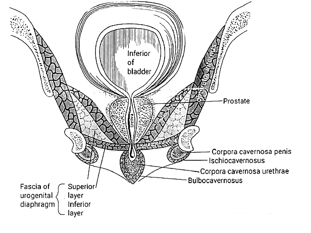
Perineal Spaces (Superficial and Deep)


Superficial Perineal Space

Deep Perineal Space

Bartholin's Gland Cysts
- Positioned within the superficial perineal pouch of the urogenital triangle, the Bartholin's glands function to produce a small quantity of mucus-like fluid.
- Typically, Bartholin's glands are not discernible during routine physical examination. Nevertheless, obstruction of the duct can lead to their enlargement, resulting in the formation of fluid-filled cysts.
- These cysts have the potential to become infected and inflamed, a condition referred to as bartholinitis.
- Infections are most frequently caused by bacteria such as Staphylococcus spp. and Escherichia coli.
Question for Applied AnatomyTry yourself: What is the function of the Bartholin's glands?View Solution
Pelvic Diaphragm

The pelvic diaphragm consists of muscle fibers from the levator ani and the coccygeus muscle.
Levator ANI
- The right and left levator ani lie almost horizontally in the pelvic floor, separated by a narrow gap transmitting the urethra, vagina, and anal canal.
- The levator ani is commonly divided into three parts: pubococcygeus, puborectalis, and iliococcygeus.
- The pubococcygeus, the primary component of the levator, extends backward from the body of the pubis toward the coccyx and may be affected during childbirth. Some fibers are inserted into the prostate, urethra, and vagina.
- The right and left puborectalis unite behind the anorectal junction to form a muscular sling, with some considering them as part of the external anal sphincter.
- The iliococcygeus, the most posterior part of the levator ani, is often poorly developed.
Coccygeus
The coccygeus, located behind the levator ani and frequently more tendinous than muscular, stretches from the ischial spine to the lateral margin of the sacrum and coccyx.
Pelvic Diaphragm - Functions
- It plays a crucial role in offering support to pelvic organs such as the bladder, intestines, and, in females, the uterus.
- It contributes to the maintenance of continence, serving as a component of the urinary and anal sphincters.
- It aids in the process of childbirth by resisting the descent of the presenting part, inducing forward rotation of the fetus to navigate through the pelvic girdle.
- It assists in sustaining optimal intra-abdominal pressure.

Thoracoabdominal Diaphragm
- The diaphragm is a curved musculofibrous dome-shaped sheet that serves as a partition between the thoracic and abdominal cavities.
- Its predominantly convex upper surface is oriented toward the thorax, while its concave inferior surface faces the abdomen.
- It is a primary muscle involved in respiration.
Parts of the Diaphragm

Vasculature of the Diaphragm

Structures passing through the Diaphragm

Congenital Diaphragmatic Hernia
Hiatal Hernia

Question for Applied AnatomyTry yourself: Which muscle is the primary component of the levator ani and extends backward from the body of the pubis toward the coccyx?View Solution
Applied Anatomy - Repeats
- Describe in brief the anatomy and applied significance of inguinal canal. Add a note on descent of testis (1995).
- Describe the formation of inguinal canal. Add a note on inguinal hernias (2000).
- Describe the boundaries of inguinal canal. Add a note on the anatomy of different types of inguinal hernias. (2014)
- Write a short note on pelvic diaphragm. (1995)
- Describe pelvic diaphragm. Add a note on supports of uterus (2001).
- Describe pelvic diaphragm; write in brief about its applied anatomy (2007).
- Describe be the gross anatomy of thoracic-abdominal diaphragm. Mention the structures that pass through it (2009).
- Write short notes on Ischio-Rectal abscess (2001)
- Discuss boundaries and contents of superficial and deep perineal spaces (pouches). Add a note on their applied Anatomy (2004).
The document Applied Anatomy | Medical Science Optional Notes for UPSC is a part of the UPSC Course Medical Science Optional Notes for UPSC.
All you need of UPSC at this link: UPSC
|
7 videos|219 docs
|
FAQs on Applied Anatomy - Medical Science Optional Notes for UPSC
| 1. What is the inguinal canal? |  |
Ans. The inguinal canal is a passage that exists in both males and females, located in the lower abdomen. It is formed by the lower abdominal muscles and serves as a pathway for the spermatic cord in males and the round ligament of the uterus in females.
| 2. What are the boundaries of the ischiorectal fossa? |  |
Ans. The ischiorectal fossa is a space located in the perineum, between the skin and the pelvic diaphragm. It is bounded by the pelvic diaphragm superiorly, the skin and subcutaneous tissue inferiorly, the ischium and its ramus laterally, and the anal canal medially.
| 3. What is the function of the pelvic diaphragm? |  |
Ans. The pelvic diaphragm is a muscular structure that forms the floor of the pelvic cavity. Its main functions include providing support to the pelvic organs, maintaining continence of the urinary and anal sphincters, and assisting in the process of childbirth.
| 4. What are the perineal spaces and what are their anatomical divisions? |  |
Ans. The perineal spaces are anatomical spaces located in the perineum, which is the region between the thighs and the buttocks. There are two main perineal spaces: the superficial perineal space, which is located between the perineal membrane and the superficial fascia, and the deep perineal space, which is located between the perineal membrane and the pelvic diaphragm.
| 5. What is a congenital diaphragmatic hernia? |  |
Ans. A congenital diaphragmatic hernia is a birth defect where there is an abnormal opening in the diaphragm, which allows abdominal organs to move into the chest cavity. This can lead to compression of the lungs and cause breathing difficulties in newborns. It requires surgical intervention to repair the diaphragm and reposition the organs back into the abdomen.

|
Explore Courses for UPSC exam
|

|
Signup for Free!
Signup to see your scores go up within 7 days! Learn & Practice with 1000+ FREE Notes, Videos & Tests.
Related Searches


