Cell-Cell Interactions | Zoology Optional Notes for UPSC PDF Download
Overview of Cell Signaling
- Common Nature: Communication between cells is ubiquitous in multicellular organisms.
- Mechanism: Cells influence each other through cell signaling.
- Signal Molecules: Various molecules serve as signals, including peptides, proteins, amino acids, nucleotides, steroids, lipids, and even gases like nitric oxide (NO).
Diversity of Signaling Molecules:
- Range of Signals: Signals attached to cell surfaces, secreted through plasma membranes, or released by exocytosis.
- Examples: Nitric oxide (NO) involved in male erections; Viagra stimulates NO release.
Cell Surface Receptors:
- Constant Signal Exposure: Cells are exposed to a continuous stream of signals.
- Specific Responses: Cells respond only to specific signals, determined by receptor proteins.
- Receptor Variety: Receptors can recognize peptides, proteins, or other molecules.
- Selective Response: Each cell responds to signals that match its set of receptor proteins, ignoring others.
Mechanism of Signal Reception:
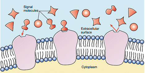
- Receptor Proteins: Located on or within the cell.
- Shape Compatibility: Receptor proteins have a specific three-dimensional shape that matches a signal molecule's shape.
- Binding and Response: When a signal molecule binds to a receptor, it induces a change in the receptor's shape, triggering a cellular response.
Challenges in Studying Receptor Proteins:
- Technical Challenge: Receptor proteins are relatively scarce in cells (constituting less than 0.01% of total protein mass).
- Purification Difficulty: Isolating them is comparable to finding a specific grain of sand in a sand dune.
Techniques for Receptor Protein Study:
- Monoclonal Antibodies: Antibodies bind specifically to molecules; monoclonal antibodies can isolate receptor proteins.
- Gene Analysis: Mutant studies and gene isolation help identify genes encoding various receptor proteins.
Advances in Receptor Analysis:
- Receptor Families: Techniques reveal that numerous receptor proteins can be grouped into a few families.
- Gene Identification: Advances in gene analysis allow the identification and isolation of genes encoding receptor proteins.
- Remarkable Discovery: Despite the vast number of receptors, they can be classified into a handful of families.
Cell signaling is a crucial mechanism in multicellular organisms, allowing cells to communicate and influence each other's behavior. Receptor proteins play a pivotal role by selectively responding to specific signals. Despite the challenges posed by the scarcity of receptor proteins, advanced techniques such as monoclonal antibodies and gene analysis have enabled significant progress in the study of cell signaling and the identification of receptor families. Further exploration promises to unveil more about the intricate world of cell communication.
Types of Cell Signaling
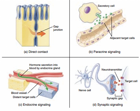
Cells employ various mechanisms for communication, with the choice depending on the distance between signaling and responding cells. Additionally, some cells engage in autocrine signaling, releasing signals that bind to receptors on their own plasma membranes, reinforcing developmental changes.
1. Direct Contact:
- Description: In close proximity, molecules on adjacent cell membranes can bind.
- Cell Interaction: Important interactions in early development occur through direct contact between cell surfaces.
- Focus: Contact-dependent interactions are explored further in this chapter.
2. Paracrine Signaling:
- Nature of Signals: Cells release signal molecules that diffuse through extracellular fluid.
- Local Influence: Molecules have short-lived, local effects, influencing neighboring cells.
- Restrictions: Limited influence as signals may be taken up by nearby cells, destroyed by enzymes, or quickly removed from the extracellular fluid.
3. Endocrine Signaling:
- Signal Molecules: Released molecules remain in the extracellular fluid.
- Circulatory System: Signals may enter the circulatory system and travel widely throughout the body.
- Longevity: Longer-lived signal molecules, known as hormones, affect cells at a distance from the releasing cell.
- Detail: Further discussed in Chapter 58; extensively used in both animals and plants.
4. Synaptic Signaling:
- Nervous System Cells: Cells of the nervous system provide rapid communication.
- Signal Molecules: Neurotransmitters are the signaling molecules.
- Travel Mechanism: Unlike hormones, neurotransmitters do not travel through the circulatory system.
- Release Location: Nerve cell extensions release neurotransmitters close to the target cells.
- Chemical Synapse: The narrow gap between cells in this process is called a chemical synapse.
- Persistence: Neurotransmitters persist briefly, providing rapid and localized signaling.
Autocrine Signaling:
- Self-Signaling: Cells release signals that bind to specific receptors on their own plasma membranes.
- Role: Thought to play a crucial role in reinforcing developmental changes.
Cell communication occurs through diverse mechanisms, each suited to specific distances between signaling and responding cells. From direct contact to long-distance endocrine signaling and rapid synaptic signaling, these mechanisms enable precise coordination of cellular activities. Autocrine signaling adds another layer, with cells influencing themselves. Understanding these signaling types is fundamental to unraveling the complexities of cellular communication in various biological processes.
Intracellular Receptors
In cell signaling pathways, common elements include a chemical signal transmitted between cells and a receptor that receives the signal in or on the target cell. This section focuses on the nature of receptors, specifically intracellular receptors.
1. Types of Receptors:
- Nature of Signals: Many signals are lipid-soluble or small molecules that easily traverse the plasma membrane.
- Receptor Location: Some bind to cytoplasmic protein receptors, while others pass through the nuclear membrane, binding to nuclear receptors.
- Responses: Intracellular receptors can initiate diverse cellular responses based on the specific receptor involved.
Cell Surface Receptors
- Nature of Signal Molecules: Most signal molecules are water-soluble, such as neurotransmitters, peptide hormones, and growth factors.
- Cell Membrane Permeability: Water-soluble signals cannot diffuse through cell membranes.
- Receptor Location: These signals bind to receptor proteins on the cell surface.
- Conversion of Signal: Cell surface receptors convert extracellular signals into intracellular ones, initiating changes within the cell's cytoplasm.
- Prevalence: The majority of a cell's receptors are cell surface receptors.
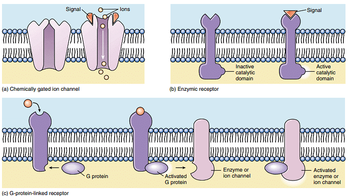
- Receptor Superfamilies: Almost all cell surface receptors belong to one of three superfamilies: chemically gated ion channels, enzymic receptors, and G-protein-linked receptors.
Understanding receptors is crucial in comprehending cell signaling processes. While lipid-soluble signals interact with intracellular receptors, water-soluble signals necessitate cell surface receptors to mediate responses. This diversity in receptor types reflects the intricate ways in which cells receive and interpret signals, leading to various cellular responses.
G-Protein-Linked Receptors
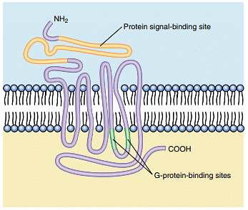
G-protein-linked receptors represent a class of cell surface receptors that act indirectly on enzymes or ion channels in the plasma membrane with the assistance of a guanosine triphosphate (GTP)-binding protein, known as a G protein.
Key Points:
G Protein Function:
- G proteins serve as mediators that initiate a diffusible signal in the cytoplasm.
- They create a transient link between the cell surface receptor and the signal pathway within the cytoplasm.
Signal Transmission Mechanism:
- When a signal molecule binds to the receptor, the G-protein-linked receptor undergoes a change in shape.
- This alteration in receptor shape leads to the twisting of the G protein, prompting it to bind to GTP.
- The activated G protein, now bound to GTP, can separate from the receptor and initiate various cellular events.
Activation Dynamics:
- The activation is short-lived due to the relatively short lifespan of GTP (seconds to minutes).
- This transient activation allows G proteins to activate multiple pathways in a brief period.
Regulation of Pathway Activity:
- Continuous extracellular signals are necessary for a pathway to remain active.
- When the rate of external signal diminishes, the pathway shuts down, ensuring a dynamic response to changing signal levels.
G-protein-linked receptors provide an intricate mechanism for transmitting signals from the extracellular environment to the cell's interior. The involvement of G proteins in transient activations allows for precise regulation of cellular pathways, emphasizing the dynamic nature of cell signaling processes.
Amplifying the Signal: Protein Kinase Cascades
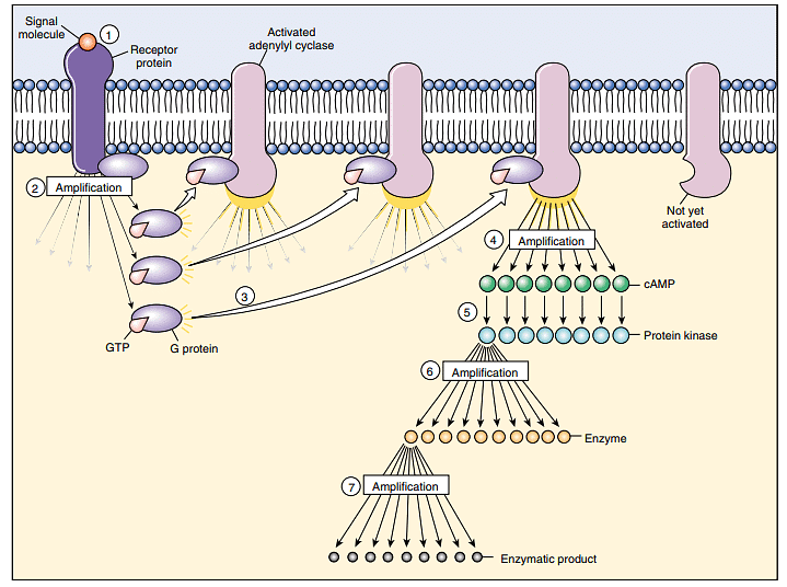
Both enzyme-linked and G-protein-linked receptors receive signals at the cell surface, but the actual cellular response often occurs in the cytoplasm or nucleus. To facilitate this process, signals are relayed through second messengers, influencing the activity of enzymes or genes. However, since many signaling molecules exist in low concentrations, their diffusion across the cytoplasm would be slow without signal amplification. Enzyme-linked and G-protein-linked receptors commonly employ a series of protein messengers to amplify the signal during transmission to the nucleus.
Signal Amplification Mechanism:
Phosphorylation Activation:
- The signal begins with the activation of a stage-one protein, typically through phosphorylation. The receptor either directly adds a phosphate group or activates a G protein, which subsequently activates a second protein responsible for phosphorylation.
Cascade Effect:
- Each activated stage-one protein then triggers the activation of numerous stage-two proteins.
- Similarly, each stage-two protein activates a large number of stage-three proteins, creating a cascade effect.
Amplification Potential:
- This cascade of protein activation results in signal amplification. The number of activated proteins increases exponentially, similar to a relay race where each runner tags multiple new runners at the end of each stage.
Overall Impact:
- A single cell surface receptor can initiate a complex cascade of protein kinase activations, greatly amplifying the original signal.
Analogy:
- The relay race analogy illustrates the sequential activation of proteins, analogous to passing a baton in a relay where each runner multiplies the number of participants.
Protein kinase cascades serve as an efficient mechanism for amplifying signals received at the cell surface. This amplification ensures a rapid and robust cellular response to extracellular signals, even when the signaling molecules are present in low concentrations.
The Expression of Cell Identity
In multicellular organisms, the development of specialized groups of cells called tissues is a hallmark of life. Despite originating from a single fertilized cell with identical genetic information, cells within tissues perform unique functions. The key question is how cells sense their location and "know" their tissue identity. The answer lies in tissue-specific identity markers and the acquisition of unique cell surface molecules during development.
Tissue-Specific Identity Markers:
Glycolipids:
- Most tissue-specific cell surface markers are glycolipids, which are lipids with carbohydrate heads.
- Glycolipids on the surface of cells, such as red blood cells, play a role in distinguishing between blood types (A, B, O).
- As cells divide and differentiate, the population of cell surface glycolipids undergoes significant changes.
MHC Proteins (Major Histocompatibility Complex):
- The immune system utilizes cell surface markers, including MHC proteins, to distinguish between "self" and "nonself" cells.
- MHC proteins serve as distinctive identity tags for individuals, as each person produces a unique set of MHC proteins.
- MHC proteins and other self-identifying markers are single-pass proteins anchored in the plasma membrane, often belonging to the immunoglobulin superfamily.
Immune System and Identity Recognition:
- Cells of the immune system continually inspect other cells encountered in the body.
- The immune system can trigger the destruction of cells displaying foreign or "nonself" identity markers, ensuring the recognition of self and nonself cells.
- The immune system's ability to distinguish self from nonself is particularly detailed in vertebrates.
Remarkable Distinction in Immune Systems:
- Vertebrates, as detailed in chapter 57, exhibit an exceptional ability to distinguish self from nonself through their complex immune systems.
- Other vertebrates and even some simpler animals, such as sponges, also possess mechanisms for distinguishing self from nonself, despite lacking a sophisticated immune system.
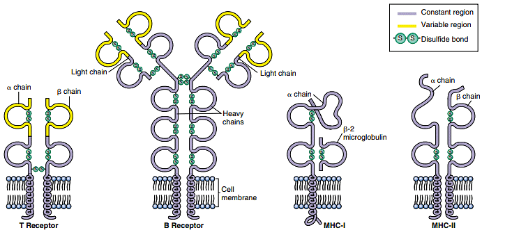
The acquisition of tissue-specific identity markers, particularly glycolipids and MHC proteins, is crucial for cells to communicate their identity to neighboring cells. These markers play a fundamental role in maintaining tissue integrity and facilitating the immune system's ability to recognize and respond appropriately to foreign entities.
Anchoring Junctions
Anchoring junctions play a crucial role in mechanically attaching the cytoskeleton of a cell to the cytoskeletons of other cells or to the extracellular matrix. These junctions are particularly prevalent in tissues that undergo mechanical stress, such as muscle and skin epithelium.
Cadherin and Intermediate Filaments: Desmosomes
Desmosomes are a type of anchoring junction that connects the cytoskeletons of adjacent cells, providing structural integrity and resistance to mechanical stress. Hemidesmosomes, on the other hand, anchor epithelial cells to a basement membrane. The key players in the formation of desmosomes are cadherins and intermediate filaments.
Cadherins:
- Cadherins are proteins, most of which are single-pass transmembrane glycoproteins.
- They create a crucial link between adjacent cells, facilitating the connection of their cytoskeletons.
- Cadherins form a strong binding interaction between cells, contributing to the structural stability of tissues.
Intermediate Filaments:
- Intermediate filaments are structural proteins present in the cytoskeleton.
- Various attachment proteins link the short cytoplasmic end of a cadherin to the intermediate filaments.
- The connection between cadherins and intermediate filaments strengthens the mechanical linkage between cells.
Strength of Connections:
- Connections between proteins anchored to intermediate filaments are more secure compared to connections involving free-floating membrane proteins.
- Proteins tethered to intermediate filaments provide a robust structural framework, enhancing the stability of cell-cell interactions.
- In contrast, untethered membrane proteins may be more susceptible to displacement due to weaker interactions with the lipid bilayer.
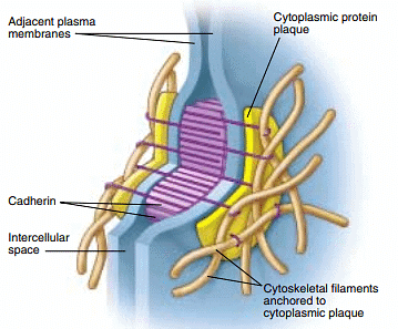
Anchoring junctions, particularly desmosomes, exemplify the importance of mechanical stability in tissues subjected to stress. The interplay between cadherins, intermediate filaments, and attachment proteins ensures not only the physical connection between cells but also the resilience needed to withstand mechanical forces. This molecular architecture contributes to the integrity and functionality of tissues in dynamic biological environments.
Communicating Junctions
Cells often communicate directly with neighboring cells through specialized structures known as communicating junctions. These junctions facilitate the direct transfer of chemical signals or small molecules between adjacent cells. In animals, these structures are called gap junctions, while in plants, they are referred to as plasmodesmata.
Gap Junctions in Animals:
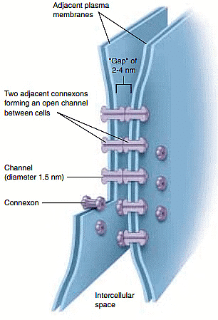
- Structure: Gap junctions are composed of connexons, which are complexes formed by six identical transmembrane proteins.
- Channel Formation: Connexons create channels through the plasma membrane, allowing for the direct exchange of small molecules and ions between the cytoplasm of two connected cells.
- Alignment: Gap junctions form when connexons from two cells align, establishing an open channel that spans both plasma membranes.
- Passageway Size: These junctions permit the passage of small substances like simple sugars and amino acids while restricting larger molecules such as proteins.
- Dynamic Nature: Gap junction channels are dynamic and can open or close in response to various factors, including the presence of ions such as Ca++ and H+.
- Protective Function: Gap junctions can close in response to cell damage, preventing the spread of damage to neighboring cells by isolating the affected cell.
Plasmodesmata in Plants:
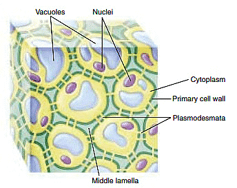
- Cell Walls: In plants, cell walls separate individual cells, and cell-cell junctions occur at openings or gaps in these walls.
- Plasmodesmata Structure: Plasmodesmata are cytoplasmic connections that form across touching plasma membranes of adjacent plant cells.
- Abundance: The majority of living cells in higher plants are connected by plasmodesmata.
- Functional Similarity to Gap Junctions: Plasmodesmata function similarly to gap junctions in facilitating the direct exchange of molecules between connected plant cells.
- Complex Structure: Unlike gap junctions, plasmodesmata are lined with plasma membrane and contain a central tubule that connects the endoplasmic reticulum of the two cells.
Communicating junctions, exemplified by gap junctions in animals and plasmodesmata in plants, are essential for direct cell-to-cell communication. These structures play critical roles in coordinating cellular activities, responding to environmental cues, and maintaining the overall integrity and functionality of tissues in both animal and plant organisms.
|
198 videos|351 docs
|


