Doc: Female External Genitalia | Additional Study Material for NEET PDF Download
VULVA
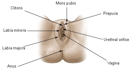
Vulva means external genitalia of female. They include mons veneris, labia majora, labia minora, clitoris, vestibule & related perineum.
 Fig: Female External Genitalia
MONS VENERIS (mons pubis) :-
It is a pad of subcutaneous connective tissue, lying in front of pubis & is covered by pubic hairs in adult female.
LABIA MAJORA :-
Vulva is bounded on each side by the elevation and folds of skin & subcutaneous tissue. Its inner surface is hairless.
Outer surface is covered by sebaceous gland, Sweat gland & hair follicles. It is homologous with the scrotum in the male.
LABIA MINORA :-
They are two thin folds of skin present just within the labia majora. Lower portion of minora fuses across the midline & form a fold of skin called fourchette.
CLITORIS :-
Small cylindrical & erectile body made by fusion of two labia minora, situated in the most anterior part of vulva. Clitoris is a homologous to the penis in the male. It is also made up of two erectile bodies (corpora cavernosa).The skin which covers the glans of clitoris is called prepuce.
At the terminal part of vagina the urethra opens separately, so they form a common chamber called vaginal vestibule or urinogenital sinus. Vagina opens outside through a slit like aperture or triangular space called vestibule. The vulva has following openings :-
(a) Urethral opening – Lies on anterior end
(b) Vaginal orifice – Lies on posterior end.
It is incompletely closed by a septum of mucous membrane called hymen, but it may not be a true sign of virginity.
(c) Openings of Bartholin; duct on either side
BARTHOLIN GLANDS :-
It is homologous to Cowper gland of male
In rabbit 1 pair is found on lateral side of vagina. It also opens into vagina.
It secretes slimy alkaline, watery fluid which makes alkaline media in vaginal passage.
In human female it is one pair on each side. These are also known as greater and lesser vestibular gland. These glands are situated on lateral side of vagina.
Histology of Oviduct :-
I. Serosa or the adventitia :- It is the outermost layer of visceral-peritoneum (Perimetrium)
II. Muscle-layer :- The middle layer of the oviduct is made up of unstripped-muscle. In uterus, thick smooth muscle bundles are found, these are called as myometrium.
III Mucous membrane :- It is the innermost layer. Mucosa consists of simple columnar epithelium.
- Epithelium contains both ciliated cells & secretory cells. The secretory cells produce viscous liquid film that provides nutrition & protects the ovum.
- Mucosa of Uterus is called endometrium, it contains tubular glands, many fibroblasts & blood vessels.
- In the uterus, the embryo is attached to endometrium. Longest unstripped muscles of the body are found in the walls of uterus (During pregnancy).
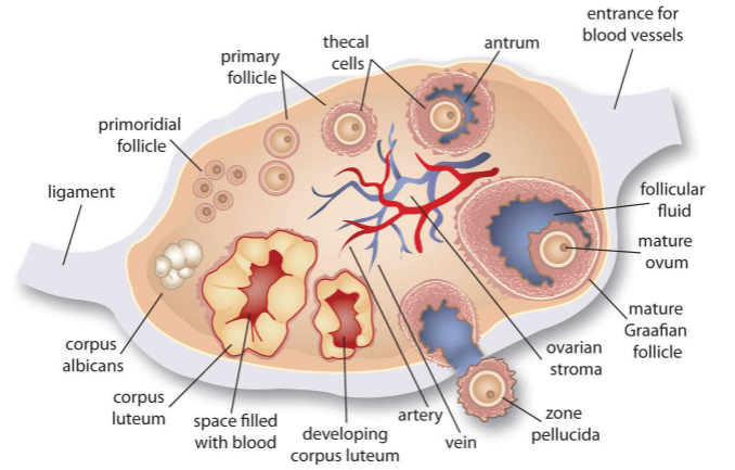
 Fig: Section of Human ovary
- Outermost layer of ovary is called germinal epithelium while the inner layer called T. albuginea is made up of white fibrous connective tissue. The inner part of ovary is called as stroma.
- It is differentiated into 2 parts, outer peripheral part is cortex & inner part is called medulla.
- Stroma consists of follicular cells, connective tissues, blood vessels & lymphatics.
- Numerous oogonial are found in cortical region in intrauterine life. In early stage of intrauterine life, they proliferate by mitosis, after which meiosis starts in them and proceeds upto prophase stage & halts there itself up to puberty (when the ovulation starts).
- Now the halted meiosis process restarts at puberty causing primary oocyte to convert into secondary oocyte just before ovulation. With this the Ist meiotic division completes and first polar body is formed.
- In secondary oocyte immediately begins the second meiotic division but this division stops again at metaphase stage. It proceeds further only when a sperm penetrates the oocyte.
|
26 videos|287 docs|64 tests
|
FAQs on Doc: Female External Genitalia - Additional Study Material for NEET
| 1. What are the main parts of the female external genitalia? |  |
| 2. How does the appearance of the female external genitalia vary among individuals? |  |
| 3. What is the function of the clitoris in the female external genitalia? |  |
| 4. How do Bartholin's glands contribute to the female external genitalia? |  |
| 5. Can the female external genitalia undergo any changes or abnormalities? |  |

|
Explore Courses for NEET exam
|

|


