NCERT Exemplar: Locomotion & Movement - 2 | Biology Class 11 - NEET PDF Download
Short Answer Type Questions
Q1. With respect to the rib cage, explain the following:
(a) Bicephalic ribs
(b) True ribs
(c) Floating ribs
Ans:
 Rib Cage
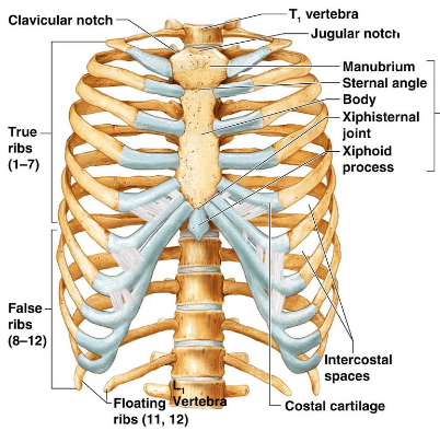
(a) Bicephalic ribs: Each rib is a thin flat bone connected dorsally to the vertebral column and ventrally to the sternum. It has two articulation surfaces on its dorsal end and is hence called bicephalic.
(b) True ribs: First seven pairs of ribs are called true ribs. Dorsally, they are attached to the thoracic vertebrae and ventrally connected to the sternum with the help of hyaline cartilage.
(c) Floating ribs: Last 2 pairs (11th and 12th) of ribs are not connected ventrally and are, therefore, called floating ribs.
Q2. In old age, people often suffer from stiff and inflamed joints. What is this condition called? What are the possible reasons for these symptoms?
Ans: In old age, people suffer from stiff and inflamed joints, it is due to rheumatoid arthritis (autoimmune disorder)
Causes:
- Inflammation of the synovial membrane
- Genetic factors (50% cases).
- Smoking
- Vitamin-D deficiency
Q3. The exchange of calcium between bone and extracellular fluid takes place under the influence of certain hormones
(a) What will happen if more of Ca++ is in extracellular fluid?
(b) What will happen if very less amount of Ca++ is in the extracellular fluid?
Ans:
(a) If more of Ca++ is in the extracellular fluid then it will be accumulated on the bones under the influence of thyrocalcitonin (TCT).
(b) If very less amount of Ca++ is in the extracellular fluid then parathyroid hormone (PTFI) acts on bones and stimulates the process of bone resorption (dissolution/demineralisation). PTH also stimulates reabsorption of Ca2+ by the renal tubules and increases Ca2+ absorption from the digested food.
Q4. Name at least two hormones which result in fluctuation of Ca++ level
Ans: Thyrocalcitonin (TCT) which lowers the calcium and potassium levels in the blood and Parathyroid hormone which increased level of this cause the bones to release their Ca+ into the blood.
Q5. Rahul exercises regularly by visiting a gymnasium. Of late he is gaining weight. What could be the reason? Choose the correct answer and elaborate.
(a) Rahul has gained weight due to accumulation of fats in body.
(b) Rahul has gained weight due to increased muscle and less of fat.
(c) Rahul has gained weight because his muscle shape has improved.
(d) Rahul has gained weight because he is accumulating water in the body.
Ans: Rahul has gained weight due to increased muscle and less of fat.
Rahul has gained weight due to increased muscle and less fat because exercising regularly can cause an increase in the amount of sarcoplasm that is the thickness of myofibrils increases along with an increase in protein synthesis and mitochondria
Q6. Radha was running on a treadmill at a great speed for 15 minutes continuously. She stopped the treadmill and abruptly came out. For the next few minutes, she was breathing heavily/fast. Answer the following questions.
(a) What happened to her muscles when she did strenuously exercised?
(b) How did her breathing rate change?
Ans:
(a) Repeated activation of the muscles can lead to the accumulation of lactic acid due to anaerobic breakdown of glycogen in them, causing fatigue.
(b) During strenuous exercise demand of oxygen also increases so the breathing rate has changed.
Q7. Write a few lines about Gout.
Ans: When metabolic waste-uric acid crystals are accumulated in bones, then it results in inflammation of bone and joints thereby causing pain. This disorder of the skeletal system is called gout.
Q8. What is the source of energy for muscle contraction?
Ans: The source of energy for muscle contraction is ATP (Adenine Triphosphate). An enzyme called myosin ATPase present on the head of the myosin molecule breaks down into ADP and inorganic phosphate in the presence of magnesium and calcium ions and releases energy in the head of the myosin.
i.e., ATP → ADP + Pi + Energy
Q9. What are the points for the articulation of Pelvic and Pectoral girdles?
Ans: The components of the pelvic girdle are ilium, ischium and pubis. It articulates with, femur through the acetabulum. The components of the pectoral girdle are the scapula and clavicle. It is the glenoid cavity of the pectoral girdle in which the head of the humerus articulates.
Long Answer Type Questions
Q1. Calcium ion concentration in blood affects muscle contraction. Does it lead to tetany in certain cases? How will you correlate fluctuation in blood calcium with tetany?
Ans: Muscle contraction is initiated by a signal sent by the central nervous system (CNS) via a motor neuron. A neural signal reaching this junction releases a neurotransmitter (acetylcholine) which generates an action potential in the sarcolemma. This spreads through the muscle fibre and causes the release of calcium ions into the sarcoplasm. An increase in Ca++ level leads to the binding of calcium with a subunit of troponin on actin filaments and thereby remove the masking of active sites for myosin. Utilising the energy from ATP hydrolysis, the myosin head now binds to the exposed active sites on actin to form a cross-bridge. This pulls the attached actin filaments towards the centre of ‘A’ band. The ‘Z’ line attached to these actins are also pulled inwards thereby causing a shortening of the sarcomere, i.e., contraction. The process continues till the Ca++ ions are pumped back to the sarcoplasmic cisternae resulting in the masking of actin filaments.
Tetany: Rapid spasms (wild contractions) in muscle due to low Ca in body fluid.
Q2. An elderly woman slipped into the bathroom and had severe pain in her lower back. After X-ray examination doctors told her it is due to a slipped disc. What does that mean? How does it affect our health?
Ans: Displacement of the intervertebral disc from’ their normal position is called a slipped disc. Effects:
- Neck or lower back pain
- Muscular weakness
- Paralysis
- Sciatica
Q3. Explain sliding filament theory of muscle contraction with neat sketches.
Ans: Mechanism of muscle contraction: Mechanism of muscle contraction is best explained by the sliding filament theory which states that contraction of a muscle fibre takes place by the sliding of the thin filaments over the thick filaments. Muscle contraction is initiated by a signal sent by the Central Nervous System (CNS) via a motor neuron. A motor neuron along with the muscle fibres connected to it constitute a motor unit. The junction between a motor neuron and the sarcolemma of the muscle fibre is called the neuromuscular junction or motor-end plate. A neural signal reaching this junction releases a neurotransmitter (acetylcholine) which generates an action potential in the sarcolemma. This spreads through the muscle fibre and causes the release of calcium ions into the sarcoplasm. An increase in Ca++ level leads to the binding of calcium with a subunit of troponin on actin filaments and thereby remove the masking of active sites for myosin. Utilising the energy from ATP hydrolysis, the myosin head now binds to the exposed active sites on actin to form a cross-bridge.
 Sliding-filament Theory of Muscle Contraction (Movement of the thin Filaments and the Relative Size of the I Band and H Zones)This pulls the attached actin filaments towards the centre of ‘A’ band. The ‘Z’ line attached to these actins are also pulled inwards thereby causing a shortening of the sarcomere, i.e., contraction. It is clear from the above steps, that during shortening of the muscle, i.e., contraction, the ‘I’ bands get reduced, whereas the ‘A’ bands retain the length. The myosin, releasing the ADP and P, goes back to its relaxed state. A new ATP binds and the cross-bridge is broken. The ATP is again hydrolysed by the myosin head and the cycle of cross-bridge formation and breakage is repeated causing further sliding. The process continues till the Ca++ ions are pumped back to the sarcoplasmic cistemae resulting in the masking of actin filaments. This causes the return of ‘Z’ lines back to their original position, i.e., relaxation.
Q4. How does a muscle shorten during its contraction and return to its original form during relaxation?
Ans: As per sliding filament theory, muscle contraction takes place because of the sliding of actin and myosin filaments towards each other. When actin and myosin filaments slide away from each other, relaxation of muscles happens. In striated muscles, the striations appear because of alternate bands of actin and myosin. The band of actin is light in colour and is called the I band. The myosin band is darker in colour and is called the A band. The actin filaments are held in the middle by an elastic band called Z line. The myosin filaments are held in the middle by an elastic band called M line. During contraction, the position of Z line changes in relation to M line and muscle fibre becomes shorter. During relaxation, the actin filaments move to their original position and the muscle fibre appears longer.
Q5. Discuss the role of Ca2+ ions in muscle contraction. Draw neat sketches to illustrate your answer.
Ans: Muscle contraction is initiated by a neural signal, which after reaching neuromuscular junction or motor end plate releases a neurotransmitter, as a result an action potential in the sarcolemma is generated. Action potential spreads through muscle fibre and causes the release of calcium ions into the sarcoplasm. An increase in Ca2+ level leads to the binding of calcium with a subunit of troponin on actin filaments and thereby removes the masking of active sites for myosin. Utilising the energy from ATP hydrolysis, the myosin head now binds to the exposed active site on actin to form a cross-bridge. This pulls the attached actin filaments towards the centre of ‘A’ band. The ‘Z’ line attached to these actins are also pulled inwards thereby causing shortening of the sarcomere, i.e., contraction.
 Stages in Cross-bridge Formation, Rotation of Head and Breaking of Cross-bridge
A new ATP binds to the myosin head and the cross-bridge is broken. The ATP is again hydrolysed by the myosin head and the cycle of cross-bridge formation and breakage is repeated causing further sliding. The process continues till the Ca++ ions are pumped back to the sarcoplasmic cistemae resulting in masking of actin filaments and breakage of all cross-bridges. This cause the return of ‘Z’ lines along with filaments back to their original position, i.e., relaxation.
Q6. Differentiate between Pectoral and Pelvic girdle.
Ans: Pectoral and pelvic girdle help in the articulation of upper and lower limbs respectively. Each girdle is made of two equal halves. Each half of a pectoral girdle consists of a clavicle and scapula. A scapula is a large triangular flat bone. There is a glenoid cavity at the joint of the scapula, clavicle and acromian process, which articulates with the head of the humerus to form the shoulder joint. Each half of the pelvic girdle is formed by three bones—ilium, ischium and pubis. At the point of their fusion; there is a cavity called acetabulum to which the head of femur articulates.
|
181 videos|361 docs|148 tests
|
FAQs on NCERT Exemplar: Locomotion & Movement - 2 - Biology Class 11 - NEET
| 1. What is locomotion and movement? |  |
| 2. How does locomotion occur in animals? |  |
| 3. What are the types of skeletal systems in animals? |  |
| 4. How do muscles contribute to locomotion? |  |
| 5. Can plants exhibit locomotion? |  |

|
Explore Courses for NEET exam
|

|


