Respiratory System | Zoology Optional Notes for UPSC PDF Download
Salient Features of the Respiratory System in Vertebrates
Definition of Respiration:
- Respiration involves the exchange of oxygen and carbon dioxide between the organism and the environment.
- External respiration refers to the gas exchange between the environment and the blood through a respiratory surface.
Types of Respiratory Organs:
- Vertebrate respiratory organs can be classified into two types:
- Gills: Evagination with the respiratory surface turned out.
- Lungs: Invagination with the respiratory surface turned in.
- Vertebrate respiratory organs can be classified into two types:
Ventilation/Breathing:
- Vertebrates use gills or lungs, along with the skin in some cases, for breathing.
- Ventilation involves the active movement of the respiratory medium (water or air) across the respiratory surface.
Types of Ventilation:
- Non-directional: Water or air flows past the respiratory surface in an unpredictable pattern.
- Unidirectional: Water or air enters the respiratory surface at one point and exits at another.
- Tidal: Water or air moves in and out from one point only.
Efficiency Requirements for Respiratory Organs:
- Renewal of Medium: Provision for renewing the supply of oxygen-containing medium (water or air) and removing carbon dioxide.
- Large Surface Area: The respiratory organs must have a large surface area with an ample capillary network for efficient gas exchange.
- Thin and Moist Membrane: The respiratory surface should be thin and moist to facilitate the passage of gases.
Structure of Gills in Aquatic Vertebrates:
- Gills serve as the respiratory organs for aquatic vertebrates.
- They have an evagination structure, and their functioning is essential for oxygen uptake and carbon dioxide elimination.
- The gill structure allows efficient gas exchange with the surrounding water.
- The gill membrane is thin and moist, providing an ideal surface for gas diffusion.
- The capillary network associated with gills ensures a large surface area for effective respiratory exchange.
Understanding these features is crucial for appreciating the diversity of respiratory adaptations seen in vertebrates, reflecting their habitat and evolutionary history.
Respiration by Gills in Vertebrates
General Gill Structure:
Composition:
- Gills are the primary respiratory organs in fishes and some aquatic amphibians.
- Composed of numerous gill filaments or gill lamellae, thin-walled extensions of the epithelial surface.
- Each gill contains a vascular network, bringing blood close to the respiratory surface for efficient gas exchange.
Gill Cavity:
- Gills are enclosed in a protective gill cavity, allowing efficient water flow over them.
Gill Arch Arrangement:
- Fish gills consist of several gill arches on either side, separating opercular and buccal cavities.
- Two rows of gill filaments extend from each gill arch, forming a sieve-like structure for water flow.
Gill Filament Structure:
Epidermal Membrane:
- Fish gills are covered by a thin epidermal membrane that folds to form plate-like lamellae.
- Lamellae increase the surface area for respiratory gas exchange.
Water Flow Direction:
- Water flows over the branched gills through the mouth.
- The flow of water is opposite to the direction of blood movement.
Variation in Gill Area:
- The area of gills varies among fishes based on their activity levels.
- More active fishes typically have larger gill areas.
Types of Gills:
External Gills:
- Develop from the integument covering branchial/visceral arches, protruding into surrounding water.
- Found in fish and larvae of vertebrates, including lung fishes and amphibians.
- Branched, filamentous structures from ectoderm.
Internal Gills:
- Associated with pharyngeal slits and pouches.
- Composed of parallel gill lamellae, may be filamentous.
- Two types:
- Primitive (Elasmobranchs): Well-developed interbranchial septa extending beyond hemibranchs.
- Advanced (Bony Fishes): Reduced interbranchial septa with hemibranchs protruding into branchial chamber.
Respiratory Process:
- In fishes with internal gills, water with dissolved oxygen enters through the mouth, passing through gill slits into gill clefts.
- As water moves over gill lamellae, oxygen is extracted, and carbon dioxide is released.
- Water then exits through external gill slits to the outside.
Understanding the diversity in gill structure and function is essential for comprehending the respiratory adaptations in aquatic vertebrates.
Respiratory System of Cyclostomes and Fishes
Cyclostomes (Hagfishes and Lampreys):
Hagfishes (Myxini):
- Number of gill openings varies (1 to 15) on each side.
- Water flows unidirectionally. Enters through a single nasal opening, passes through the pharynx, gill pouches, and exits through numerous gill slits.
- Myxine has a single gill opening on each side, located midventrally.
Lampreys (Petromyzontida):
- Seven pairs of internal gill slits open into gill clefts.
- Tidal ventilation in adults (water enters and exits through gill slits), unidirectional in larvae.
- Single nasal opening connected to the pituitary via a duct, forming a nasohypophysial pouch.
- Pharynx divided into dorsal esophagus and ventral respiratory tube.
- Gill pouches with gill lamellae open externally through gill slits.
- Operculum, velum, and muscles aid in tidal ventilation.
Fishes' Respiratory System:
Cartilaginous Fishes (Elasmobranchii):
- Large volume of water sucked into buccal cavity and spiracles.
- Buccal expansion increases water volume; mouth and spiracles close, forcing water past gills and out through gill slits.
- Countercurrent flow enhances oxygen extraction from water.
Bony Fishes (Teleosts):
- Five gill slits covered by a bony operculum.
- Buccal pump and opercular suction drive water over gills.
- Swim bladder aids in buoyancy.
- Opercular movement assists in water expulsion.
Swim Bladder (Teleosts):
- Hydrostatic organ providing buoyancy.
- Adjusts gas volume for neutral buoyancy.
- Present in most pelagic fishes.
Lung Fishes:
Polypterus (Actinopterygian):
- Bilobed lung symmetrically developed.
- Single pair of external integumentary gills.
- Ventral duct from lung to pharynx.
Neoceratodus and Others:
- Lungs used when oxygen tension is low.
- Surface furrows in lung epithelium increase contact with air.
- Buoyancy maintained by swim bladder or modified lungs.
Accessory Respiratory Structures in Freshwater Fishes:
- Freshwater fishes evolved accessory organs due to the risk of their environments drying up.
- Some fishes have evolved additional respiratory structures to breathe air in addition to gills.
- The lack of water in branchial chambers and mucus accumulation can affect gill function when exposed to air.
Respiration in Amphibian Larvae Using Gills
Amphibians undergo a remarkable transition from larval life in water to adulthood on land. During the larval stage, they employ various respiratory structures, including external gills, skin, buccal cavity, internal gills, and later, lungs.
External Gills in Amphibian Larvae:
Appearance and Development:
- Three pairs of external gills emerge at hatching, connected to blood vessels from aortic arches.
- Folds from the hyoid arches cover the gills as operculum.
- Gills are eventually resorbed as the larva develops.
Blood Supply:
- Blood vessels from aortic arches provide vascular connections to external gills.
Internal Gills in Amphibian Larvae:
Formation and Structure:
- Internal gills develop with the establishment of the mouth opening.
- Branchial filaments form ventral to external gills on the branchial arches, projecting into the opercular cavity.
- Third, fourth, and fifth visceral arches have two rows of filaments, while the sixth arch has one row on its anterior side.
Water Flow:
- Water passes from the mouth to the pharynx, then through gill slits into the opercular chamber.
- It exits through the opercular aperture.
Metamorphosis:
- Internal gills disappear during metamorphosis.
Transition to Lungs:
- In some urodeles (salamanders), gills are retained throughout life, but in most urodeles and all tailless amphibians, gills disappear during metamorphosis.
- Lungs, newly developed during metamorphosis, take over the respiratory function.
Analogous Structures in Reptiles, Birds, and Mammals:
- In reptiles, five pairs of pharyngeal pouches develop during embryonic life.
- In birds and mammals, typically only four pairs develop, with the fifth remaining rudimentary and attached to the fourth pair.
- Pharyngeal pouches usually do not break through to the outside, but in rare cases, they may, leading to the formation of branchial cysts and fistulae.
Respiration by Lungs
The transition from water breathing to air breathing marked a crucial evolutionary shift in vertebrates. Lungs, the primary respiratory organs in terrestrial animals, are elastic bags designed for breathing air.
Lung Development:
Embryonic Origin:
- Lungs develop embryologically from outpocketing of endoderm from the pharynx.
- The outpocketing divides into lung buds, destined to become bronchi, and the lungs proper.
Structure:
- Lungs are elastic and expand during inhalation, contracting during exhalation.
Trachea and Bronchi:
- The original duct connecting lungs to the pharynx is known as the trachea.
- The trachea typically bifurcates into two bronchi, each leading to one lung.
- Bronchi further branch into bronchioles, which end in air sacs or alveoli.
Lung Anatomy:
Bronchial Tree:
- The bronchi resemble a tree structure, branching into smaller bronchioles.
- Bronchioles lead to alveoli, where gas exchange occurs.
- Humans have approximately 600 million alveoli, providing a large surface area with ample capillary network for gas exchange.
Ventilation:
- Lungs, being invaginated organs with a narrow entry/exit point, are ventilated tidally, i.e., bidirectionally (in-and-out).
- Tidal ventilation creates dead space in major air-conducting passages.
Dead Space:
- The trachea, bronchi, bronchioles, and alveoli collectively form a significant volume of air.
- During exhalation, most air is expelled, but some remains in the passageways, constituting the dead space.
- Tidal volume, the total inhaled air in a breath, is reduced by the dead space volume.
Adaptations in Different Vertebrate Groups:
- Snakes:
- Some snakes may have a reduced or absent lung due to their slender bodies.
Key Respiratory Parameters:
Tidal Volume:
- The total volume of air inhaled in a single breath.
- Normal human tidal volume at rest is around 500 ml.
Dead Space:
- The volume of air remaining in the respiratory passageways after exhalation.
- Approximately 150 ml (30% of tidal volume) in humans.

Understanding the structure and adaptations of lungs across different vertebrate groups provides insights into how respiratory systems have evolved to suit diverse environments.
Respiratory System in Amphibians
Amphibians, including anurans (frogs), urodeles (salamanders), and caecilians, exhibit a multifaceted respiratory system that includes cutaneous and pulmonary respiration.
Cutaneous Respiration:
Importance:
- Amphibians rely significantly on cutaneous respiration, especially when submerged in water.
- The skin, being membranous with a vast capillary network, facilitates gas exchange.
Skin Adaptations:
- Skin is moist and scaleless, containing mucus glands that secrete glycopeptides and glycerol.
- Mucus helps keep the skin moist, aiding in gas exchange.
Gas Exchange:
- Oxygen and carbon dioxide exchange occurs between atmospheric gases and deoxygenated blood in capillaries.
- Oxygenated blood returns to the heart via the cutaneous vein.
Pulmonary Respiration:
Respiratory Tract in Frogs:
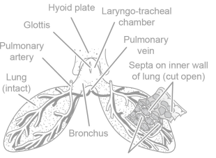
- Frog respiratory tract includes external nostrils, nasal chambers, internal nostrils, bucco-pharyngeal cavity, glottis, laryngo-tracheal chamber, and bronchi.
Lung Structure:
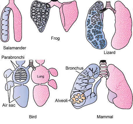
- Amphibian lungs are typically two simple sacs, elongated in urodeles and bulbous in anurans.
- Lungs are located in the pleuriperitoneal cavity along with other viscera.
Internal Lining:
- The internal lining of amphibian lungs may be smooth or have folds and septa, increasing the surface area for respiration.
Pulmonary Respiration Process:
- Amphibians lack ribs and a diaphragm, so inspiration occurs in two stages.
- First Stage:
- Air is drawn into the buccal cavity through external nostrils, functioning as a suction pump.
- Buccopharyngeal respiration occurs, involving gaseous exchange in the buccal cavity.
- Glottis opens, releasing air from the lungs into the buccal cavity.
- Second Stage:
- Nostrils and mouth close, raising the floor of the buccal cavity, increasing pressure.
- Air enters the laryngo-tracheal chamber through the glottis.
- Elastic lung walls dilate, allowing air entry and gas exchange in the alveoli.
- During expiration, elastic lung walls recoil, expelling air to the buccal cavity and then outside through nostrils.
Adaptations in Different Amphibians:
- Urodeles may have poorly vascularized lungs, while more active species like Hyla (tree frog) exhibit elaborate lungs with increased capillary meshes.

- Urodeles may have poorly vascularized lungs, while more active species like Hyla (tree frog) exhibit elaborate lungs with increased capillary meshes.
The respiratory system of amphibians showcases a dynamic interplay between cutaneous and pulmonary respiration, adapting to their aquatic and terrestrial lifestyles.
Respiratory System in Reptiles
Reptiles, the first vertebrates adapted to terrestrial life, predominantly rely on lungs for gas exchange due to their dry, scaly skin. Their respiratory system, including lungs with numerous compartments, exhibits adaptations to support terrestrial life.
Cutaneous Respiration:
- Reptiles have reduced cutaneous respiration to negligible levels due to their dry and scaly skin.
Lung Structure:
- Unlike the simple sac-like lungs of amphibians, reptile lungs are composed of many small compartments, resembling a sponge.
- Septa within the lungs form partitions that increase the respiratory surface area.
- Faveoli, the air compartments, are not found at the end of a highly branched tracheal system like alveoli in mammalian lungs.
Respiratory Mechanism:
- Reptile lungs are aspiratory, involving the suction of air by changing the size and pressure within the lungs.
- Expansion of the rib cage during inhalation reduces lung pressure, drawing air in.
- Expiration occurs through contraction of trunk muscles and elastic recoil of lungs during a period of apnea.
Variations in Lung Structure:
- In some reptiles, the left lung may be rudimentary or absent.
- Snakes and limbless lizards have elongated lungs functionally divided into vasculated and avascular zones.
- Marine reptiles may have multi-chambered lungs, and in higher lizards, crocodilians, and turtles, septa form numerous large chambers.
Adaptations for Specific Functions:
- Reptiles may have a relatively larger lung volume but a smaller surface area compared to mammals.
- Some species, like the puffing adder, have specialized lung structures for specific behaviors, such as inflation for defense.
- Aquatic reptiles often have smooth avascularized air sacs for buoyancy and maintaining breath-holding ability.
Metabolic Adaptations:
- Reptiles have a lower oxygen requirement, with a standard metabolic rate only 10 to 20 percent of that in homeotherms.
- They tolerate greater changes in blood circulatory components, allowing them to survive in low-oxygen conditions for extended periods.
Species-Specific Respiratory Features:
- Lizards have a trachea supported by cartilaginous rings and exhibit rib movements for inspiration.
- Crocodiles have a muscular diaphragm and use the hepatic piston method for lung expansion.
- Turtles, with a rigid carapace, utilize forelimbs and pectoral girdle for forcing air in and out.

Reptiles showcase diverse respiratory adaptations, allowing them to thrive in terrestrial environments while exhibiting unique features based on their ecological niches.
Respiratory System in Birds
The respiratory system in birds is uniquely adapted for the high metabolic demands of flight, featuring extensive air sacs, one-way airflow, and efficient gas exchange. Here are the key features of the avian respiratory system:
Adaptations for Flight:
- The avian respiratory system occupies about 20% of the body volume, reflecting the high metabolic requirements of flight (compared to 5% in humans).
- Birds have highly modified lungs with extensive air sacs and intricate air passages.
Air Sacs:
- Birds have nine interconnected air sacs, including cervical, anterior thoracic, posterior thoracic, abdominal sacs (paired), and an unpaired interclavicular sac.
- These air sacs invade various parts of the body, including bones, forming pneumatic cavities.
- The air sacs extend into the bones, replacing bone marrow and allowing some birds to respire through bones if trachea is blocked.
Lung Structure:
- Bird lungs are small, spongy, and paired organs with little elasticity.
- Air enters the lungs through bronchi, leading to mesobronchi, dorsobronchi, and parabronchi.
- Unlike mammals, where tertiary branches end in sac-like alveoli, bird tertiary branches form tube-like air capillaries for continuous airflow.
- Gas exchange primarily occurs in air capillaries.
One-Way Airflow:
- Birds exhibit a unidirectional flow of air through the respiratory system, enhancing the efficiency of gas exchange.
- Valves ensure that the airflow remains one way, promoting a more efficient oxygen concentration.
Respiration Cycle:
- The respiration cycle involves two breath cycles: inhalation and exhalation.
- During inhalation, air moves from the bronchi into posterior air sacs.
- Exhalation moves the previously inhaled air from posterior air sacs into parabronchi of the lungs.
- The second inhalation moves this air into anterior air sacs, and the second exhalation passes air through bronchi and out of the system.
- Oxygen exchange occurs during both inhalation and exhalation.
Sternum Movement:
- The sternum of birds is raised when at rest and lowered during flight, actively decreasing the size of the body cavity for exhalation.
Metabolic Adaptations:
- Small passerine birds with higher metabolic rates have more efficient lungs compared to gliding birds with lower energy demands.
- Some birds, like fowls and emus, which do not fly extensively, may have lower pulmonary blood diffusion capacity.

The avian respiratory system is a marvel of adaptation, allowing birds to meet the metabolic demands of flight while efficiently extracting oxygen from the air.
Respiratory System in Mammals
Mammals possess a well-developed respiratory system with multi-chambered lungs enclosed within the thoracic cavity. Here are the key features of the mammalian respiratory system:
Thoracic Cavity:
- The bony framework of the thoracic cavity consists of thoracic vertebrae, ribs, and the sternum.
- Mammalian lungs are multi-chambered and typically divided into lobes, with the right side having more lobes than the left side.
Respiratory Pathway:
- Air enters through external nostrils and nasal passages into the pharynx.
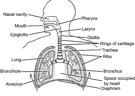
- From the pharynx, it passes through the glottis into the trachea, a long tube that traverses the neck and lies ventral to the gullet.
- The trachea bifurcates into two primary bronchi, each entering a lung and further branching into secondary and tertiary bronchi, and then into bronchioles.
- Terminal bronchioles lead into thin-walled, delicate alveolar ducts, which end in clusters of alveoli where gas exchange occurs.
Lung Protection:
- The lungs are protected and cushioned by the pleura, consisting of two layers: visceral pleura (adhered to the lungs) and parietal pleura (lining the chest wall).
- The pleural cavity between the layers contains pleural fluid, allowing smooth movement during lung inflation and deflation.
Respiration Process:
- In mammals, the buccal cavity plays no role in respiration.
- During inhalation, rib muscles (external intercostal muscles) and the diaphragm contract, increasing the size of the thoracic cavity and lowering the pressure, allowing air to enter the lungs (inspiration).
- Expiration is a passive process, driven by the relaxation of intercostal muscles and the diaphragm, bringing the thoracic cavity back to its normal size and forcing air out.
Alveoli Characteristics:
- Alveoli are separated by thin-walled, vascularized septa.
- The thickness of alveolar septa varies among species; for example, shrews and bats have thin septa due to their high metabolic rate, while sirenians (manatees and dugongs) have thicker septa.
Gas Exchange:
- Gas exchange occurs across the alveolar surface, primarily involving Type I alveolar cells.
 The respiratory system of humans
The mammalian respiratory system relies on the coordinated action of muscles, including the diaphragm and intercostal muscles, to facilitate the exchange of gases in the alveoli.
|
181 videos|346 docs
|

|
Explore Courses for UPSC exam
|

|


