Revision Notes: Structural Organization in Animals | NCERT Exemplar & Revision Notes for NEET PDF Download
Epithelial Tissues
- An epithelium is a tissue composed of one or more layers of cells that cover the body surface and lines its various cavities.
- It serves for protection, secretion and excretion.
- The word ‘epithelium’ was introduced by Ruysch.
- Epithelial tissue evolved first in animal kingdom.
- It originates from all the three primary germ layers. e.g. Epidermis arises from ectoderm, Coelomic epithelium from the mesoderm and epithelial lining of alimentary canal from the endoderm.
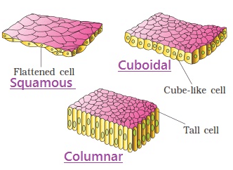
- Types of Epithelium
Glands
- Multicellular exocrine glands are classified by structure, using the shape of their ducts and the complexity (branching) of their duct system as distinguishing characteristics.
- Shape include tubular and alveolar (Sac like).
- Simple exocrine glands e.g. intestinal glands, mammalian sweat glands, cutaneous glands of frog etc. have only one duct leading to surface.
- Compound exocrine glands have two or more ducts e.g. liver, salivary glands etc.
- Structural classification of exocrine glands:
| Type | Example |
| Simple tubular | Intestinal glands, crypts of Lieberkuhn in ileum. |
| Simple coiled tubular | Sweat glands in man |
| Simple branched tubular | Gastric (stomach) gland, and Uterine gland. |
| Simple alveolar | Mucous gland in skin of frog, Poison gland of toad and seminal vesicle. |
| Simple branched alveolar | Sebaceous glands |
| Compound tubular | Brunner’s gland, bulbourethral gland and liver. |
| Compound alveolar | Sublingual and submandibular parotid salivary gland |
| Compound tubulo alveolar | Parotid salivary glands, Mammary gland and Pancreas. |
Important Tips
- Study of tissue outside the body in a glass tube is known as in vitro, while study of living tissue in situ is known as in vivo.
- Among epithelium, simple epithelium were first to evolve.
- Transitional epithelium also called plastic epithelium or urothelium. It lacks basement membrane
- False epithelium derived from mesenchymal a diffuse network of tissue derived from embryonic mesoderm) and lining the synovial cavities.
- Mammary glands without teats are present in prototheria.
- A malignant tumors arising from an epithelium is called a carcinoma. If it arises from a squamous epithelium it is a squamous cell carcinoma and if it arises from glandular epithelium it is called an adenoma.
- The epithelial lining of brain ventricles and central canal of spinal cord is known as ependyma.
- Stereocilia are elongated membrane outgrowths found in certain parts of male reproductive tract.
Muscle Tissues
- Muscle cells are highly contractile (contracting to 1/3 or 1/2 the resting length).
- Muscle cells lose capacity to divide, multiply and regenerate to a great extent. Study of muscle is called myology.
- About 40% to 50% of our body mass is of muscles.
- The muscle cells are always elongated, slender and spindle-shaped, fibre-like cells, These are, therefore called muscle fibres.
- These possess large numbers of myofibrils formed of actin and myosin.
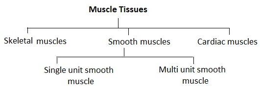
Difference between three types muscle fibres
| S.No. | Feature | Striated or Striped or Skeletal or Voluntary muscle fibres | Non-striated or Unstriped or Smooth or Visceral or Involuntary muscle fibres | Cardiac muscle fibres |
| 1. | Shape | Long cylindrical | Fusiform (thick in middle tapering at ends) (0.02 nm to 0.2 nm long) | Network of fiber |
| 2. | Stripes | Dark A bands and light I bands present | Absent | Present |
| 3. | Nucleus | Many (syncytial) at periphery | Single at the centre of each cell | Many nuclei between successive and plates central position |
| 4. | Unit | Sarcomeres, cylindrical long myofibrils placed end to end forming cylindrical myofibrils | Fusiform cells with inconspicuous borders | Oblique cross-connecting fibres make this muscle an interconnected bundle of myofibrils |
| 5. | Attachment | To bones | To soft organs or viscera | Not attached to other organs except major blood vessels which are isolated and covered by pericardium |
| 6. | Sarcolemma | Distinct | Absent | Absent |
| 7. | Sarcoplasmic Reticulum | Well developed | Less extensive | Poorly formed |
| 8. | Blood supply | Rich | Poor | Rich |
| 9. | Contraction | Quick, fatigue fast | Slow, sustained contraction | Rhythmic, contractions originate in heart (pace maker immune to fatigue) |
| 10. | Location | Generally peripheral, tongue, proximal part of oesophagus | Central, in hollow visceral organs, iris of the eye, dermis of the skin | Only in heart |
| 11. | Intercalated discs | Absent | Absent | Present |
| 12. | T-tubule system | Well developed | Lacking | Well developed |
| 13. | Innervated nerves | Motor nerves from central nervous system (neurogenic) | Nerves from autonomic nervous system (neurogenic) | Nerves from central and autonomic nervous system (myogenic) |
| 14. | Fibres | Unbranched | Unbranched | Fibres join by short oblique bridges |
| 15. | Action | Voluntary | Involuntary | Involuntary |
Connective Tissues
- It connects and supports all the other tissues, the intercellular elements predominating.
- The cellular element is usually scanty. In function this tissue may be mechanical, nutritive and defensive.
- It is a tissue made up of matrix (abundant intercellular substance or ground substance) and living cells that connects and support different tissues.
- Connective tissue was called mesenchyme by Hertwig (1893).
- Types of connective tissues
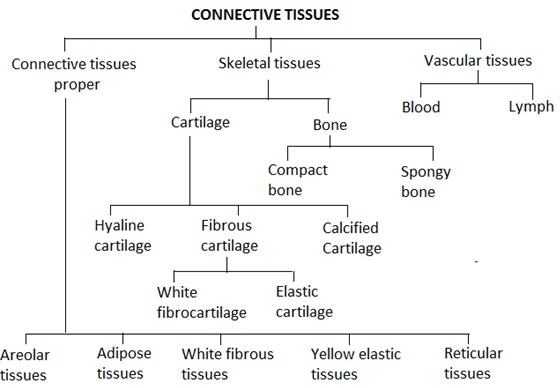
(1) On the basis of their texture:
The bones are divided into two categories spongy or cancellous or tubecular bones and compact or periosteal bones
| Bone | Cartilage |
| 1. Matrix is composed of a tough, inflexible material, the ossein. | 1. Matrix is composed of a firm, but flexible material, the chondrin. |
| 2. Matrix is always impregnated with calcium salts. | 2. Matrix may be free or impregenated with calcium salts. |
| 3. Bone cells lie in lucunae singly. | 3. Cartilage cells lie in lacunae singly or in groups of two or four. |
| 4. Osteocytes are irregular and give off branching processes in the developing bone. | 4. Chondroblasts are oval and devoid of processes. |
| 5. Lacunae give off canaliculi. | 5. Lacunae lack canaliculi. |
| 6. There are outer and inner layers of special bone forming cells, the osteoblasts, that produce new osteocytes, which secrete new lamellae of matrix. | 6. There are no special cartilage-forming cells. Cartilage grows by division of all chondroblasts. |
| 7. Matrix occurs largely in concentric lamellae. | 7. Matrix occurs in a homogenous mass. |
| 8. Bone is highly vascular. | 8. Cartilage in nonvascular. |
| 9. Bone may have bone marrow at the centre. | 9. No such tissue is present. |
(2) On the basis of origin of bone:
Ossification or osteogenesis is the process of bone formation. A bone is classified into four categories.
| Characters | Spongy bone | Compact bone |
| Arrangement of lamellae | There is no regular Haversian system so have spongy texture. | Have regular Haversian system |
| Occurrence | In skull bones, ribs, centrum of vertebrae and epiphyses of long bones | In the shaft (diaphysis) of long bones |
| Marrow cavity | Broad | Narrow |
| Type of bone marrow | Red marrow in the spaces between lamellae | Yellow marrow in marrow cavity |
| Function | Marrow forms RBCs and Granular WBCs | Marrow stores fats |
(3) On the basis of treatment:
These are of two types :-
| Characters | Dried bone | Decalcified bone |
| Type of treatment | Subjected to high temperature. | Subjected to dilute solution of HCl. |
| Nature of matter left | With only mineral matter. | With only organic matter. |
| Marrow cavity | Empty. | With bone-marrow. |
| Fate of cells | Periosteum, endosteum, osteoblasts and osteocytes are absent being killed by high temperature. | Periosteum, endosteum, osteoblasts and osteocytes all are present. |
| Lacunae | Lacunae present. | Lacunae absent. |
(6) Number of RBC: The number of RBCs is counted by instrument haemocytometer. The total number of RBC per cubic mm of blood is called RBC count. RBC count is slightly lower in women than a man and number of RBC is more in people who live on mountains because there is less oxygen. RBC are absent in cockroach.
| S.No. | Organism | Number of RBCs |
| 1. | Male | 5 – 5.4 million / cubic mm of blood |
| 2. | Female | 4.5 – 5 million / cubic mm of blood |
| 3. | Infants | 65 – 70 lacs/ cubic mm of blood |
| 4. | Embryo | 85 lacs/ cubic mm of blood |
| 5. | Rabbit | 70 lacs / cubic mm of blood |
| 6. | Frog | 4 lacs / cubic mm of blood |
(7) Life span of RBC: The life span of red blood corpuscles circulating in the blood stream varies in different animals. RBCs have longest life span in blood. The mammalians RBC have short life span due to absence of nucleus, which is disappeared during development.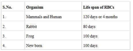
(8) Function of RBCs: The major function of erythrocytes is to receive O2 of respiratory surfaces and then transport and readily deliver it to all cells of body. This important function is performed by haemoglobin which has a great ability to combine loosely and reversibly with O2 and is, hence, called “respiratory pigment”. Haemoglobin, in annelids, is dissolved in the plasma because of absence of red blood corpuscles. In mollusc and some arthropods, etc., a different respiratory pigment, haemocyanin is found dissolved in the plasma. This pigment is bluish due to presence of copper in place of iron.
(9) Comparison Between Blood and Lymph
Important Points
- Argentaffin cells which produce a precursor of serotonin, a potent vasoconstrictor hormone, occurs in intestinal cells.
- The brown adipose tissue in human is restricted till third month of post natal life.
- White fibres yield gelatin on boiling and are digestible with enzyme pepsin but yellow (elastic) fibres are not digestible by enzyme trypsin.
- The fat in the globules is stored in the form of triglycerides.
- The Cytoplasmic granules basophils contain histamine.
- Sprain – Excessive pulling of ligaments.
- Plasma cells are also called as “Cart wheel cells”.
- Collagen constitutes about 33% of total body protein.
Nervous Tissues
- A most complex tissue in the body, composed of densely packed interconnected nerve cells called neurons (as many as 1010 in the human brain).
- It specialized in communication between the various parts of the body and in integration of their activities.
- Nervous tissue is ectodermal (from neural plate) in origin.
- Difference between axon and dendron

Classification of nervous tissues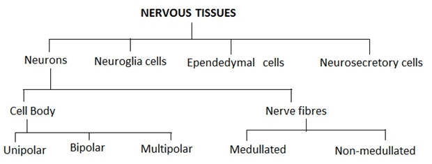
FAQs on Revision Notes: Structural Organization in Animals - NCERT Exemplar & Revision Notes for NEET
| 1. What is the structural organization in animals? |  |
| 2. How are cells organized in animals? |  |
| 3. What are the primary organ systems in animals? |  |
| 4. How do organ systems interact in animals? |  |
| 5. What is the importance of structural organization in animals? |  |


