Techniques of Cell Biology | Botany Optional for UPSC PDF Download

Immunofluorescence Microscopy
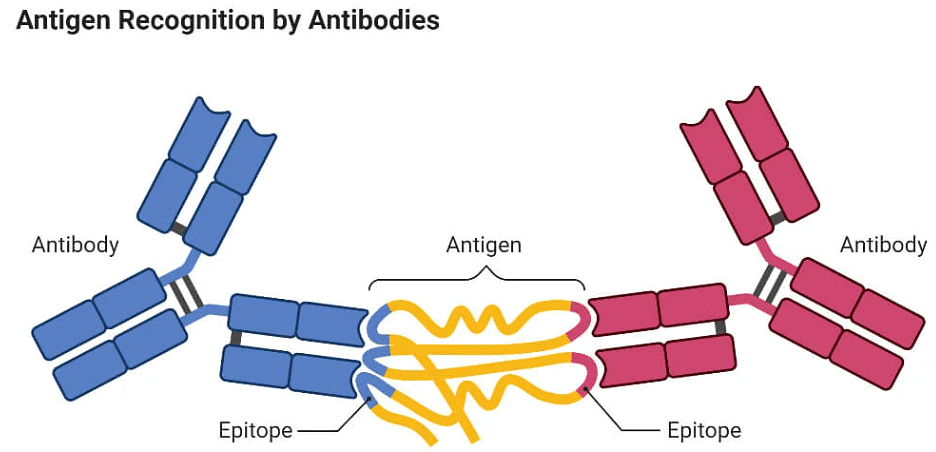
- Immunofluorescence serves as a powerful technique for pinpointing the whereabouts of antigens or antibodies within tissue samples or smears by employing specific antibodies or antigens that are tagged with a fluorescent label. This method is founded on the interaction between antigens and antibodies, where antibodies selectively bind to specific antigens.
- In the Direct Fluorescent Antibody (DFA) Method, the antibody forms a coat around the antigen, such as a bacterial cell. This bond is resilient and impervious to removal through elution or washing. After the removal of all non-antibody globulins, the antibody remains firmly attached to the cell. Notably, the antibody is made fluorescent through a process called conjugation, involving the attachment of substances like fluorescein or other dyes. Consequently, the bacterial cell's outline, enveloped by the fluorescent antibody, can be effortlessly observed using a specialized microscope.
- Conversely, the Indirect Fluorescent Antibody (IFA) Method entails a multi-step approach. Initially, the specific antibody interacts with the antigen, and the non-antibody globulin is subsequently removed through washing. Subsequently, a labeled antibody is introduced, binding to the specific antibody already attached to the antigen. For example, if the specific antibody was generated in a rabbit, it is treated with fluorescein-labeled anti-rabbit globulin. This results in a combination of the labeled antibody with the rabbit immunoglobulin that is already connected to the antigen.
- The utility of fluorescent antibody studies extends to the detection of a wide array of bacterial, viral, fungal, and protozoan infections, as well as the identification of numerous microscopic tissue components. This capacity to precisely pinpoint and identify antigens or antibodies within biological specimens has ushered in a new era of research and diagnostics in the realm of medicine and beyond.
Ion-Exchange Chromatography
Principle of Ion Exchange Chromatography
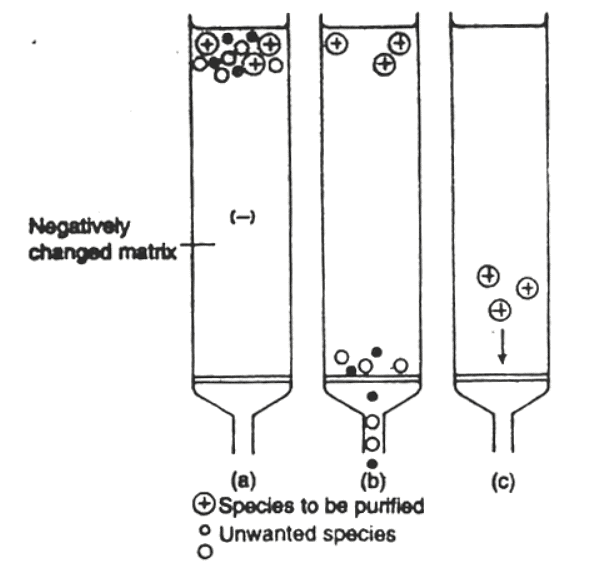
- The foundation of ion exchange chromatography lies in the distinct affinities of ions for an ion exchange resin packed into a column. This principle forms the basis for processes like protein purification, water softening, and metal separation. It is also instrumental in separating organic compounds like amino acids. Ion exchange chromatography encompasses two main types:
- Cation Exchange Chromatography: This technique specializes in separating cations. For instance, elements like zirconium, hafnium, niobium, and tantalum have been effectively separated using cation exchange chromatography.
- Anion Exchange Chromatography: In contrast, anion exchange chromatography is employed for the separation of anions.
Procedure of Ion Exchange Chromatography
The procedure for ion exchange chromatography typically involves the following steps:
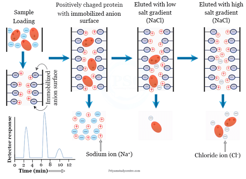
- Sample Loading: An impure protein sample is introduced into the ion exchange chromatography column.
- Binding: Positively charged protein molecules within the sample bind to the negatively charged groups present on the resin molecules.
- Elution: A salt gradient, varying in concentration, is employed to elute the protein from the resin. Lower salt gradients result in the elution of only a few positively charged protein molecules, whereas higher gradients release most of the protein from the resin.
Several factors influence the elution process, such as particle size, pH levels, and the protein's isoelectric point. The latter is crucial, as amino acids within the protein do not migrate in an electric field at their isoelectric point.
Exploring the Ion Exchange Chromatography Principle
- The ion exchange chromatography principle draws parallels with ionic crystalline solids. For instance, in ionic crystals like potassium chloride (KCl), each potassium ion (K+) is surrounded by eight chloride ions (Cl−). When a medium with a high dielectric constant, such as water, is introduced, the attractive forces between ions decrease, leading to ion exchange in the solution.
- Ion Exchange Resin: Ion exchange resins, integral to this technique, are high molecular weight, insoluble polymers or electrolytes. They contain functional groups like HSO3H, COOH, and OH that facilitate ion exchange. Two main types of resins are used:
- Cation Exchange Resin: This synthetic resin is typically produced by polymerizing styrene with a small amount of divinylbenzene, followed by sulfonation. In this resin, hydrogen ions (H+) are exchangeable cations. When treated with sodium ions (Na+), the H+ ions are replaced by sodium ions.
- Anion Exchange Resin: Anion exchange resins contain amines or quaternary ammonium groups within the polymeric network. In this case, chloride ions serve as exchangeable ions.
Instrumentation of Ion Exchange Chromatography
- The instrumentation for ion exchange chromatography involves:
- Columns constructed from inert materials like Kel-F, Teflon, and polypropylene.
- Introduction of the sample at the top of the column.
- Utilization of a double piston pump for precise liquid flow control.
- Specially manufactured ion exchange resin within the column.
- Implementation of microprocessor technology for conductivity detection of ions at nano concentrations.
- Passage of eluted samples through a suppressor column to release ions with high conductivity.
- Recording and analysis of ion concentration from the elution profile.
Applications of Ion Exchange Chromatography
- Ion exchange chromatography finds extensive applications across various domains:
- Analytical Chemistry: Widely used for separating and purifying cations and anions.
- Biological Chemistry: Crucial for protein and amino acid separation and purification.
- Medicine: Used in sugar and amino acid analysis, as well as blood component purification.
- Chemical Industries: Employed for impurity analysis and quality control.
- Agriculture: Valuable in micronutrient analysis in soil samples.
- Petrochemical Industry: Utilized for sulfur compound determination.
- Environmental Chemistry: Vital for detecting and separating toxic pollutants in aquatic and atmospheric environments.
Affinity Chromatography
 Affinity chromatography is a powerful technique employed in the field of biochemistry for the separation and purification of complex biochemical mixtures. This method is celebrated for its exceptional specificity and efficiency in isolating target molecules. In this article, we delve into the principles, components, and applications of affinity chromatography, exploring its advantages and disadvantages.
Principles of Affinity Chromatography
- At its core, affinity chromatography relies on the specific affinity between the stationary phase and the analyte, often referred to as a ligand. This interaction can be driven by various forces, including electrostatic attraction, hydrophobic interactions, or hydrogen bonding. The selection of an appropriate stationary phase and mobile phase is paramount and depends on the nature of the interaction between the target molecule and the ligand.
- The fundamental principle involves covalently attaching an immobilized biochemical, known as an affinity ligand, to a solid support matrix. When a sample is passed through the chromatographic column, only the solute that selectively binds to the complementary ligand is retained. This "lock and key" binding mechanism ensures the specific capture of the target molecule, while other components in the sample elute without retention. Subsequently, the retained solute(s) can be eluted from the column by altering the mobile phase conditions.
Components of Affinity Chromatography
Affinity chromatography systems consist of two primary components: the stationary phase and the mobile phase.
- Stationary Phase: The stationary phase in affinity chromatography typically comprises a solid matrix and the affinity ligand. The ligand is an immobilized chemically insoluble matrix that selectively adsorbs specific molecules from the analyte mixture. Various types of ligands are available, each tailored for specific applications. Some common ligand types include:
- Antibodies: Monoclonal or polyclonal antibodies are used for highly specific binding.
- DNA: DNA can be employed for capturing DNA-binding proteins, polymerases, helicases, and restriction enzymes.
- Biometric Dyes: These are suitable for capturing proteins.
- Peptides: Peptides find use in capturing various biomolecules.
- Additionally, a spacer arm, composed of carbon and/or other atoms, positions the ligand away from the solid matrix, making it more accessible and less restricted by steric hindrance. The solid matrix itself must meet specific criteria, including being mechanically and chemically stable, resistant to microorganisms, and inert to prevent non-specific interactions.
- Mobile Phase: The mobile phase used in affinity chromatography can be either nonpolar or polar, depending on the specific requirements of the separation.
- Instrumentation Affinity chromatography involves several key steps, including the introduction of the sample, adsorption of the analyte, removal of impurities, and elution of the sample. The following components play essential roles in the process:
- Injection-Pump System: This automated delivery system introduces the sample onto the high-performance bioaffinity matrix.
- Affinity Column: The column houses the ligand, spacer, and solid matrix, facilitating the separation and purification process.
- Online Monitoring: Effluent is continuously monitored and assayed for specific biological properties.
- Post-Collection Monitoring: Ensuring the quality of the collected analyte.
Applications of Affinity Chromatography
One noteworthy example of affinity chromatography is the purification of antibodies using immunoaffinity chromatography. This technique employs immobilized antigens as ligands, allowing the isolation and purification of specific antibodies from complex mixtures, such as blood serum.
Advantages and Disadvantages
- Advantages:
- Exceptional specificity.
- Rapid and reversible interaction.
- Faster separation.
- High resolution and efficiency.
- Stable under pressure.
- Well-defined column conditions.
- Simple detection and analysis.
- Suitable for small sample sizes.
- Reduced purification steps.
- Minimal protein denaturation.
- Applicable to large-scale processes.
- Disadvantages:
- Potential for irreversible adsorption.
- Slower kinetics in some cases.
In conclusion, affinity chromatography stands as a cornerstone technique in the field of biochemistry, offering remarkable specificity and efficiency in the separation and purification of target molecules. While it is not without its limitations, its advantages far outweigh its disadvantages, making it an indispensable tool in the arsenal of biochemists and researchers.
Partition and Adsorption Chromatography
Chromatography: A Versatile Separation Technique
Chromatography is a highly versatile analytical technique used for the separation, purification, and identification of substances within mixtures, serving both qualitative and quantitative analysis purposes. This sophisticated methodology hinges on various factors, including hydrophobic interactions, polarity, enzymes, and net charges, to segregate and scrutinize compounds effectively. At its core, chromatography stands as a physical means of separating complex mixtures into their individual components. This article delves into the diverse types of chromatography methods, providing a comprehensive overview of their principles and applications.
Categorizing Chromatographic Techniques
Chromatography techniques can be broadly categorized into three main groups:
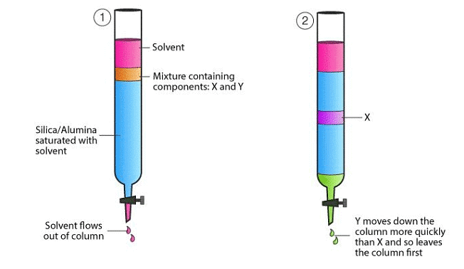
- Adsorption Chromatography: Adsorption chromatography is a method that separates components within a mixture based on their differential adsorption to a stationary phase housed in a chromatographic column. The varying rates at which mixture components traverse the column stem from disparities in their non-covalent interactions with the stationary phase. Typically, this technique employs a solid stationary phase known as an adsorbent, with the mobile phase being either a liquid or a gas. Adsorbents themselves can be polar or non-polar molecules.
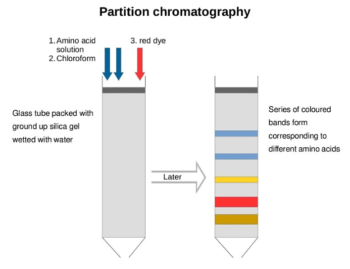
- Partition Chromatography: Partition chromatography distributes mixture components into two phases due to differences in their partition coefficient (Kd), representing the ratio of solute concentrations in the two phases. The key driving force behind this separation is solubility differences. In partition chromatography, the stationary phase takes on a liquid form, while the mobile phase can be either liquid or gas. This technique can be further subdivided into two types:
- Normal-Phase Partition Chromatography: In this type, the stationary phase is more polar than the mobile phase. The elution process commences with the least polar analyte and culminates with the most polar one.
- Reversed-Phase Partition Chromatography: Conversely, this technique features a mobile phase that is significantly more polar than the stationary phase. As a result, the most polar solutes are the first to elute, while the least polar ones exit the column last.
- Size Exclusion Chromatography: Size exclusion chromatography, also known as molecular sieve chromatography, separates molecules based on their size and shape. A column matrix filled with porous gel beads, composed of insoluble and hydrated polymers like polyacrylamide, dextran, or agarose, serves as the stationary phase. Two primary types of size exclusion chromatography are Gel Permeation Chromatography and Gel Filtration Chromatography, differing in their choice of mobile solvent—organic for the former and aqueous for the latter.
- Affinity Chromatography: Affinity chromatography is a specialized technique tailored for the purification of biomolecules concerning their biological function or specific chemical structure. This method hinges on the specific and reversible adsorption of the target substance to a ligand immobilized within a chromatographic bed material. The workflow of affinity chromatography encompasses multiple stages, including the selection of an appropriate ligand, ligand immobilization onto a support matrix, binding of the molecules of interest with the ligand, removal of non-specifically bound molecules, and the elution of purified molecules.
- High-Performance Liquid Chromatography (HPLC): HPLC represents a column chromatography variant wherein the mobile phase consists of a liquid, while the stationary phase can be either solid or liquid. This technique operates across various chromatographic modes, such as size exclusion, adsorption, ion-exchange, and partition. The distinction lies in the application of high pressure to force the mobile phase through the column, resulting in superior performance and speed compared to traditional column chromatography.
- Ion Exchange Chromatography: Ion exchange chromatography is employed for the separation of charged molecules. In this type of chromatography, ionic solutes engage in reversible electrostatic interactions with a charged stationary phase, typically composed of covalently attached anions or cations within an insoluble matrix known as ion exchangers. Two main categories exist within ion exchange chromatography:
- Cation Exchanger: Also referred to as an acidic ion exchanger, it facilitates the separation of cations.
- Anion Exchanger: Alternatively known as a basic ion exchanger, it enables the separation of anions.
- Gas Chromatography: In gas chromatography, a carrier gas serves as the mobile phase while the stationary phase can be either a solid adsorbent (gas-solid chromatography) or a liquid on an inert support (gas-liquid chromatography). This technique is primarily employed for the analysis of volatile substances in the gas phase, distinguished by its unique feature of not utilizing the mobile phase for interacting with the analyte.
In conclusion, chromatography is a multifaceted analytical tool with a wide array of applications across various scientific disciplines. Understanding the different types of chromatography and their underlying principles is crucial for scientists and researchers aiming to separate, purify, and analyze complex mixtures effectively. Whether investigating the composition of chemical compounds or purifying biomolecules, chromatography remains an indispensable asset in the laboratory toolkit.
Gel Filtration Chromatography
Gel filtration chromatography is an indispensable tool in the realm of biomolecule purification. It is a technique that enables the separation of biomolecules based on their molecular weight or size. In this article, we will delve into the intricacies of gel filtration chromatography, exploring its principles, various types, components, procedural steps, as well as its manifold applications. Furthermore, we will discuss the advantages and limitations of this technique to provide a comprehensive understanding of its utility in the scientific domain.
Principle of Gel Filtration Chromatography
- Gel filtration chromatography operates on the principle of segregating biomolecules according to their size. It is noteworthy for being one of the simplest and mildest chromatography techniques available. Unlike other methods that rely on factors like charge, hydrophobicity, or biorecognition, gel filtration primarily distinguishes molecules based on their dimensions.
- To initiate the process, a gel filtration medium is tightly packed into a column, forming a packed bed. This medium comprises porous spherical particles chosen for their chemical and physical stability. These particles remain inert, meaning they do not react with the substances passing through them. The packed bed is equilibrated with a buffer solution, which saturates the pores of the matrix and the interstitial spaces between the particles. This buffer-filled matrix constitutes the stationary phase, while the fluid outside the particles acts as the mobile phase.
- The stationary phase consists of a porous polymer matrix, with its pores entirely filled by the mobile phase solvent. Subsequently, the sample containing biomolecules is pumped through specialized columns packed with this microporous gel. The separation hinges on the fact that molecules above a certain size are completely excluded from the pores, while smaller molecules gain access, either partly or wholly. Consequently, as the mobile phase flows through the column, larger molecules pass through unimpeded, while smaller ones are delayed, contingent on their penetration into the gel matrix. This disparity in passage times leads to the separation of the biomolecules.
Types of Gel Filtration Chromatography
Gel filtration chromatography manifests in two main types:
- Group Separations: This type classifies components of a sample into two major groups based on their size range. It is useful for tasks such as removing high or low molecular weight contaminants or desalting and exchanging buffers from the sample.
- High-Resolution Fractionation of Biomolecules: In this type, components of a sample are separated according to differences in their molecular size. It serves various purposes, including isolating specific components, separating monomers from aggregates, determining molecular weight, and conducting molecular weight distribution analyses.
Steps in Gel Filtration Chromatography
The procedure for gel filtration chromatography encompasses the following steps:
- Packing the Column: Spherical gel filtration medium particles are packed into a column.
- Sample Application: The sample is applied to the column.
- Column Elution: Buffer (the mobile phase) and the sample move through the column.
- Molecular Diffusion: Molecules diffuse in and out of the matrix pores, effectively partitioning between the mobile and stationary phases.
- Separation by Size: Smaller molecules infiltrate the matrix pores more deeply and thus remain in the column for a longer time.
- Continuous Buffer Flow: As the buffer continuously traverses the column, molecules larger than the pore size cannot diffuse into the matrix and, therefore, pass through the column without hindrance.
- Detection of Components: Separation occurs at distinct intervals, followed by the detection of individual components.
Applications of Gel Filtration Chromatography
Gel filtration chromatography finds extensive utility in various scientific applications:
- Purification of Biomolecules: It plays a pivotal role in purifying enzymes, polysaccharides, nucleic acids, proteins, and other biological macromolecules.
- Protein Refolding: Gel filtration aids in the refolding of denatured proteins by precisely controlling buffer conditions.
- Protein Fractionation: It is instrumental in protein fractionation experiments.
- Molecular Weight Determination: Gel filtration is used for determining the molecular weights of compounds.
- Size-Based Separation: It separates substances like sugars, proteins, peptides, and rubbers based on their size.
- Quaternary Structure Analysis: Gel filtration is employed to determine the quaternary structure of purified proteins.
Advantages of Gel Filtration Chromatography
Gel filtration chromatography boasts several advantages:
- Robustness: It is well-suited for handling biomolecules sensitive to pH, metal ion concentration, and environmental conditions.
- Flexibility: Conditions can be adjusted to suit specific samples or purification requirements without compromising separation.
- Compatibility: It can be performed in the presence of essential ions, detergents, and various conditions.
- No Binding: Unlike other chromatography methods, molecules do not bind to the medium, eliminating buffer composition effects on resolution.
- Efficiency: Gel filtration offers short analysis times, well-defined separation, narrow bands, good sensitivity, no sample loss, and minimal mobile phase requirements.
Limitations of Gel Filtration Chromatography
While gel filtration chromatography is a versatile technique, it does have limitations:
- Peak Resolution: It can resolve only a limited number of peaks within a short timeframe.
- Filtration Requirements: Filtrations must precede instrument use to prevent particulate interference.
- Broad Peaks: For molecules with closely spaced molecular masses, gel filtration may yield broad peaks with limited differentiation.
In conclusion, gel filtration chromatography is a vital tool in the purification and analysis of biomolecules. Understanding its principles, types, components, steps, applications, as well as its advantages and limitations is essential for scientists and researchers seeking to employ this technique effectively in their work.
Radioactive Tracer Technique
- The most effective method for tracking cellular processes and their localization within cells is the use of radioactive isotopes. In biological research, several commonly used isotopes include H3, C14, P32, S35, I125, and I131. These isotopes emit high-energy electrons or beta particles during their radioactive decay.
- Radioactive decay is a spontaneous process, and its rate varies depending on the source. The number of atoms undergoing decay at any given time is proportional to the number of atoms present in the isotope at that moment. This process is conveniently quantified using the concept of half-life, which is defined as the time required for the radioactive activity to decrease by half. The table 8.3 provides information about the half-life of some important isotopes.
- In the International System of Units (SI), the unit of radioactivity is the Becquerel (Bq), representing one disintegration per second. However, the most commonly used unit is the curie (Ci), which measures the number of nuclear disintegrations per second compared to that of 1 gram of Radium, equivalent to 3.7 x 10¹⁰ disintegrations per second.
- In biological research, particularly with biological materials, microcuries (µCi) and millicuries (mCi) are used to measure radioactivity. The disintegrations are quantified as "counts" using a counter.
- To study cellular macromolecules, researchers label them with radioactive compounds by exposing tissues or cells to these compounds. The fate of the radioactive compound can then be monitored. For instance, the study of DNA synthesis in various cell types or tissues often requires the use of H3 (tritium).
- Various organic and inorganic compounds labeled with Tritium are available from the Bhabha Atomic Energy Research Centre in Mumbai. Tritiated Thymidine and Uridine are commonly used to study DNA and RNA, respectively.
- In experimental settings, the radioactive substance is added to a solution containing cells or tissues, and these are exposed for a specific period. Samples are either fixed at regular intervals or aliquots are collected at various time points to measure radioactivity.
- Radioactivity can be measured using a Liquid Scintillation Counter, or the distribution of radioactivity within different cellular locations can be observed through Autoradiography, a type of Radioactive Tracer Technology. In Autoradiography, labeled cells are fixed, placed on a slide, and covered with a thin layer of special autoradiographic stripping film, or sometimes photographic emulsion. These slides are then stored in the dark. During storage, beta particles emitted by the radioactive substance activate silver halide crystals in the film or emulsion. After a few weeks, the slides are developed, similar to photographic film, revealing activated silver crystals as black spots when examined under a light microscope.
- Autoradiographic techniques are also applicable to electron microscopy, where cells or tissues are placed on a grid. In this case, silver grains are opaque and electron-dense, making them visible against the electron-transparent background.
- One important application of radioactive tracer techniques is to study metabolic pathways within cells using a Pulse-chase experiment with a radioactive precursor. During this experiment, tissues or cells are exposed to radioactive compounds for a specific time period, referred to as the "Pulse." Subsequently, cells are transferred to a medium free of radioactivity after washing, referred to as the "Chase." By fixing cells or taking aliquots at different time points and employing techniques like autoradiography or scintillation counting, researchers can observe biochemical transformations and the movement of the precursor (labeling) within the cell.
Radioimmunoassay (RIA)
Defining RIA: An Introduction
- At its core, RIA is an in vitro technique that revolves around the interaction between antigens and antibodies. The distinguishing feature of RIA is its use of radioisotopes, instead of enzymes, as labels for conjugation with antigens or antibodies. This ingenious approach enables the highly sensitive detection of the antigen-antibody complex.
- The origins of RIA date back to 1960 when Solomon Berson and Rosalyn Yalow of the Veterans Administration Hospital in New York first described this method. Initially developed for measuring endogenous plasma insulin, RIA has since found widespread use in various scientific and medical fields.
The Principle of RIA: Competitive Binding
- The classical RIA methods rely on the principle of competitive binding. In this mechanism, an unlabeled antigen competes with a radiolabeled antigen for binding to an antibody with specific affinity. Consequently, when mixtures of radiolabeled and unlabeled antigens are incubated with the corresponding antibody, the amount of free radiolabeled antigen directly correlates with the quantity of unlabeled antigen present in the mixture.
- The competitive binding or competitive displacement reaction in RIA provides specificity to the assay, ensuring that only the intended antigen binds to the antibody.
The Procedure: How RIA Works
The procedure for conducting RIA is a meticulous series of steps designed to yield accurate results:
- Preparation: Radiolabeled antigens, often referred to as "hot antigens," are labeled with gamma-ray emitting isotopes such as I-125 or beta-ray emitting isotopes like Tritium. Specific antibodies are also essential, and they are needed in smaller quantities than the antigens. Unlabeled antigens, known as "cold antigens," are collected from the sample.
- Microtitre Plate: A 96-well microtitre plate is used as the reaction vessel.
- Washing: A wash buffer, typically 1% Trifluoroacetic acid, is employed to carefully wash the microtitre plate, removing any unbound antigens.
- Incubation: The microtitre plate is loaded with known concentrations of hot antigens, and this is followed by the addition of cold antigens from the sample. During this incubation period, unlabeled antigens compete with radiolabeled antigens for binding to specific antibodies.
- Radio Emission Measurement: Once the incubation is complete, the radio emission of the antigen-antibody complex is measured. This involves quantifying the gamma rays emitted by the radiolabeled antigen.
- Data Analysis: By comparing the amount of free radiolabeled antigen to the bound unlabeled antigen, a binding curve is generated. This curve serves as a reference to determine the concentration of the antigen in the patient's sample.
Interpreting RIA Results
- Interpreting RIA results hinges on the measurement of radioactivity. Initially, the radioactivity is at its peak when the labeled antigens bind to the antibodies. However, as specific antigens in the sample bind to the antibodies, they displace labeled antigens, leading to a decrease in radioactivity. A decrease in radioactivity signifies the presence of the antigen of interest in the sample, while unchanged radioactivity indicates a negative test result.
- A standard curve, obtained by plotting radioactivity (in percentage) against the concentration of unlabeled antigens, facilitates the quantification of antigen concentrations in patient samples.
Applications of RIA
The versatility of RIA extends to various fields:
- Peptide Hormone Detection: RIA was initially used for detecting peptide hormones.
- Viral Antigens: It is employed in the detection of different viral antigens.
- Hormones and Drugs: RIA is instrumental in measuring various hormones and drugs.
- Hepatitis B Surface Antigens: It plays a crucial role in the detection of Hepatitis B surface antigens.
- Mycotoxins: RIA is utilized for mycotoxin detection.
- Cancer Screening: In the early stages of cancer detection, RIA is a valuable tool.
Advantages and Limitations of RIA
RIA offers several advantages:
- High Specificity: It is highly specific, ensuring accurate results.
- High Sensitivity: RIA can detect minute amounts (nanograms) of antigens or antibodies.
However, it comes with its set of limitations:
- Radioactive Materials: Working with radioactive substances can be risky.
- Disposal Challenges: Proper disposal of radioactive substances can be problematic.
- Costly Equipment and Reagents: The equipment and reagents required for RIA can be expensive.
- Limited Shelf-life: Radiolabeled substances used have a short shelf-life.
In conclusion, Radioimmunoassay (RIA) is a sophisticated and sensitive technique that plays a pivotal role in the precise determination of antigens and antibodies. Its applications are vast, spanning various scientific and medical domains. While it offers high specificity and sensitivity, the handling of radioactive materials and associated costs pose certain challenges. Nonetheless, RIA remains an indispensable tool in modern scientific research and diagnostics.
Enzyme Immunoassay
 Enzyme Immunoassay (EIA), also known as Enzyme-Linked Immunosorbent Assay (ELISA), is a well-established laboratory technique that skillfully merges the realms of immunology and enzymology. It serves the primary purpose of identifying the presence and quantifying the concentration of specific molecules, such as antigens or antibodies, within a given sample. In this article, we will delve into the intricacies of EIA, its fundamental principles, diverse applications, advantages, limitations, and comparisons with other assay methods.
Definition and Purpose
- Definition of Enzyme Immunoassay (EIA): Enzyme Immunoassay is a sophisticated laboratory methodology that marries immunological principles with enzymatic reactions to discern and quantify the existence of particular molecules, such as antigens or antibodies, in a sample. This ingenious technique exploits the specificity of antibodies, proteins naturally produced by the immune system in response to foreign invaders, to selectively bind to the molecule of interest. The subsequent interaction between these antibodies and the target molecule sets the stage for an enzymatic reaction, generating a measurable signal indicative of the presence or concentration of the target.
- Purpose of Enzyme Immunoassay: The versatility of EIA renders it an invaluable tool in various domains, thanks to its unrivaled sensitivity and specificity. Its pivotal roles encompass:
- Clinical Diagnostics: EIA is extensively employed in medical laboratories to diagnose diseases and monitor health conditions. It has the ability to detect a myriad of biomarkers linked to diseases such as infections, autoimmune disorders, cancer, hormonal imbalances, and more.
- Research and Biotechnology: Within the realm of research, EIA plays a pivotal role by facilitating the study of molecular interactions, detection of protein expression, and analysis of biological samples. It is often the method of choice for validating the presence of specific molecules in experimental settings.
- Pharmaceutical Development: In drug development and testing, EIA is a formidable ally. It quantifies the concentration of drug compounds, antibodies, or other pertinent molecules in biological samples, aiding researchers in the evaluation of potential drug effectiveness.
- Food Safety and Environmental Monitoring: EIA comes to the rescue in detecting contaminants, pathogens, allergens, and toxins in food products and environmental samples, thereby ensuring the safety and quality of our food and evaluating environmental conditions.
- Blood Banking and Transfusion Medicine: EIA finds its place in blood typing and crossmatching procedures, allowing the identification of blood group antigens and antibodies—an imperative step in guaranteeing safe blood transfusions.
- Pregnancy Testing: Notably, home pregnancy tests rely on EIA to discern the presence of human chorionic gonadotropin (hCG), a hormone, in urine samples, conclusively indicating pregnancy.
- Allergy Testing: For allergen-specific IgE testing, EIA serves as the go-to method for identifying allergens that trigger allergic reactions in individuals.
- Hormone Assays: Clinical samples are scrutinized through EIA for hormone levels, facilitating the diagnosis and monitoring of endocrine disorders.
- Virology and Immunology: EIA's significance lies in detecting viral infections, studying immune responses, and evaluating vaccination effectiveness.
- Quality Control in Industry: In sectors such as pharmaceuticals, biotechnology, and manufacturing, EIA is implemented to ensure product quality and consistency.
Basic Principles of EIA
- Immunological Recognition: EIA rests upon the highly specific interactions between antibodies and antigens. Antibodies, the product of the immune system's response to foreign substances, known as antigens, are uniquely designed to recognize and bind to a particular antigen with an exceptional level of specificity.
- Antigen-Antibody Binding: In EIA, a solid surface, typically a microtiter plate well, is coated with either the antigen or the antibody of interest. When a sample is introduced to this coated surface, the target molecule (antigen) will bind to the immobilized antibody (or vice versa, in the case of detecting antibodies), forming the foundation of the assay.
- Enzyme Labeling: To discern the antigen-antibody interaction, an enzyme is conjugated (linked) to either the primary antibody (direct method) or a secondary antibody (indirect method). The enzyme acts as a label, amplifying the signal generated by the binding event.
- Enzymatic Reaction and Signal Generation: Following the antigen-antibody interaction, the plate is subjected to a washing step to eliminate any unbound molecules. A substrate solution containing a substrate specific to the enzyme conjugate is then introduced. The enzyme catalyzes the conversion of the substrate into a detectable product. The nature of the product varies depending on the enzyme used. For instance, in colorimetric EIA, the enzyme may facilitate a reaction that generates a colored product.
- Signal Measurement: The intensity of the signal produced, such as a color change or fluorescence, corresponds directly to the amount of the target molecule present in the sample. Appropriate instruments like microplate readers, spectrophotometers, or fluorometers are employed to measure and quantify this signal.
- Calibration Curve and Quantification: To quantify the target molecule's concentration, standards with known concentrations of the molecule are included in the assay. These standards create a calibration curve that establishes a relationship between the signal generated and the target molecule's concentration in unknown samples.
Advantages and Limitations
EIA offers several advantages, including its simplicity, speed, and reduced risk of cross-reactivity. However, it has limitations, such as limited sensitivity compared to other methods. Here is a brief comparison:
- Direct ELISA: This technique involves immobilizing the antigen directly onto a solid surface, typically a microtiter plate well. A labeled enzyme-conjugated antibody specific to the antigen is then added, binding to the immobilized antigen. The signal generated by the enzyme-conjugated antibody is directly proportional to the amount of antigen present in the sample.
- Advantages: Simplicity, speed, and reduced risk of cross-reactivity.
- Limitations: Limited sensitivity compared to other methods.
- Indirect ELISA: In the indirect ELISA, the primary antibody is immobilized, and the sample containing the antigen is added. Subsequently, a labeled secondary antibody, specific to the primary antibody, is introduced. This secondary antibody binds to the primary antibody-antigen complex, leading to signal generation.
- Competitive ELISA: Competitive ELISA is tailored for detecting small molecules or haptens that are not sufficiently immunogenic to induce antibody production. In this method, a known quantity of labeled antigen, conjugated with an enzyme, competes with the unlabeled antigen in the sample for binding to a limited amount of immobilized antibodies. The signal is inversely proportional to the concentration of the unlabeled antigen in the sample.
- Advantages: Suitable for small molecules or haptens.
- Limitations: Potential steric hindrance due to competitive binding.
- Sandwich ELISA: In the sandwich ELISA, the target antigen is directly immobilized on a solid surface (usually a microtiter plate well). A labeled enzyme-conjugated antibody specific to the antigen is then added, which binds to the immobilized antigen. The signal generated by the enzyme-conjugated antibody is directly proportional to the amount of antigen present in the sample.
- Advantages: High sensitivity and specificity.
- Limitations: None specific binding can affect accuracy.
- Blocking: To reduce non-specific binding of other molecules to the coated surface, a blocking agent, often bovine serum albumin (BSA) or milk powder, is used.
- Antibodies: Antibodies play a pivotal role in EIA. These include the capture antibody (immobilized on the solid surface), primary antibody (binds to the target antigen in the sample), and secondary antibody (conjugated to an enzyme, binding to the primary antibody).
- Microplate Reader or Detection Instrument: This equipment is employed to measure the intensity of the signal generated by the enzymatic reaction, quantifying the absorbance, fluorescence, or luminescence, which corresponds to the target molecule's concentration in the sample.
- Data Analysis Software: Specialized software aids in data interpretation and analysis, providing a comprehensive report that may include sample identifiers, concentrations, and pertinent notes.
Applications of EIA
- Clinical Diagnostics: EIA proves indispensable in diagnosing various medical conditions, encompassing infectious diseases (such as HIV and hepatitis), autoimmune disorders (including rheumatoid arthritis and lupus), and cancer (for detecting tumor markers).
- Hormone Assays: EIA quantifies hormones like insulin, thyroid hormones, and reproductive hormones, facilitating the diagnosis of endocrine disorders.
- Screening for Disease Agents: It identifies agents like HIV, hepatitis, and syphilis in donated blood, effectively preventing transmission.
- Virology and Immunology:
- Viral Load Measurement: EIA quantifies viral loads, aiding in the monitoring of viral infections and assessing the effectiveness of antiviral treatments.
- Immune Response Evaluation: It measures antibody levels, enabling the assessment of immune responses, vaccine effectiveness, and immunity status.
- Pregnancy Testing: Home pregnancy tests rely on EIA to detect low concentrations of target molecules with high specificity due to the specific antigen-antibody interactions.
- Quantitative Analysis: EIA allows for the quantitative measurement of target molecules, providing vital concentration information.
- Wide Range of Applications: EIA's versatility extends to diverse fields such as clinical diagnostics, research, food safety, and environmental monitoring.
- Multiplexing Capability: EIA can be adapted for multiplex assays, allowing for the simultaneous detection of multiple analytes, further enhancing its utility.
Challenges and Future Directions
EIA, while a potent tool, comes with its set of challenges:
- Labor-Intensive: The manual steps involved in EIA can be labor-intensive, particularly when handling large numbers of samples.
- Skill-Dependent: Proficiency in EIA necessitates meticulous technique adherence, which can pose challenges for inexperienced practitioners.
- Sample Matrix Effects: Complex samples with diverse components may interfere with antigen-antibody interactions or enzymatic reactions.
- Cost: While widely adopted, the cost of reagents, equipment, and expertise can be a limiting factor.
- Matrix Interference: Complex matrices like serum, plasma, or cell lysates can contain components that interfere with antigen-antibody interactions or enzymatic reactions, impacting accuracy.
- Antibody Availability: The success of EIA hinges on the availability of specific, high-affinity antibodies. The unavailability of these antibodies can pose a roadblock to conducting the assay.
- Non-Quantitative: While EIA is often deployed for quantitative analysis, certain variations might provide semi-quantitative or qualitative results, which may not meet the needs of all applications.
ELISA vs. EIA
ELISA (Enzyme-Linked Immunosorbent Assay) is a specific subtype of EIA, primarily distinguished by the use of enzymes as labels for detection. In essence, ELISA is a subset of EIA. Here's a quick comparison: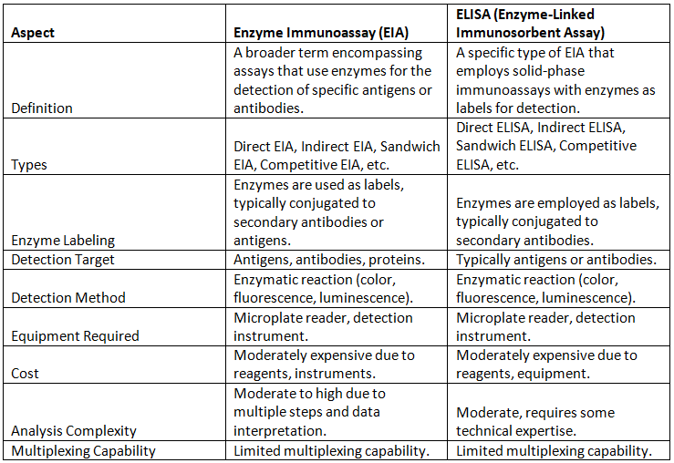
Future Directions and Potential Innovations
As technology advances, the future of EIA holds numerous possibilities:
- Integration with Digital and Wearable Technologies: The amalgamation of EIA with digital sensors or wearable devices could usher in an era of real-time biomarker monitoring for personalized health management.
- Enhanced Automation and Robotics: Advanced robotics and automated systems have the potential to streamline EIA workflows, diminish human errors, and augment assay throughput.
- Enhanced Signal Detection Techniques: Developing more sensitive and efficient detection methods could lead to heightened precision in EIA.
- Enhanced Sample Preparation Techniques: Improved sample preparation methodologies could mitigate interference and matrix effects, enhancing accuracy and reliability.
- Eco-Friendly and Sustainable Approaches: Pioneering developments in assay components and materials might contribute to more environmentally friendly and sustainable EIA protocols.
Spectroscopy
- Spectroscopy encompasses a range of techniques based on the fundamental principle of studying the interaction between electromagnetic radiation and matter. This electromagnetic radiation can take various forms, including light, heat, microwaves, infrared, and X-rays, all of which travel at an average speed of 3 x 10⁸ meters per second.
- These electromagnetic waves consist of two components: an electric field and a magnetic field, both oscillating perpendicular to each other. Light exhibits both wave-like and particle-like properties.
- In the lowest energy state, known as the ground state, electrons within an atom are typically found. When an atom is exposed to light or electromagnetic radiation, its electrons transition from the ground state to an excited state, absorbing energy in the process. Upon returning to the ground state, excited electrons emit radiation of specific wavelengths or energies, a phenomenon harnessed in spectroscopic analysis.
- In certain substances, energy emission can occur spontaneously, without the need for external radiation. Additionally, when molecules are exposed to electromagnetic radiation, some substances' atoms undergo changes in their electronic or nuclear properties.
- The study of absorption and emission properties, along with the examination of changes in nuclear structure, provides valuable insights into various macromolecules.
Nuclear Magnetic Resonance Spectroscopy (NMR)
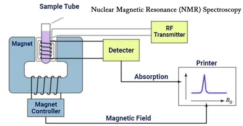
- Nuclear Magnetic Resonance (NMR) spectroscopy is a method used to detect interactions between atomic nuclei and the magnetic field of electromagnetic radiation. Protons and neutrons within an atom possess spin properties. In cases where protons and neutrons exist in pairs within the atomic nucleus, there is no net spin.
- However, if there are unpaired protons, these individual protons will exhibit a magnetic moment and can interact with an applied magnetic field. As a result, the nuclei can absorb energy and exist in either a low-energy state (with nuclear spin parallel to the field) or a higher-energy state (with nuclear spin antiparallel to the field).
- This interaction of unpaired protons with a magnetic field forms the fundamental principle of NMR spectroscopy. When exposed to a magnetic field, these nuclei absorb radio wave radiation, leading to a phenomenon called nuclear magnetic resonance.
- In biological research, NMR studies typically involve unpaired nuclei such as H1, C13, N14, O17, and P31. NMR spectra are constructed by plotting the absorbed energy against the strength of the magnetic field. A radio wave frequency of 40 MHz is commonly used to induce resonance in the H1 nucleus. NMR spectrometer readings are typically in units of tesla (T).
- The basic components of an NMR instrument include:
- A radiation source.
- A receiver to detect energy absorption.
- A magnetic field.
- An oscilloscope or recorder.
- NMR is employed to investigate various aspects, such as the molecular structure of organic molecules, the effects of antibiotics and drugs on living systems, alterations in the structure of molecules within the plasma membrane, and the impact of cholesterol on erythrocyte membranes. Advanced high-field NMR instruments, with frequencies like 750 MHz or even gigahertz (GHz) range, have been developed for exploring the structure and dynamic properties of proteins in solution.
Optical Rotatory Dispersion (ORD) and Circular Dichroism
- Optical Rotatory Dispersion (ORD) and Circular Dichroism (CD) are techniques used to study the three-dimensional structure of macromolecules in solution by observing their interactions with polarized light.
- Plane polarized light consists of light waves oscillating in a single plane and can be obtained by passing light through a Nicol prism or a polarizing screen. When plane polarized light passes through a substance, its polarization plane can rotate at a certain angle, depending on the structure of the substance. This rotation is also found to be dependent on the wavelength of light. Therefore, the rate of rotation change with respect to the wavelength of light is measured, and this is known as Optical Rotatory Dispersion (ORD).
- Certain optically active substances absorb polarized light differently, resulting in the differential absorption of right (R) and left (L) circularly polarized light. Circular Dichroism Spectroscopy (CD) is a spectroscopy technique developed to investigate how polarized light interacts with samples.
- While both CD and ORD serve similar purposes, CD analysis has become superior due to the relative simplicity of CD spectra and the higher resolution of CD bands.
- Circularly polarized light is obtained by superimposing two plane polarized lights of the same wavelength, which are passed through the monochromator and Nicol prism. This superimposed light can be split into Right (R) and Left (L) waves. Certain substances selectively absorb R and L waves differently and exhibit refraction with elliptically polarized light.
- ORD and CD are valuable tools for studying the secondary structure of macromolecules, especially proteins and amino acids in solution. CD spectra are also useful for examining the binding of substrates and inhibitors to enzymes.
- The helical structures of DNA and proteins can be explored using CD spectra. CD spectra are highly sensitive to any structural changes in macromolecules, making them suitable for studying protein-nucleic acid interactions by observing changes in CD spectra. Additionally, CD spectrophotometers can be used to investigate transitions between double-stranded and single-stranded nucleic acids.
Infra-red (IR) Spectrophotometry
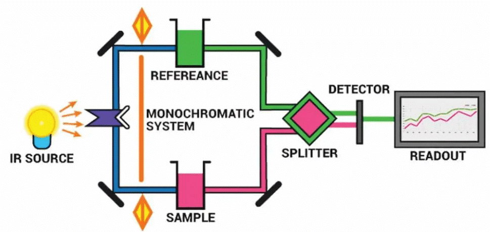
- In the infrared (IR) region of the electromagnetic spectrum, which ranges from 103 to 104 nanometers (nm), molecules exhibit vibrational spectra. This means that molecules exposed to infrared radiation display distinct vibrational energy levels. These vibrational energy levels vary depending on the bonding characteristics of the compound. For example, the vibrational modes of C-H, -CH2, and CH3 groups will exhibit differences.
- Similarly, different functional groups present in molecules, such as methyl, carbonyl, and amide groups, will produce distinct IR spectra. As a result, IR spectroscopy finds significant utility in biochemistry, particularly for the examination of macromolecules and cell membranes. It is also valuable for drug identification and for studying the secondary structure of proteins, including the determination of the number of helical structures present in a protein.
Atomic Absorption/ Flame Spectrometry
- When atoms in a compound are energized, either through exposure to a flame or electrothermal means, they absorb or emit specific wavelengths of light. An emission flame spectrophotometer is employed to measure the emission of specific wavelengths of atoms in a flame. This instrument is used to assay various elements present in biological samples.
- On the other hand, an atomic absorption spectrophotometer detects the absorption of a particular wavelength by atoms in a sample when it is heated, either in a flame or by other means. The flameless method of atomic absorption spectrophotometry is more sensitive compared to flame spectrophotometry. It is particularly useful for quantifying the amount of heavy metals or other toxic metals present in biological samples.
- The primary components of the flame spectrophotometer include:
- Nebulizer or Atomizer, which converts a small volume of the sample into fine droplets and then passes these droplets into the flame with the help of forced air pressure.
- Monochromator, which selects the specific wavelength of light.
- Detector equipped with a photocell to measure the intensity of the selected wavelength.
- In the case of the atomic absorption spectrophotometer, additional components include a source of white light (cathode discharge lamp) and a double monochromator, in addition to a detector and atomizer.
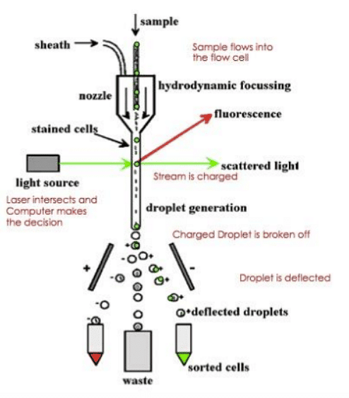
Flow Cytometer
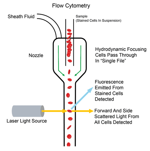
- The Flow Cytometer operates based on the principle that individual cells suspended in a fluid are passed through an illuminated field, and each cell is analyzed quantitatively by either staining them with fluorescent dyes or measuring the scattering of light by the cells.
- Here is a breakdown of the procedure and operation of the Flow Cytometer:
- Sample Preparation: Cell suspensions are initially placed in a flow cell that is equipped with a mechanism to create a liquid jet.
- Flow of Cells: The cells travel through the center of the liquid jet at a rapid rate, typically between 5 to 10 meters per second.
- Illumination: As the cells pass through an area of intense light, any fluorochromes present in the cells emit fluorescence. This fluorescence is detected using various optical components, including lenses, beam splitters, and photomultiplier tubes.
- Operation of the Flow Cytometer: Different fluorochromes used to stain the cells emit different types of fluorescence. These fluorescence signals are then quantified and stored as histograms in the computer's memory.
- Droplet Formation: During the flow process, droplets are formed using a piezoelectric crystal within the flow cell.
- Droplet Deflection: At the end of the instrument, there is a droplet deflector assembly. This assembly directs specific cells, as quantified by the computer, into a collection vessel, such as a microtiter plate or tube. Undesirable cells are directed into a waste container.
- Fluorochromes: Different types of fluorochromes are used for staining different types of cells. This allows for the sorting of cells with various characteristics, such as those containing two types of nuclei (heterokaryons).
Cell Sorter
Flow Cytometers and Cell Sorters have a wide range of applications in various fields of biology and research.
Here are some of the key applications:
- Somatic Hybrid and Cybrid Cell Selection: Flow Cytometers are crucial for selecting somatic hybrid and cybrid cells using fluorescence labeling techniques. This is especially important in hybridoma technology and in creating novel cell lines.
- Characterization of Protoplasts: Flow Cytometers are used to analyze the physical and physiological characteristics of plant protoplasts. This is significant in molecular biology and the manipulation of plant protoplasts for genetic and breeding studies.
- Cell Cycle Studies: These instruments are valuable tools for studying the cell cycle. They can help researchers understand cell cycle progression, identify cell cycle phases, and assess factors that influence cell cycle regulation.
- Chromosome Isolation: Flow Cytometers can aid in the isolation of chromosomes. This is useful for various genetic and cytogenetic studies, including karyotyping and chromosome analysis.
- Cell Sorting: Flow Cytometers can be used for cell sorting. By temporarily charging fluid-containing cells and then deflecting charged cells of interest in an electric field, researchers can selectively collect specific cell types, including mitochondria, nuclei, chromosomes, protoplasts, pollen, and more.
- DNA Content Measurement: Flow Cytometry is employed to measure the DNA content of cells and chromosomes. This is particularly useful for determining the ploidy level of organisms or cell populations in a relatively short amount of time.
- Cell-Fusion Studies: Flow Cytometers enable the rapid sorting of heterokaryons in cell-fusion studies. This is important for understanding cell fusion processes and their applications in various fields.
- Genetic Engineering: In genetic engineering studies, Flow Cytometers are used to sort and isolate transformed or hybrid cells. This is essential for generating genetically modified organisms and conducting genetic engineering experiments.
Overall, Flow Cytometers and Cell Sorters are versatile instruments with applications spanning cell biology, genetics, molecular biology, and genetic engineering. They play a crucial role in understanding cellular processes and manipulating cells for various research purposes.
Non-invasive Scanning of Soft Tissues
Non-invasive scanning of soft tissues has become increasingly important in medical diagnostics. Several methods have been developed for examining soft tissues, particularly the brain, without the need for injecting any contrast substances. These methods include:
- Computer Tomography Scan (CT Scan): CT scans use the differential absorption of X-rays by brain tissue to construct a three-dimensional image (3D image). CT scans require a very low dose of X-rays. They are particularly useful for detecting the location of tumors, sites of hemorrhage, and intracranial bleeding.
- Magnetic Resonance Imaging (MRI): MRI utilizes a magnetic field to detect differences in the vibrations of protons (H+) in water molecules present in body tissues. The vibration of water molecules depends on the chemical environment of the tissue. MRI can also detect the vibrations of other atoms like fluorine, sodium, phosphorus, and nitrogen. MRI is valuable for investigating metabolic processes in the brain and other organs. Unlike CT scans, MRI does not use X-rays.
- Microscopic Magnetic Resonance Imaging (mMRI): mMRI is a technically complex technique that involves placing the specimen inside a strong magnetic field. It is capable of imaging large and opaque specimens, including living ones. mMRI can digitally record anatomical information from intact specimens and apply unique contrast mechanisms to highlight different specimen features.
- Positron Emission Tomography (PET): PET scans rely on the use of positrons, which are positively charged particles similar to electrons. PET scans provide 3D images that reveal the locations of molecules. They are used to measure blood flow in the brain, glucose utilization, oxygen consumption, and diagnose various conditions such as psychiatric disorders, brain tumors, epilepsy, and degenerative changes associated with Alzheimer's disease.
These advanced imaging techniques have revolutionized medical diagnosis by enabling non-invasive visualization of soft tissues and providing valuable insights into the structure and function of organs, including the brain. These methods offer enhanced diagnostic capabilities and contribute to improved patient care.
|
179 videos|143 docs
|


video lectures
,Objective type Questions
,Techniques of Cell Biology | Botany Optional for UPSC
,Extra Questions
,mock tests for examination
,Exam
,Semester Notes
,shortcuts and tricks
,Free
,Previous Year Questions with Solutions
,Sample Paper
,Techniques of Cell Biology | Botany Optional for UPSC
,study material
,MCQs
,Viva Questions
,Techniques of Cell Biology | Botany Optional for UPSC
,past year papers
,ppt
,Important questions
,Summary
,practice quizzes
;


Techniques of Cell Biology Free PDF Download
Importance of Techniques of Cell Biology
Techniques of Cell Biology Notes
Techniques of Cell Biology UPSC Questions
Study Techniques of Cell Biology on the App
|
© EduRev
|
Education Revolution
|


|


