The Female Reproductive System | Biology Class 12 - NEET PDF Download
The female reproductive system is a fascinating and complex network of organs and structures that allows women to bear children. One of the most incredible aspects of this system is the ability of the uterus to expand up to 500 times its normal size during pregnancy.
As we read more about the female reproductive system further in this document, we'll discover even more amazing facts and functions that make this system truly remarkable.
The female reproductive system consists of the pair of ovaries, uterine tubes, uterus, vagina, and external genitalia. The breasts, or mammary glands, are structurally and functionally integrated to support the processes of ovulation, fertilization, pregnancy, birth, & childcare.
 The Female Reproductive System
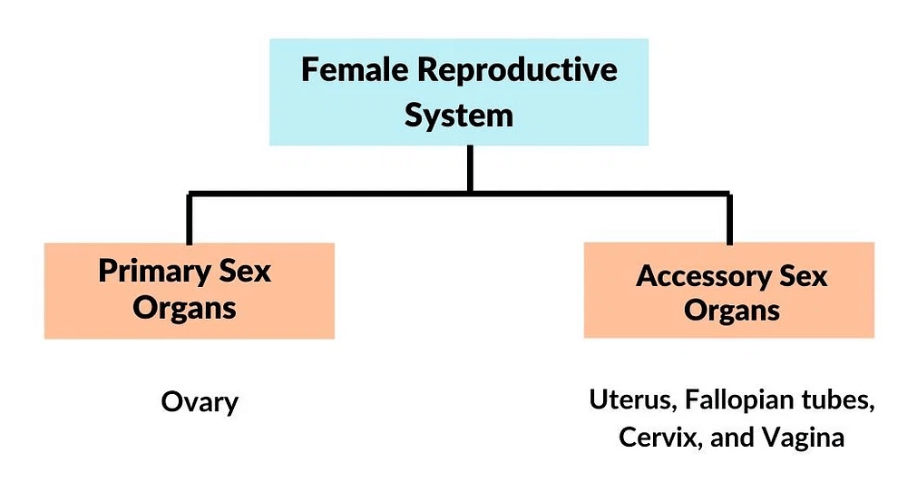
Female Reproductive System is classified into two categories: Primary Sex Organs and Accessory Sex Organs.

1. The Ovaries
The ovaries are a vital part of the female reproductive system, responsible for producing and storing the eggs that are necessary for fertilization and pregnancy. It's a commonly known fact that women are born with all the eggs they will ever have, usually around 1-2 million, and these eggs are stored within the ovaries.
- In females, the ovaries are the primary sex organs.
- Ovaries are located one on each side of the lower abdomen.
- Each ovary is about 2 to 4 cm long and shaped like an unshelled almond.
- The ovarian ligament connects the ovary to the uterus.
- Each ovary has a thin epithelium that encloses the ovarian stroma. The stroma is split into two sections: the peripheral cortex and the inner medulla.
- The ovaries, as we will see later, are in charge of producing female sex hormones and ova.
 Internal Structure of Ovary
The ovary is made up of the following parts:
(i) The ovary is covered by a layer of cubical epithelium called the germinal epithelium. The germinal epithelium is covered by the visceral peritoneum.
(ii) Beneath the epithelium is the tunica albuginea- a layer of connective tissue and underlying it is the ovarian stroma.
(iii) The medulla is a dense, inner layer of the stroma, while the cortex is a less dense, outer layer of the stroma.
Cortex: In cortex region of ovaries, there are little things called follicles that are growing up. First, they start as oocytes and then they grow into primary follicles, which then turn into secondary follicles. After that, they become tertiary follicles and eventually, they become fully mature graafian follicles. And finally, they turn into something called corpus luteum which is a yellow body after which they become corpus albicans, which is nothing but a white body and this process of growth of follicles is called folliculogenesis.
Medulla: Deep inside the ovary, there's a special part called the medulla. It's kind of like the "core" of the ovary. Inside the medulla, you can find a bunch of important stuff like blood vessels, connective tissue, fibrous sheaths, ligaments, and even smooth muscles.
(iv) No more oogonia are formed and added after birth.
- Oogonia (egg mother cells) divide by mitosis forming primary oocytes.
- Each primary oocyte then gets surrounded by a layer of granulosa cells called primary follicles.
- A large number of these follicles degenerate during the phase from birth to puberty.
- Therefore, at puberty, only 60,000-80,000* primary follicles are left in each ovary.
- The primary follicles are surrounded by more layers of granulosa cells and called secondary follicles.
- The secondary follicle soon changes into a tertiary follicle which is characterized by a fluid-filled cavity called follicular antrum.
- The tertiary follicle is further converted into a mature follicle or Graafian follicle.

Cross-section of Ovary
(v) Interspersed throughout the cortex are many ovarian follicles (also called Grafian follicles) in different stages of development. The ovarian follicle comprises the following parts:
- A follicle consists of an oocyte covered by a homogenous membrane the zona pellucida.
- When the surrounding cells form a single layer they are called follicular cells. Later in development when they form several layers, they are referred to as granulosa cells.
- The surrounding cells nourish the developing oocyte and begin to secrete estrogens called membrana granulosa.
- The follicle has an eccentric follicular cavity or follicular antrum filled with a fluid, the follicular fluid or liquor folliculi.
- It projects into the follicular cavity (follicular antrum).
- Later, the granulosa cells lying in close vicinity of the oocyte and zona pellucida, become elongated to form the corona radiata.
- The membrana granulosa is surrounded by the theca interna (theca - cover) and theca externa.
 A mature follicle
 - The total number of follicles in each ovary of a normal young adult woman is about 60,000 to 80,000.
- Many ovarian follicles (during the primary oocyte stage) undergo degeneration.
- This degenerative process of follicles is called follicular atresia and such follicles are known as atretic follicles.
- The release of secondary oocytes from the ovary is called ovulation.
- It occurs due to the rupturing of the ovarian follicle and the wall of the ovary.
 Process of Ovulation
 - Generally, one secondary oocyte is released in each menstrual cycle (average duration 28 days) by alternate ovaries.
- Only about 450 secondary oocytes (ova) are produced by a human female over the entire span of her reproductive life which lasts about 40-50 years of age (in some cases 45-55 years).
- In addition to releasing an oocyte, the follicle also produces hormones.
- While the follicle is maturing, some of the follicular cells produce estrogens, mainly estradiol.
(vi) After ovulation many of the follicular cells remain in the collapsed follicle on the surface of the ovary.
- The antrum (cavity) of the collapsed follicle fills with a partially clotted fluid.
- The follicular cells enlarge and fill with a yellow pigment, lutein.
- Such a follicle is called a corpus luteum- literally, yellow body.
- The lutein cells secrete a small amount of estradiol hormone and a significant amount of progesterone hormone.
- The Corpus luteum also secretes the relaxin hormone.Question for The Female Reproductive SystemTry yourself:After ovulation Graafian follicle regresses intoView Solution

(vii) Degenerated part of the corpus luteum is called corpus albicans, literally meaning white body. In fact, it is a white scar-like area.
 Degeneration of Corpus Luteum into Corpus Albicans
Functions of Ovaries:
i) Ovaries produce female sex hormones that regulate menstrual cycles, breast development, and other changes during puberty.
ii) The ovaries also produce ova, or eggs, which are released during ovulation and can potentially be fertilized by sperm to start a pregnancy.
How many Eggs ovaries release in females in general?
In each menstrual cycle, which is around 28 days long on average, one egg cell is released by one of the ovaries. These eggs are called secondary oocytes, and they are released by each ovary in turn.
You might be wondering, how many eggs a woman produces in her lifetime?
- Well, it turns out that over the course of her reproductive years, which usually lasts from around 40-50 years old (sometimes longer), a woman will only produce about 450 eggs in total. That might sound like a lot, but when you consider that a woman releases one egg per cycle, it's really not that many.
As we know,
Two Fallopian tubes (oviducts), uterus and vagina constitute the female accessory ducts.
2. Fallopian Tubes
They consist of the following parts:
(i) The infundibulum is a dilated trumpet-like portion opening into the peritoneal cavity. The end of the tube has finger-like projections called fimbriae. It extends from the end of the fallopian tubes in the female reproductive system and are responsible for sweeping the released egg from the ovary into the fallopian tube. The fimbriae create a wave-like motion to help sweep the egg into the tube. Without them, the egg might not be able to make it into the fallopian tube and could be lost.
(ii) The ampulla is the widest and longest part of the Fallopian tube.
(iii) The isthmus is the short, narrow thick-walled portion that follows the ampulla.
(iv) The uterine part passes through the uterine wall and communicates with the uterine cavity.
(v) Uterus is single and known as Womb.
 Image showing uterine tube
Functions of Fallopian Tubes: The Fallopian tube conveys the ovum from the ovary to the uterus. It is done by peristalsis. Fertilization of the ovum generally takes place in the upper portion of the Fallopian tube (ampulla).
3. Uterus (Metra or Hystera or Womb)
- The uterus is a hollow muscular and inverted pear-shaped structure. It lies in the pelvic cavity between the urinary bladder and the rectum.
- It comprises three parts:
(i) The fundus is the upper dome-shaped part of the uterus above the openings of the uterine parts of the Fallopian tubes.
(ii) The body (corpus) is the main part that is narrowest inferiorly where it continues with the cervix. It is the usual site of implantation of blastocyst.
(iii) The cervix is the part that joins the anterior wall of the vagina and opens into it. The cavity of the cervix is called the cervical canal. The cervix communicates above with the body of the uterus by an aperture called internal os and with the vagina below by an opening, the external os.

- The uterine walls are made up of three layers of tissue.
(i) The perimetrium is the peritoneum's thin outer covering.
(ii) The myometrium is a thick layer of smooth muscle fibres in the middle of the uterus that contracts strongly during childbirth.
(iii) The endometrium is the uterine cavity's inner glandular layer. During the menstrual cycle, the endometrium undergoes cyclical changes.
 Differences between Endometrium and Myometrium
Functions of Uterus: After puberty, the uterus goes through the menstrual cycle. If the fertilization has taken place, the embryo gets attached to the uterine wall where it is nourished and protected. At the end of the gestation period, labour begins and concludes when the child is born known as Parturition.
During childbirth, the uterus contracts to push the baby out through the cervix and into the vagina.
If fertilization does not occur, the uterus sheds its lining during menstruation, which is a normal part of the menstrual cycle.
4. Vagina
The vagina is an elastic, muscular canal that connects the uterus to the outside of the body. It is an important part of the female reproductive system.
- The vagina is a tube, about 10 cm long, that extends from the cervix to the outside of the body.
- It is easily stretched.
- The opening of the vagina, called the vaginal orifice (vaginal opening), is partially covered by a membrane called the hymen.
Functions of Vagina: It provides a passageway for the menstrual flow, serves as the receptacle for sperm during intercourse, and forms part of the birth canal during labour.
The hymen is often torn during the first coitus (intercourse). However, it can also be broken by a sudden fall or jolt, active participation in some sports like horseback riding, bicycling, etc. In some women, the hymen remains even after coitus. In fact, the presence or absence of a hymen is NOT a reliable indicator of virginity.
5. External Genitalia (Vulva)
Vulva means external genitalia of the female. It includes mons veneris, labia majora, labia minora, clitoris, hymen, vestibule & related perineum.
(i) Mons veneris (mons pubis): It is a cushion of fatty tissue or of subcutaneous connective tissue, lying in front of the pubis & is covered by pubic hairs in the adult female.
(ii) Labia majora: Vulva is bounded on each side by the elevation and fleshy folds of skin & subcutaneous tissue. Its inner surface is hairless. The outer surface is covered by the sebaceous gland, Sweat gland & hair follicles. It is homologous with the scrotum in the male.
(iii) Labia minora: They are two thin folds of skin present just within the labia majora. The lower portion of the minora fuses across the midline & form a fold of skin called a fourchette.
(iv) Clitoris: It is a tiny finger-like structure that lies at the upper junction of the two labia minora above the urethral opening. It is made up of two erectile bodies (corpora cavernosa). The skin which covers the glans of the clitoris is called a prepuce. At the terminal part of the vagina, the urethra opens separately, so they form a common chamber called the vaginal vestibule or urogenital sinus.
The vulva has the following openings:
(a) Urethral opening: Lies on the anterior end
(b) Vaginal orifice: Lies on the posterior end.
It is incompletely closed by a septum of mucous membrane called the hymen, but it may not be a true sign of virginity.
(c) Opening of Bartholin's ducts: These are opening of one pair of bartholin's/greater vestibular glands situated on the lateral side of the vagina. They secrete alkaline fluid during sexual excitement.

(v) Perineum: It is the area that extends from the fourchette to the anus.
 Lateral View of Female Reproductive System
6. Breasts
- The mammary glands are paired structures (breasts) that contain glandular tissue and a variable amount of fat.
- The glandular tissue of each breast is divided into 15-20 mammary lobes containing clusters of cells called alveoli.
- The cells of alveoli secrete milk, which is stored in the cavities (lumens) of alveoli.
- The alveoli open into mammary tubules.
- The tubules of each lobe join to form a mammary duct.
- Several mammary ducts join to form a wider mammary ampulla which is connected to the lactiferous duct through which milk is sucked out.
 Mammary gland

- Mammary glands produce a nutritive fluid, milk for the nourishment of young ones.
- Milk protects the young ones from various infections up to some months after birth.
- The mammary glands of the female undergo differentiation during pregnancy and start producing milk towards the end of pregnancy by the process called lactation.
- This helps the mother in feeding the newborn.
- Breast-feeding during the initial period of infant growth is recommended by doctors for bringing up a healthy baby.
Human milk consists of water and organic and inorganic substances. Its main constituents are fat (fat droplets), casein (milk protein), lactose (milk sugar), mineral salts (sodium, calcium, potassium, phosphorous, etc.) and vitamins. Milk is poor in iron content. Vitamin C is present in very small quantities in milk. The process of milk secretion is regulated by the nervous system. It is also influenced by the psychic state of the mother. The process of milk production is also influenced by hormones of the pituitary gland (already mentioned), the ovaries and other endocrine glands. A nursing woman secretes 1 to 2 litres of milk per day. Milk contains an inhibitory peptide. If the mammary glands are not fully emptied the peptide accumulates and inhibits milk production.
Female Reproductive System Hormonal Control
- The hypothalamus secretes GnRH, which stimulates the anterior lobe of the pituitary gland to secrete luteinizing hormone (LH) and FSH.
- FSH promotes the growth of ovarian follicles as well as the development of egg/oocytes within the follicle to complete meiosis I and form secondary oocytes.
- FSH also promotes the production of estrogens.
- The corpus luteum is stimulated by LH to secrete progesterone.
- Rising progesterone levels inhibit GnRH release, which in turn inhibits the production of FSH, LH, and progesterone.

The Onset of Puberty in the Human Females
- Females reach puberty around the age of thirteen.
- The pituitary gland starts producing follicle-stimulating hormones around this time (FSH).
- FSH stimulates the development of the ovaries, which results in the production of the hormone estrogen.
- This hormone is in charge of the development of female secondary sex characteristics such as voice change and the development of external genitalia, breasts, body hair, pubic hair, and feminine shape.
- This shape is characterized by a widening of the pelvis and fat deposits in the thighs, buttocks, and face.
Functions of Female Reproductive System
1. Germinal epithelial cells of the ovary produce ova (oogenesis).
2. Fertilization takes place in the Fallopian tube (ovíduct).
3. After puberty the uterus goes through the menstrual cycle.
4. Implantation and prenatal growth take place in the uterus.
5. The vagina receives the seminal fluid during copulation.
6. Parturition (the process of the birth of a child) is also an important function of the female reproductive system.
7. Mammary glands of the female secrete milk after parturition.
Which Reproductive System is more complex?
Now that you know both the reproductive systems i.e. female & male both.
And, if you think it is the male reproductive system, then you may be wrong. Because it is the Female Reproductive System which is more complex than the male reproductive system because it not only produces eggs but also supports the growth and development of a baby.
|
87 videos|294 docs|185 tests
|
FAQs on The Female Reproductive System - Biology Class 12 - NEET
| 1. What are the main organs of the female reproductive system? |  |
| 2. What is the hormonal control of the female reproductive system? |  |
| 3. How does the onset of puberty occur in human females? |  |
| 4. What are the functions of the female reproductive system? |  |
| 5. Is the female reproductive system more complex than the male reproductive system? |  |

|
Explore Courses for NEET exam
|

|


