Superficial Lymph Nodes | Animal Husbandry & Veterinary Science Optional for UPSC PDF Download
Importance of Superficial Lymph Nodes
- Knowing about the lymph nodes in oxen is crucial for veterinarians.
- It helps diagnose various diseases in animals.
Normal Characteristics:
- Understanding the normal size, location, and arrangement of lymph glands is vital.
- This knowledge aids in identifying potential health issues.
Easily Detectable Lymph Nodes:
- Some lymph nodes are easily felt on the surface of the ox's body.
- These are important for veterinarians to check during examinations.
Clinical Significance:
- Even deeply located lymph nodes are discussed due to their importance in diagnosing diseases.
- This comprehensive understanding helps veterinarians provide effective care.
Highlighted Nodes:
- Emphasis on a few specific lymph nodes that are both on the surface and deep, making them clinically relevant.
- This information assists in practical veterinary work and contributes to better animal healthcare.
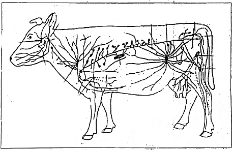
 Fig: Diagram showing the lymph nodes of the ox
Genito-Urinary Organs, Mammary Glands and Bovine Superficial Lymph Glands
Mandibular Lymph Node:
- Located between the sternocephalic muscles and the ventral part of the mandibular salivary gland.
- Dimensions: Approximately 3-4.5 cm in length and 2-3 cm in width.
- Positioned dorsally to the linguofacial vein (external maxillary vein).
Parotid Lymph Node:
- Situated ventral to the temporomandibular joint, on the caudal part of the masseter muscle.
- Dimensions: 6-9 cm in length and 2-3 cm in width.
- Has a flat, oval shape and can be palpated on the edge of the jaw and the surface of the masseter muscle.
- Remains connected to the tissues of the parotid gland at slaughter.
Lateral Retropharyngeal Lymph Node (Atlantal Lymph Node):
- Located under the free border of the wing of the atlas, partly covered by the upper end of the mandibular gland.
- Flat, oval node with a length of 4-5 cm.
Medial Retropharyngeal Lymph Node (Parapharyngeal Lymph Node):
- Lies medial to the stylohyoid bone on the pharyngeal muscles.
- Dimensions: 3-6 cm in length, oval-shaped.
Superficial Cervical Lymph Node (Prescapular):
- Positioned at the cranial border of the supraspinatus muscle.
- Covered by the brachiocephalic and omotransversarius muscles.
- Dimensions: 7-9 cm in length and 1-2 cm in width, elongated.
Deep Cervical Lymph Node:
- a) Cranial Deep Cervical Lymph Node:
- 4-6 nodes with a size ranging from 1-1 cm.
- Found near the thyroid gland along the course of the carotid artery.
- b) Middle Deep Cervical Lymph Node:
- Variable number (1-7 nodes) with a size of 0.5-3.0 mm.
- Located on the middle-third of the cervical part of the trachea.
- c) Caudal Deep Cervical Lymph Nodes:
- 2-4 nodes with a size of 1-6 cm.
- Immediately in front of the first rib, near the thoracic inlet, dorsal and ventral to the common jugular vein.
- a) Cranial Deep Cervical Lymph Node:
Caudal Mediastinal Lymph Node:
- Comprises several nodes situated in the caudal mediastinum.
- Usually, one node is very long (15-25 cm), lying dorsally against the esophagus.
- Enlarged nodes can cause partial closure of the thin-walled esophagus.
Subiliac Lymph Node (Prefemoral):
- Dimensions: 6-11 cm with a size of 1.5-2.5 cm.
- Located in front of the cranial border of the tensor fasciae latae muscle, approximately 12 to 15 cm dorsal to the patella.
Superficial Inguinal Lymph Node:
- a) Mammary Lymph Node in Females:
- 1-3 mammary lymph nodes with a size of 6 to 10 cm in length.
- Located above the caudal border of the base of the udder.
- b) Scrotal Lymph Node in Males:
- Superficial inguinal nodes in males are referred to as scrotal lymph nodes.
- The number varies from one to four, with dimensions of 3-6 cm in length and 2-4 cm if only one node is present.
- Situated below the prepubic tendon and covered, in part, by the retractor muscle of the prepuce.
- a) Mammary Lymph Node in Females:
Paralumbar Lymph Node:
- One or two small subcutaneous nodes occurring inconsistently in the middle of the flank, behind the last rib, near the ends of the transverse processes of the lumbar vertebrae.
Coxal Lymph Node:
- Situated cranial to the proximal part of the quadriceps femoris muscle.
- Present in the majority of individuals with a size of 3 cm.
Tuberal Lymph Node:
- Located subcutaneously immediately medial to the tuber ischii and medial to the insertion of the broad sacrotuberal ligament.
- Frequently removed with subcutaneous fat while skinning the carcass.
- Constant size of 2-3 cm in length.
Popliteal Lymph Node:
- In bovines, superficial popliteal lymph nodes are absent.
- The deep popliteal lymph node is situated deeply in a mass of fat on the gastrocnemius muscle between the gluteobiceps and semitendinosus muscles.
- Positioned 7-9 cm from the posterior edge of the semitendinosus and gluteobiceps muscles, with dimensions of 3-4 cm by 2-3 cm.
FAQs on Superficial Lymph Nodes - Animal Husbandry & Veterinary Science Optional for UPSC
| 1. What are superficial lymph nodes and why are they important for the genito-urinary organs, mammary glands, and bovine superficial lymph glands? |  |
| 2. How do superficial lymph nodes contribute to the health of genito-urinary organs? |  |
| 3. What role do superficial lymph nodes play in the health of mammary glands? |  |
| 4. How are bovine superficial lymph glands connected to the overall health of cattle? |  |
| 5. Can abnormalities in superficial lymph nodes indicate underlying health issues in genito-urinary organs, mammary glands, and bovine superficial lymph glands? |  |


