


All Exams >
MCAT >
MCAT Biological and Biochemical Foundations >
All Questions
All questions of Tissues (BIO) for MCAT Exam
Mucus cells (Goblet cells) :-- a)Unicellular gland
- b)Multicellular glands
- c)Endocrine glands
- d)Parietal cells of gastric glands
Correct answer is option 'A'. Can you explain this answer?
Mucus cells (Goblet cells) :-
a)
Unicellular gland
b)
Multicellular glands
c)
Endocrine glands
d)
Parietal cells of gastric glands
|
|
Rajeev Saxena answered |
The main role of goblet cells is to secrete mucus in order to protect the mucous membranes where they are found. Goblet cells accomplish this by secreting mucins, large glycoproteins formed mostly by carbohydrates. ... Secretion may be stimulated by irritants such as dust and smoke, especially in the airway.

Stratified squamous epithelium found in :-- a)Tonsil
- b)Payer's patch
- c)Appendix
- d)Spleen
Correct answer is option 'A'. Can you explain this answer?
Stratified squamous epithelium found in :-
a)
Tonsil
b)
Payer's patch
c)
Appendix
d)
Spleen
|
|
Vijay Bansal answered |
The luminal surface of the tonsils are covered with a stratified squamous epithelium (in common with the oral epithelia). ... The epithelial cells are able to phagocytose bacteria, and transfer them to macrophages, which then present the foreign antigens to B-cells, which are activated (with the help of T cells).
Lining of brain ventricle & central canal of spinal cord is called as:-- a)Ependyma
- b)Endothelium
- c)Mesothelium
- d)Neurosensory
Correct answer is option 'A'. Can you explain this answer?
Lining of brain ventricle & central canal of spinal cord is called as:-
a)
Ependyma
b)
Endothelium
c)
Mesothelium
d)
Neurosensory
|
|
Rohan Singh answered |
The ependyma is made up of ependymal cells called ependymocytes, a type of glial cell. These cells line the CSF-filled ventricles in the brain and the central canal of the spinal cord. These are nervous tissue cells with a ciliated simple columnar shape much like that of some mucosal epithelial cells.
Inner lining of gut, stomach & liver is made up of :-- a)Simple squamous
- b)Simple cuboidal
- c)Simple columnar
- d)Pseudo stratified epithelium.
Correct answer is option 'C'. Can you explain this answer?
Inner lining of gut, stomach & liver is made up of :-
a)
Simple squamous
b)
Simple cuboidal
c)
Simple columnar
d)
Pseudo stratified epithelium.
|
|
Naresh Kumar answered |
Simple columnar cells are found In major organs like stomach as it plays a very important role in secretion and absorption.
Lining of larynx is :-- a)Stratified ciliated columnar Epithelium
- b)Stratified squamous Epithelium
- c)Stratified cuboidal Epithelium
- d)Stratified columnar Epithelium
Correct answer is option 'A'. Can you explain this answer?
Lining of larynx is :-
a)
Stratified ciliated columnar Epithelium
b)
Stratified squamous Epithelium
c)
Stratified cuboidal Epithelium
d)
Stratified columnar Epithelium
|
|
Jyoti Kapoor answered |
Larynx is lined by ciliated pseudostratified columnar epithelium except vocal cords (lined by stratified squamous epithelium). The wall of larynx is reinforced by cartilage tissue. Large cartilages (cartilago thyroidea, cartilago cricoidea) and part of cartilago arytenoidea are composed of hyaline cartilage.
Cells of Peritoneum comprise :-- a)Ciliated Epithelium
- b)Glandular Epthelium
- c)Columnar Epithelium
- d)Squamous Epithelium
Correct answer is option 'D'. Can you explain this answer?
Cells of Peritoneum comprise :-
a)
Ciliated Epithelium
b)
Glandular Epthelium
c)
Columnar Epithelium
d)
Squamous Epithelium
|
|
Ananya Das answered |
- If the epithelium is composed of one layer of cells, it is referred to as simple squamous epithelium and if it possesses multiple layers, it is referred to as stratified squamous epithelium.
- The peritoneum is the serous membrane that forms the lining of the abdominal cavity or coelom. It covers most of the intra-abdominal (or coelomic) organs and is composed of a layer of mesothelium supported by a thin layer of connective tissue. The mesothelium is a membrane composed of simple squamous cells that form the lining of several body cavities like peritoneum.
Hence the correct option is D.
Term tissue is coined by (for animal anatomy) :-- a)Bichat
- b)Mayer
- c)Malpighi
- d)Hertwig
Correct answer is option 'A'. Can you explain this answer?
Term tissue is coined by (for animal anatomy) :-
a)
Bichat
b)
Mayer
c)
Malpighi
d)
Hertwig
|
|
Meera Singh answered |
The tissue is an ensemble of similar cells from the same origin that together carry out a specific function. Organs are then formed by the functional grouping together of multiple tissues.
Marie François Xavier Bichat was a French anatomist and physiologist, who is best remembered as the father of modern histology and descriptive anatomy. Despite working without a microscope, he was the first to introduce the notion of tissues as distinct entities, and maintained that diseases attacked tissues rather than whole organs or the entire body, causing a revolution in anatomical pathology.
Pseudostratified epithelium is present in :-- a)Nephron & Neuron
- b)Larynx & Pharynx
- c)Trachea & Bronchi
- d)Urinary Bladder & Intestine
Correct answer is option 'C'. Can you explain this answer?
Pseudostratified epithelium is present in :-
a)
Nephron & Neuron
b)
Larynx & Pharynx
c)
Trachea & Bronchi
d)
Urinary Bladder & Intestine
|
|
Gaurav Kumar answered |
Ciliated pseudostratified columnar epithelia are found in the linings of the trachea as well as the upper respiratory tract. Non-ciliated pseudostratified columnar epithelia are located in the membranous part of male vas deferens. Most of the respiratory passageways, from the nasal cavity through the bronchi, are lined by ciliated, pseudostratified columnar epithelium with goblet cells.
Stretchable & Water proof Epithelium :-- a)Simple cuboidal
- b)Simple squamous
- c)Simple Columnar
- d)Transitional
Correct answer is option 'D'. Can you explain this answer?
Stretchable & Water proof Epithelium :-
a)
Simple cuboidal
b)
Simple squamous
c)
Simple Columnar
d)
Transitional
|
|
Vijay Bansal answered |
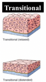
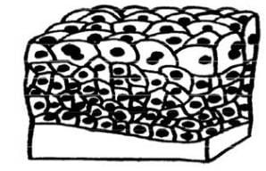
Transitional epithelium is a type of tissue consisting of multiple layers of epithelial cells that contract and expand. This tissue structure type is found in urothelium, including that of the urinary bladder, the ureters and the superior urethra and gland ducts of the prostate. Transitional epithelium is stretchable and waterproof. The cells of transitional epithelium are highly keratinized and contain tight junctions or virtually impenetrable junctions, that seal together the cellular membranes of neighboring cells. This barrier prevents reabsorption of toxic wastes and pathogens by the bloodstream.
Inner lining of Blood vessels and heart is tesselleted Epithelium. Which is :-- a)Simple squamous due to wavy appearance
- b)Simple squamous due to tile like appearance
- c)Simple cuboidal due to wavy appearance
- d)Simple columnar Epithelium
Correct answer is option 'A'. Can you explain this answer?
Inner lining of Blood vessels and heart is tesselleted Epithelium. Which is :-
a)
Simple squamous due to wavy appearance
b)
Simple squamous due to tile like appearance
c)
Simple cuboidal due to wavy appearance
d)
Simple columnar Epithelium
|
|
Vijay Bansal answered |
Tessellated epithelium is another term used for simple squamous epithelium also called as pavement epithelium due to it's flat appearance. It is mainly present in the alveoli and blood capillaries and performs the function of diffusion.
Compound squamous epithelium occurs in :-a)Stomachb)Pharynxc)Intestined)TracheaCorrect answer is option 'B'. Can you explain this answer?
|
|
Vijay Bansal answered |
The compound epithelial tissue consists of two or more layers of cells. They form stratified layers. In the skin, they have a protective function. They do not have much of a role in absorption and secretion. Their function is to provide protection against any form of mechanical or chemical stress. The compound epithelium can be found on the dry surface of the skin, buccal cavity, pharynx, the lining of the salivary glands ducts and pancreatic ducts.
What are cuboidal or columnar cells called when they bear cilia?- a)Ciliated epithelium
- b)Flagellated epithelium
- c)Convoluted epithelium
- d)Brush border epithelium
Correct answer is option 'A'. Can you explain this answer?
What are cuboidal or columnar cells called when they bear cilia?
a)
Ciliated epithelium
b)
Flagellated epithelium
c)
Convoluted epithelium
d)
Brush border epithelium
|
|
Advait Das answered |
Ciliated Epithelium
Ciliated epithelium refers to a type of epithelial tissue that consists of cuboidal or columnar cells with cilia. Cilia are hair-like structures present on the surface of these cells that play a crucial role in various physiological processes.
Structure of Ciliated Epithelium
- Cell Shape: The cells of ciliated epithelium are typically cuboidal or columnar in shape.
- Cilia: These cells bear numerous cilia, which are microtubule-based structures extending from the cell surface. Cilia are composed of a central pair of microtubules surrounded by nine pairs of microtubules in a ring-shaped structure called the axoneme.
- Basal Body: Each cilium arises from a basal body, which is a modified centriole located at the base of the cilium.
- Function: The cilia in ciliated epithelium have rhythmic beating movements that facilitate the movement of various substances across the epithelial surface.
Functions of Ciliated Epithelium
Ciliated epithelium serves several important functions in different parts of the body:
- Mucociliary Escalator: In the respiratory tract, ciliated epithelium lines the airways and helps in the clearance of mucus and foreign particles. The coordinated beating of cilia propels the mucus upward, away from the lungs, preventing the accumulation of debris and pathogens.
- Oocyte Transport: In the female reproductive system, ciliated epithelium in the fallopian tubes helps in the transport of oocytes from the ovaries to the uterus. The beating of cilia creates fluid currents that aid in moving the oocytes towards their destination.
- Cerebrospinal Fluid Circulation: Ciliated epithelium lines the ventricles of the brain and the central canal of the spinal cord. The rhythmic beating of cilia in these regions facilitates the circulation of cerebrospinal fluid, which helps in maintaining the brain and spinal cord environment.
- Smell and Taste Sensation: Cilia on the olfactory receptor cells in the nasal cavity and taste receptor cells in the taste buds help in detecting and transmitting sensory signals related to smell and taste.
In conclusion, cuboidal or columnar cells with cilia are called ciliated epithelium. These cells play vital roles in various physiological processes such as mucociliary clearance, oocyte transport, cerebrospinal fluid circulation, and sensory perception.
Stereocilia present in :-- a)Epididymis
- b)Seminalvesicle
- c)Ureter
- d)Kidney
Correct answer is option 'A'. Can you explain this answer?
Stereocilia present in :-
a)
Epididymis
b)
Seminalvesicle
c)
Ureter
d)
Kidney
|
|
Rohit Shah answered |
Epididymitis care involves rest for 1 – 2 days with the scrotum raised if possible. The aim is to get the inflamed area above the level of the heart. This helps blood flow, which lowers swelling and pain, and helps with healing. Putting ice on the scrotum now and then can also help.
Epidermis of skin of vertebrates comprises :-- a)Simple Epithelium
- b)Stratified Epithelium
- c)Transitional Epithelium
- d)Columnar Epithelium
Correct answer is option 'B'. Can you explain this answer?
Epidermis of skin of vertebrates comprises :-
a)
Simple Epithelium
b)
Stratified Epithelium
c)
Transitional Epithelium
d)
Columnar Epithelium
|
|
Rohan Singh answered |
A stratified squamous epithelium consists of squamous (flattened) epithelial cells arranged in layers upon a basal membrane. Only one layer is in contact with the basement membrane; the other layers adhere to one another to maintain structural integrity.
Germinative layer of Keratinized st. sq. Epithelium :-- a)Cuboidal
- b)Squamous
- c)Pseudo stratified
- d)Transitional
Correct answer is option 'A'. Can you explain this answer?
Germinative layer of Keratinized st. sq. Epithelium :-
a)
Cuboidal
b)
Squamous
c)
Pseudo stratified
d)
Transitional
|
|
Meera Singh answered |
The basal layer of the stratified squamous epithelium is the cuboid layer which forms the other layer known as germinativum layer.
Transitional Epithelium is found in :-- a)Renal pelvis & Ureter
- b)Urinary bladder
- c)Upper part of male urethra
- d)All of above
Correct answer is option 'D'. Can you explain this answer?
Transitional Epithelium is found in :-
a)
Renal pelvis & Ureter
b)
Urinary bladder
c)
Upper part of male urethra
d)
All of above
|
|
Lavanya Menon answered |
Transitional epithelium is a special type of epithelium which has the property of stretchability, it is found in places which requires strechibility as a function such as urinary bladder,ureter etc.
Columnar Epithelium with microvilli or Brush Border is present in :-- a)Intestine
- b)Stomach
- c)Appendix
- d)Pharynx
Correct answer is option 'A'. Can you explain this answer?
Columnar Epithelium with microvilli or Brush Border is present in :-
a)
Intestine
b)
Stomach
c)
Appendix
d)
Pharynx

|
Maheshwar Saini answered |
A brush border (striated border or brush border membrane) is the microvilli-covered surface of simple cuboidal epithelium and simple columnar epithelium cells found in certain locations of the body.
The most important brush border enzymes are dextrinase and glucoamylase, which further break down oligosaccharides. Other brush border enzymes are maltase, sucrase, and lactase. Lactase is absent in most adult humans and for them lactose, like most poly-saccharides, is not digested in the small intestine.
The most important brush border enzymes are dextrinase and glucoamylase, which further break down oligosaccharides. Other brush border enzymes are maltase, sucrase, and lactase. Lactase is absent in most adult humans and for them lactose, like most poly-saccharides, is not digested in the small intestine.
Which of the following is not a type of simple epithelial tissue?- a)Squamous epithelium
- b)Cuboidal epithelium
- c)Columnar epithelium
- d)Compound epithelium
Correct answer is option 'D'. Can you explain this answer?
Which of the following is not a type of simple epithelial tissue?
a)
Squamous epithelium
b)
Cuboidal epithelium
c)
Columnar epithelium
d)
Compound epithelium
|
|
Mayank Gupta answered |
Simple Epithelial Tissue
Simple epithelial tissue is a type of tissue that is composed of a single layer of cells. These cells are tightly packed together and cover a surface or line a cavity. Simple epithelial tissue is classified based on the shape of the cells.
Types of Simple Epithelial Tissue
a) Squamous epithelium - This type of simple epithelial tissue is composed of flat, thin cells that allow for the rapid diffusion of substances. Squamous epithelium is found in areas where rapid diffusion is necessary, such as the alveoli of the lungs.
b) Cuboidal epithelium - This type of simple epithelial tissue is composed of cube-shaped cells that are involved in secretion and absorption. Cuboidal epithelium is found in the glands and tubules of the kidney.
c) Columnar epithelium - This type of simple epithelial tissue is composed of tall, elongated cells that are involved in absorption and secretion. Columnar epithelium is found in the lining of the stomach and intestines.
d) Compound epithelium - This is not a type of simple epithelial tissue. Compound epithelium is composed of multiple layers of cells and is found in areas that require protection, such as the skin.
Conclusion
Simple epithelial tissue is an important type of tissue that plays a crucial role in the functioning of many organs and tissues in the body. It is classified based on the shape of the cells and includes squamous, cuboidal, and columnar epithelium. Compound epithelium is not a type of simple epithelial tissue.
Ends of two long bones are 'connected' by[MP PMT. 1997, CPMT 2000]- a)Cartilage
- b)Muscles
- c)Ligaments
- d)Tendons.
Correct answer is option 'C'. Can you explain this answer?
Ends of two long bones are 'connected' by
[MP PMT. 1997, CPMT 2000]
a)
Cartilage
b)
Muscles
c)
Ligaments
d)
Tendons.

|
Addala Srilakshmi answered |
Ends of two long bones connected by ligaments.
But muscle and bone are connected by tendons.so option C.
But muscle and bone are connected by tendons.so option C.
Ciliated Epithelium occurs in frog :-- a)Oviduct & Buccal cavity
- b)Stomach & urinaryBladder
- c)Blood vessels & Lymph vessels
- d)Kidney & stomach
Correct answer is option 'A'. Can you explain this answer?
Ciliated Epithelium occurs in frog :-
a)
Oviduct & Buccal cavity
b)
Stomach & urinaryBladder
c)
Blood vessels & Lymph vessels
d)
Kidney & stomach

|
Abhishek Desai answered |
The free surface may have microvilli. They are present in the lining of the stomach and intestine. Its functions include absorption and secretion. Ciliated Epithelium – When the columnar epithelial tissues have cilia, then they are called the ciliated epithelium.
Epithelial lining of cornea is composed of :-- a)Startified squamous nonkeratinised
- b)Transitional
- c)Simple cuboidal
- d)Simple squamous
Correct answer is option 'A'. Can you explain this answer?
Epithelial lining of cornea is composed of :-
a)
Startified squamous nonkeratinised
b)
Transitional
c)
Simple cuboidal
d)
Simple squamous
|
|
Pooja Mehta answered |
The corneal epithelium is the outermost layer of the cornea. It is composed of a single layer of basal cells and 4-5 cell layers of nonkeratinized, stratified squamous epithelial cells, which are held together by tight junctions, to form an effective barrier against fluid loss and pathogen penetration.
Term Epithelium coined by :-- a)Ruysch
- b)Mayer
- c)Bichat
- d)Marcellomalpighi
Correct answer is option 'A'. Can you explain this answer?
Term Epithelium coined by :-
a)
Ruysch
b)
Mayer
c)
Bichat
d)
Marcellomalpighi
|
|
Anjali Iyer answered |
The term epithelium was introduced in 18th century by Duth Anatomist Ruysch to refer to the fact that these tissue grow upon other tissues.
Basement membrane is absent in :-- a)Transitional Epithelium
- b)Sq. Epithelium
- c)Columnar Epithelium
- d)Simple cuboidal Epithelium
Correct answer is option 'A'. Can you explain this answer?
Basement membrane is absent in :-
a)
Transitional Epithelium
b)
Sq. Epithelium
c)
Columnar Epithelium
d)
Simple cuboidal Epithelium
|
|
Vijay Bansal answered |
Transitional epithelium is a stratified tissue made of multiple cell layers, where the cells constituting the tissue can change shape depending on the distention in the organ. When the organ is filled with fluid, cells on the topmost layer of this epithelium can stretch and appear flattened. Alternately, they can also appear cuboidal with a rounded shape when the fluid pressure is low.
This epithelium is found lining the urinary bladder, ureters and urethra, as well as in the ducts of the prostrate gland.
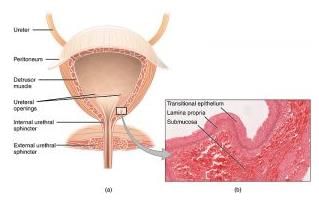
The image shows a cross section of the bladder with an inset displaying the histology of the epithelium, the underlying connective tissue (lamina propria) and submucosa.
This epithelium is found lining the urinary bladder, ureters and urethra, as well as in the ducts of the prostrate gland.
The image shows a cross section of the bladder with an inset displaying the histology of the epithelium, the underlying connective tissue (lamina propria) and submucosa.
Prothrombin, albumin and fibrinogen are synthesised by[AFMC 1997]- a)Pancreas
- b)Bone marrow
- c)Spleen
- d)Liver
Correct answer is option 'D'. Can you explain this answer?
Prothrombin, albumin and fibrinogen are synthesised by
[AFMC 1997]
a)
Pancreas
b)
Bone marrow
c)
Spleen
d)
Liver
|
|
Sanjana Singh answered |
Prothrombin is a component of blood which helps in blood coagulation .It is mainly synthesised by the liver .
Prothrombin of blood plasma is converted to thrombin in the presence of thromboplastin and calcium ion during blood clotting .
Albumin is a globular protein , produced by the liver .
Fibrinogen is a glycoprotein complex that circulate in the blood of vertebrate
Basement membrane is composed of :-- a)Hyaluronic Acid + glycoproteins
- b)Only mucopoly sacharides
- c)Endodermal cells
- d)Epidermal cells
Correct answer is option 'A'. Can you explain this answer?
Basement membrane is composed of :-
a)
Hyaluronic Acid + glycoproteins
b)
Only mucopoly sacharides
c)
Endodermal cells
d)
Epidermal cells
|
|
Pooja Mehta answered |
The basement membrane is composed of two layers, the basal lamina and the underlying layer of reticular connective tissue. The clear layer closer to the epithelium is called the lamina lucida, while the dense layer closer to the connective tissue is called the lamina densa.
The internal lining of blood vessels is called as :-- a)Mesothelium
- b)Endothelium
- c)Pavement Epithelium
- d)Stratified Epithelium
Correct answer is option 'B'. Can you explain this answer?
The internal lining of blood vessels is called as :-
a)
Mesothelium
b)
Endothelium
c)
Pavement Epithelium
d)
Stratified Epithelium
|
|
Rohan Singh answered |
Endothelium refers to cells that line the interior surface of blood vessels and lymphatic vessels, forming an interface between circulating blood or lymph in the lumen and the rest of the vessel wall. It is a thin layer of simple, or single-layered, squamous cells called endothelial cells.
Germinal Epithelium of ovary is formed of :-- a)Columnar Epithelium
- b)Squamous Epithelium
- c)Cuboidal Epithelium
- d)Stratified Epithelium
Correct answer is option 'C'. Can you explain this answer?
Germinal Epithelium of ovary is formed of :-
a)
Columnar Epithelium
b)
Squamous Epithelium
c)
Cuboidal Epithelium
d)
Stratified Epithelium
|
|
Vijay Bansal answered |
The cellular covering of internal and external surfaces of the body, including the lining of vessels and other small cavities. It consists of cells joined by small amounts of cementing substances. Epithelium is classified into types on the basis of the number of layers deep and the shape of the superficial cells. Cuboidal epithelium epithelium whose cells are of approximately the same height and width, and appear square in transverse section.
Epithelial lining of bartholins duct is composed of which type of cells :-- a)Transitional
- b)Cuboidal
- c)Columnar
- d)squamous
Correct answer is option 'B'. Can you explain this answer?
Epithelial lining of bartholins duct is composed of which type of cells :-
a)
Transitional
b)
Cuboidal
c)
Columnar
d)
squamous
|
|
Rohit Shah answered |
The gland is composed of columnar epithelium, and ducts are lined by stratified squamous epithelium and transitional cell epithelium. ... The Bartholin complex consists of a duct that is lined by squamous epithelium as it enters the distal vagina.
Epithelial tissue origined from :-- a)Ectoderm
- b)Endoderm
- c)Mesoderm
- d)All of above
Correct answer is option 'D'. Can you explain this answer?
Epithelial tissue origined from :-
a)
Ectoderm
b)
Endoderm
c)
Mesoderm
d)
All of above
|
|
Rohan Singh answered |
An epithelium is a tissue composed of one or more layers of cells covering the external and internal surfaces of various body parts. Epithelial tissue also forms glands. The term “epithelium” (sing, of epithelia) was given by a Dutch anatomist Ruysch (1638-1731) to refer to the fact that epithelial (Gr. epi- upon, thelio- grows) tissues grow upon other tissues.
The epithelial tissues occur on external and internal exposed surfaces of the body parts where they form protective covering.
Origin of Epithelial Tissue:
Epithelial tissues evolved first and are also formed first in the embryo. The epithelial tissues arise from all the three primary germ layers: ectoderm, mesoderm and endoderm, of the embryo. For example the epidermis of the skin from the ectoderm, coelomic epithelium from the mesoderm and epithelial lining of alimentary canal (= gut) from the endoderm.
Features of Epithelial Tissues:
Epithelial tissues consist of variously shaped cells closely arranged in one or more layers. There is little intercellular material between the cells. The cells are held together by intercellular junctions. The epithelial tissues usually rest on a thin non-cellular basement membrane.
Usually blood vessels are absent in epithelial tissues. However the underlying connective tissues are generally well supplied with blood vessels. Nutrients enter epithelial tissues from the underlying connective tissues by diffusing through the basement membrane.
Exoskeleton originated form (Eg feathers, nail, horn, hoofs) :-- a)Connective tissue proper
- b)Epithelium tissue
- c)Skeletal tissue
- d)Vascular tissue
Correct answer is option 'B'. Can you explain this answer?
Exoskeleton originated form (Eg feathers, nail, horn, hoofs) :-
a)
Connective tissue proper
b)
Epithelium tissue
c)
Skeletal tissue
d)
Vascular tissue
|
|
Rajat Kapoor answered |
Epithelial Tissue
Epithelial tissues are thin tissues that cover all the exposed surfaces of the body. They form the external skin, the inner lining of the mouth, digestive tract, secretory glands, the lining of hollow parts of every organ such as the heart, lungs, eyes, ears, the urogenital tract, as well as the ventricular system of the brain and central canals of the spinal cord.
The cells making up epithelia are often closely bound to one another through specialized structures called tight junctions. They are also free from blood vessels and nerves and are supported by a connective tissue called the basement membrane. They have polarity with a distinct basal domain facing the basement membrane and the other apical surface facing the lumen of an organ or the external environment.
During blood clotting, fibrin is produced by[MP PMT 2000]- a)Thrombokinase
- b)Prothrombin
- c)Liver
- d)Proteolysis
Correct answer is option 'B'. Can you explain this answer?
During blood clotting, fibrin is produced by
[MP PMT 2000]
a)
Thrombokinase
b)
Prothrombin
c)
Liver
d)
Proteolysis
|
|
C Tamil Arasan69 answered |
Thrombin converts fibrinogen into fibrin
the inactive form of thrombin is prothrombin
the inactive form of thrombin is prothrombin
Which of the following functions is not performed by epithelial tissue?- a)Protection
- b)Secretion
- c)Absorption
- d)Conduction
Correct answer is option 'D'. Can you explain this answer?
Which of the following functions is not performed by epithelial tissue?
a)
Protection
b)
Secretion
c)
Absorption
d)
Conduction
|
|
Advait Das answered |
Epithelial tissue is a type of tissue that covers the surface of the body and lines various organs and cavities. It has several functions that include protection, secretion, and absorption. However, it does not perform the function of conduction.
Protection
Epithelial tissue serves as a protective barrier against physical, chemical, and biological damage. It protects the underlying tissues and organs from injury, abrasion, and invasion by microorganisms.
Secretion
Epithelial tissue also secretes various substances such as mucus, enzymes, and hormones. For example, the epithelial cells in the stomach secrete hydrochloric acid and digestive enzymes that aid in the digestion of food.
Absorption
Epithelial tissue is involved in the absorption of nutrients, water, and other substances from the external environment. For example, the epithelial cells in the small intestine absorb nutrients from digested food and transport them to the bloodstream.
Conduction
Epithelial tissue does not perform the function of conduction. Conduction refers to the transmission of nerve impulses from one nerve cell to another. This function is performed by nervous tissue, which is a separate type of tissue.
In conclusion, epithelial tissue performs several important functions such as protection, secretion, and absorption, but it does not perform the function of conduction.
Cartilage is[Har. PMT 2000]- a)Nonvascular
- b)Poorly vascular
- c)Highly vascular
- d)Irregularly vascular.
Correct answer is option 'A'. Can you explain this answer?
Cartilage is
[Har. PMT 2000]
a)
Nonvascular
b)
Poorly vascular
c)
Highly vascular
d)
Irregularly vascular.
|
|
Ragini Shukla answered |
Cartilage is avascular it means that there is no vessels to carry the blood to the cartilage tissue. It lies in the space called as lacunae with upto eight chondrocytes located in each. Cartilage is a flexible connective tissue that differs from the bones in several ways . It is also aneural i.e. lacks nerves.
Number of WBCs per mm3 of human blood is[Pb. PMT 2000]- a)8000
- b)7000
- c)6500
- d)6000
Correct answer is option 'A'. Can you explain this answer?
Number of WBCs per mm3 of human blood is
[Pb. PMT 2000]
a)
8000
b)
7000
c)
6500
d)
6000
|
|
C Tamil Arasan69 answered |
6000 to 8000 according to ncert
Mesothelium is :-- a)Lining of coelom which originated from mesoderm
- b)Lining of coelom which originated from ectoderm
- c)Lining of heart which originated from Endoderm
- d)Lining of heart which originated from Mesoderm
Correct answer is option 'A'. Can you explain this answer?
Mesothelium is :-
a)
Lining of coelom which originated from mesoderm
b)
Lining of coelom which originated from ectoderm
c)
Lining of heart which originated from Endoderm
d)
Lining of heart which originated from Mesoderm
|
|
Rajeev Saxena answered |
The mesothelium is a membrane composed of simple squamous epithelium that forms the lining of several body cavities: the pleura (thoracic cavity), peritoneum (abdominal cavity including the mesentery), mediastinum and pericardium (heart sac).
Immature RBCs of mammals have[BHU 1999]- a)Single nucleus.
- b)Single beaded nucleus
- c)Many nuclei
- d)No nucleus
Correct answer is option 'D'. Can you explain this answer?
Immature RBCs of mammals have
[BHU 1999]
a)
Single nucleus.
b)
Single beaded nucleus
c)
Many nuclei
d)
No nucleus
|
|
Creative Hat answered |
Yes Immature RBC's are Known as reticulocytes.They are devoid oF nucleus Just like Mature RBC.
Which one is unrelated ?[RPMT 1996]- a)Keratin
- b)Elastin
- c)Dextrin
- d)Collagen
Correct answer is option 'C'. Can you explain this answer?
Which one is unrelated ?
[RPMT 1996]
a)
Keratin
b)
Elastin
c)
Dextrin
d)
Collagen
|
|
Geetika Shah answered |
Keratin is a family of fibrous structural proteins. It is the protein that protects epithelial cells from damage or stress that has potential to kill the cell.
Elastin is a highly elastic protein in connective tissue and allows many tissues in the body to resume their shape after stretching or contracting.
Dextrins are a group of low-molecular-weight carbohydrates produced by the hydrolysis of starch or glycogen.
Collagen is the main structural protein in the extracellular space in the various connective tissues in animals.
As Dextrin is a carbohydrate, whereas rest others are proteins, hence it is unrelated. Hence, option C is correct.
Mammalian pinna is supported by[MP PMT 1995]- a)Hyaline cartilage
- b)Calcified cartilage
- c)Elastic cartilage
- d)White fibrous connective tissue.
Correct answer is option 'C'. Can you explain this answer?
Mammalian pinna is supported by
[MP PMT 1995]
a)
Hyaline cartilage
b)
Calcified cartilage
c)
Elastic cartilage
d)
White fibrous connective tissue.
|
|
Ajay Yadav answered |
Pinna is composed of a thin plate of yellow elastic cartilage, which is covered with integument and connected to the surrounding parts by ligaments and muscles. Hence, option C is correct answer.
Loose conective tissue is[CPMT 1999]- a)Areolar
- b)Adipose
- c)Blood
- d)Cartilage.
Correct answer is option 'A'. Can you explain this answer?
Loose conective tissue is
[CPMT 1999]
a)
Areolar
b)
Adipose
c)
Blood
d)
Cartilage.

|
Srishti Sen answered |
Loose connective tissue is a category of connective tissue which includes areolar tissue, reticular tissue, and adipose tissue. ... Cells called fibroblasts are widely dispersed in this tissue; they are irregular branching cells that secrete strong fibrous proteins and proteoglycans as an extracellular matrix.
White fibrous tissue is[CPMT 1999]- a)Nervous
- b)Muscular
- c)Ligaments
- d)Tendons.
Correct answer is option 'D'. Can you explain this answer?
White fibrous tissue is
[CPMT 1999]
a)
Nervous
b)
Muscular
c)
Ligaments
d)
Tendons.
|
|
Tanvi Dear answered |
White fibrous connective tissue are of 2 types:
1) Regular
2) Irregular
Tendons belong to dense regular white fibrous connective tissue as it is made up of collagen fibres and having no intercellular spaces , arranged in regular manner containing macrophage nd mast cells in abundant amount .
1) Regular
2) Irregular
Tendons belong to dense regular white fibrous connective tissue as it is made up of collagen fibres and having no intercellular spaces , arranged in regular manner containing macrophage nd mast cells in abundant amount .
Epithelial tissue with thin flat cells appearing like packed tiles occurs on: [AIPMT - 94]
- a)inner lining of ovary
- b)inner lining of stomach
- c)Inner lining of fallopian tubes
- d)inner lining of cheek
Correct answer is option 'D'. Can you explain this answer?
Epithelial tissue with thin flat cells appearing like packed tiles occurs on: [AIPMT - 94]
a)
inner lining of ovary
b)
inner lining of stomach
c)
Inner lining of fallopian tubes
d)
inner lining of cheek
|
|
Niti Kumar answered |
Epithelial tissue is a type of tissue that covers the surface of the body and lines the cavities and organs within the body. It is composed of tightly packed cells that are specialized to perform specific functions. There are several types of epithelial tissue, including simple squamous, simple cuboidal, simple columnar, stratified squamous, and transitional.
Thin flat cells appearing like packed tiles are characteristic of simple squamous epithelium. This type of epithelial tissue is found in areas where rapid diffusion or filtration is necessary. The correct option in this case is option D, which is the inner lining of the cheek.
Explanation:
The inner lining of the cheek is composed of a thin layer of squamous epithelial cells that are tightly packed together. These cells are flat and appear like packed tiles when viewed under a microscope. This type of epithelial tissue is ideal for the inner lining of the cheek because it allows for rapid diffusion of nutrients, oxygen, and waste products.
The inner lining of the ovary is composed of a layer of cuboidal epithelial cells that secrete hormones and support the development of eggs. The inner lining of the stomach is composed of a layer of columnar epithelial cells that produce mucus and digestive enzymes. The inner lining of the fallopian tubes is composed of ciliated columnar epithelial cells that help move the egg from the ovary to the uterus.
In conclusion, thin flat cells appearing like packed tiles are characteristic of simple squamous epithelium, which is found in areas where rapid diffusion or filtration is necessary. The correct option in this case is the inner lining of the cheek.
Mast cells occur in[MP PMT 1997]- a)Connective tissue
- b)Epithelial tissue
- c)Skeletal tissue
- d)Nervous tissue.
Correct answer is option 'A'. Can you explain this answer?
Mast cells occur in
[MP PMT 1997]
a)
Connective tissue
b)
Epithelial tissue
c)
Skeletal tissue
d)
Nervous tissue.
|
|
Afifa Aaliya answered |
Mast cells are occur in connective tissue like blood they release histamine to dilate the blood vessels which makes them permeable for entry of WBC. They rush to site of infections. Connetive tissue are surrounding blood cells, smooth muscles, mucous etc.
So option " A " is correct answer.
So option " A " is correct answer.
Connective tissue belongs to[MP PMT 1996]- a)Ectoderm
- b)Mesoderm
- c)Endoderm
- d)Any of the above
Correct answer is option 'B'. Can you explain this answer?
Connective tissue belongs to
[MP PMT 1996]
a)
Ectoderm
b)
Mesoderm
c)
Endoderm
d)
Any of the above
|
|
Prerna Wakpaijan answered |
Here the answer is B- Connective tissue it is mesodermal in origin.Connective tissue has three main components: cells, fibers, and ground substance. Together the ground substance and fibers make up the extracellular matrix.
Which types of cells line blood vessels?- a)columnar epithelium
- b)smooth muscle tissue
- c)connective tissue
- d)squamous epithelium
Correct answer is option 'D'. Can you explain this answer?
Which types of cells line blood vessels?
a)
columnar epithelium
b)
smooth muscle tissue
c)
connective tissue
d)
squamous epithelium
|
|
Rajesh Gupta answered |
Blood vessels are lined by single-layered simple squamous epithelium tissue called the endothelium. The endothelium is the thin layer of cells that line the interior surface of blood vessels. It helps in the exchange of materials like gases and nutrients between the blood and the body tissues.
Hence, the correct option is D.
NCERT Reference: Page no. 101 of topic “7.1.1 Epithelial Tissue” of chapter 7.
NCERT Reference: Page no. 101 of topic “7.1.1 Epithelial Tissue” of chapter 7.
Blood leucocytes are[Pb. PMT. 1998]- a)Epithelial
- b)Endothelial
- c)Glandular
- d)Connective
Correct answer is option 'D'. Can you explain this answer?
Blood leucocytes are
[Pb. PMT. 1998]
a)
Epithelial
b)
Endothelial
c)
Glandular
d)
Connective
|
|
Radhika Boggarapu answered |
Blood is a fluid connective tissue as WBC r components of blood they are considered as part of connective tissue this may help u..
In which of the following places, the cuboidal epithelium is not found?- a)Ducts of glands
- b)Tubular parts of the nephron
- c)Germinal epithelium of gonads
- d)Vas deferens
Correct answer is option 'D'. Can you explain this answer?
In which of the following places, the cuboidal epithelium is not found?
a)
Ducts of glands
b)
Tubular parts of the nephron
c)
Germinal epithelium of gonads
d)
Vas deferens
|
|
Aditya Yadav answered |
Cuboidal epithelium is a type of tissue that consists of cube-shaped cells. It is found in various parts of the body where secretion, absorption, and protection are required. However, there are certain places where cuboidal epithelium is not found. One such place is the vas deferens.
The vas deferens is a part of the male reproductive system that is responsible for transporting sperm from the epididymis to the urethra. It is a muscular tube that is lined by a different type of epithelial tissue known as pseudostratified columnar epithelium.
The pseudostratified columnar epithelium is characterized by its tall column-shaped cells that appear stratified but are actually single-layered. This epithelium is well-suited for the functions of the vas deferens as it provides a larger surface area for absorption and secretion. Additionally, it contains specialized cells called cilia that help in the movement of sperm.
The presence of pseudostratified columnar epithelium in the vas deferens instead of cuboidal epithelium is essential for its function. The movement of sperm requires the coordinated action of the muscles in the vas deferens, and the pseudostratified columnar epithelium allows for efficient absorption and secretion of fluids needed for sperm transport.
In contrast, cuboidal epithelium is found in other parts of the male reproductive system, such as the ducts of glands. It is also found in the tubular parts of the nephron in the kidneys, where it plays a role in reabsorption and secretion. Additionally, cuboidal epithelium is present in the germinal epithelium of the gonads, which is responsible for the production of gametes.
In conclusion, the cuboidal epithelium is not found in the vas deferens. Instead, the vas deferens is lined by pseudostratified columnar epithelium, which is better suited for its function in sperm transport.

Which of the following do not contain ciliated epithelium?- a)Gastrointestinal tract
- b)Bronchioles
- c)Uterus
- d)Fallopian tubes
Correct answer is option 'A'. Can you explain this answer?
Which of the following do not contain ciliated epithelium?
a)
Gastrointestinal tract
b)
Bronchioles
c)
Uterus
d)
Fallopian tubes

|
Stepway Academy answered |
Ciliated epithelium present on the inner lining of uterus, fallopian tubes and bronchioles facilitate the movement of particles and mucus in a specific direction. The lining of the gastrointestinal tract contains non-ciliated epithelial tissue.
Hence, the correct option is A.
NCERT Reference: Page no. 101 of topic “7.1.1 Epithelial Tissue” of chapter 7.
NCERT Reference: Page no. 101 of topic “7.1.1 Epithelial Tissue” of chapter 7.
In which state iron is present in haemoglobin[RPMT 1998]- a)Unionic
- b)Fe2+
- c)Fe3+
- d)None of the above.
Correct answer is option 'B'. Can you explain this answer?
In which state iron is present in haemoglobin
[RPMT 1998]
a)
Unionic
b)
Fe2+
c)
Fe3+
d)
None of the above.
|
|
Puneet Shukla answered |
Hey student, In this lesson, we will cover
0:6a very important respiratory pigment,
0:9which is haemoglobin. Haemoglobin, it is a
0:13very, very important respiratory pigment,
0:15which is present in the RBC's. So, each
0:19molecule of RBC, it contains around 26
0:24crores of haemoglobin molecules in them.
0:27Now when we talk about haemoglobin, if we see its
0:30composition, we see that it has 2 parts.
0:33The first , in here, we can see is the
0:36haem.
0:38This is a haem and next to it, is the
0:43globin. So, these are the two parts of
0:48haemoglobin. Now, when we talk about
0:51haem, haem basically refers to the iron.
0:54So, haem is formed of again two
0:56components, which is iron and a
1:3porphyrin. So, porphyrine, it is a ring like
1:8structure in which the iron is present
1:11in here. Now, when we talk about the iron,
1:14we see that the iron which is present
1:16in the haemoglobin, it is present in the
1:19form of ferrous meaning fe2 plus but the
1:25haemoglobin which is present in the
1:27muscle cells, it is called myoglobin. And
1:30in myoglobin, this iron is present in
1:33the form of ferric which is Fe 3 plus. So
1:38myoglobin has fe 3 plus form of iron and
1:41haemoglobin has fe 2 plus form of iron. Now,
1:46when we are talking about the porphyrin
1:47ring, we say that, it is made up
1:50of two parts, the first is the acetic
1:56acid and it is also containing the
2:1glycine amino acid. So, we have seen
2:7that the haem part of haemoglobin, it
2:10contains iron and porphyrin. In the
2:13ring of porphyrin, iron is located in
2:15the center. So it contains this iron part
2:19and the porphyrin ring is made up of
2:22acetic acid and glycine amino acid.
2:25Now, coming to the globin part, in
2:29the globin part we see that it is the
2:31protein part. And each molecule of this
2:35protein, it is made up of four types of
2:38polypeptide chains. Now let's see what
2:42are these polypeptide chains? So, if I
2:45show you, in here, there are four types of
2:49polypeptide chain.
2:51The first one is called alpha
2:53polypeptide chain, the next is the beta
2:56polypeptide chain,
2:58next we see it is gamma and lastly is
3:2the delta. So, alpha, beta, gamma and delta
3:6polypeptide chains. They form a
3:9single molecule of protein. Now, in alpha
3:14polypeptide chain, basically, there are
3:16present 141 amino acids, whereas in
3:22all the rest three, there are present
3:25around 146 amino acid. So, this is the
3:32composition of these polypeptide chains.
3:35Now on the basis of, you know, these four
3:38polypeptide chains, there can be found
3:40three types of haemoglobin. Let's see what
3:44are they. So, the first one is the HbA
3:49meaning the adult haemoglobin.
3:53So this adult haemoglobin, it is found
3:56approximately like 95 to ninety-seven
3:59percent in a human adult and it is formed
4:3of two polypeptide chains of alpha
4:7plus two polypeptide chains of beta.
4:12So, this is the haemoglobin of an
4:15adult,
4:16okay? or we call it adult haemoglobin. So the next
4:19is HbA2, so this is the
4:25second type of a adult haemoglobin which is found
4:28to be like 2 to 3 percent only.
4:30Now, this is formed of two alpha
4:35polypeptide chains plus two Delta
4:39polypeptide chain. So this is the second
4:42type of adult haemoglobin. The third
4:45type is HbF, meaning the fetal haemoglobin.
4:49Now, this fetal haemoglobin, it is formed of
4:52two polypeptide chains of alpha plus two
4:58polypeptide chains of gamma. So we have
5:1seen, that the globins, these are the
5:4protein molecules and they can be formed
5:7of the four types of polypeptide chains.
5:9These can be HbA, HbA 2 and HbF, which
5:14are the three types of haemoglobin and
5:17these three types, we have seen that the
5:19composition is somewhat like this. Now
5:23moving forward if we talk about
5:24haemoglobin ,its function basically, so the
5:27basic function of haemoglobin is to, it
5:30carries the oxygen. So a single molecule
5:33of haemoglobin, it carries 4 molecules
5:36of oxygen and also if we see one gram of
5:42haemoglobin, we will write HB, it carries around 1.34
5:50ml of O2,
5:52okay? So one gram of haemoglobin, it carries
5:55around 1.3 ml of O2. Now, we see that
6:1hundred ml of blood, how much haemoglobin
6:7is present in hundred ml of blood ? It is
6:915 gram, approximately 15 gram, So, now
6:15can we find out that hundred of ml of
6:17blood will have how much amount of
6:19oxygen ?
6:20It will be simple, multiplying
6:251.34 into 15 grams, which
6:29will make it somewhere up to 20 ml of
6:33oxygen. So this way, we have found out
6:37that when one gram of haemoglobin carries
6:401.34 ml of oxygen, we see that hundred ml
6:45of blood carries 20ml of oxygen because
6:49hundred ml of blood has 15 gram of
6:52haemoglobin present in it . Now it is very,
6:55very important for you to note, that the
6:57haemoglobin F which is the fetal
6:59haemoglobin. It has higher affinity of
7:1oxygen towards itself as compared to
7:4the haemoglobin of an adult. So students,
7:8in this lesson you have learned that the
7:10haemoglobin has two parts, the haem and
7:13the globin. Haem is basically the iron
7:16and porphyrin and globin is the
7:18protein part. On the basis of the
7:21different polypeptide chains, there can
7:22be three types of haemoglobin and the
7:25major function of haemoglobin, as it acts
7:28like a respiratory pigment is to carry
7:31oxygen.
Learn with Videos
Chapter doubts & questions for Tissues (BIO) - MCAT Biological and Biochemical Foundations 2025 is part of MCAT exam preparation. The chapters have been prepared according to the MCAT exam syllabus. The Chapter doubts & questions, notes, tests & MCQs are made for MCAT 2025 Exam. Find important definitions, questions, notes, meanings, examples, exercises, MCQs and online tests here.
Chapter doubts & questions of Tissues (BIO) - MCAT Biological and Biochemical Foundations in English & Hindi are available as part of MCAT exam.
Download more important topics, notes, lectures and mock test series for MCAT Exam by signing up for free.
MCAT Biological and Biochemical Foundations
367 videos|157 docs|88 tests
|
|
© EduRev
|
Education Revolution
|


|


Signup to see your scores
go up
within 7 days!
within 7 days!
Takes less than 10 seconds to signup