Structural Organization in Animals Chapter Notes | Biology Class 11 - NEET PDF Download
| Table of contents |

|
| What are Animal Tissues? |

|
| Organ & Organ System |

|
| Morphology of Cockroach |

|
| Anatomy of Cockroach |

|
| Frog |

|
| Morphology of a Frog |

|
| Anatomy of a Frog |

|
| FAQs (Frequently Asked Questions) |

|
What are Animal Tissues?
Tissue comprises groups of similar cells with common functions, along with intercellular substances. These tissues organize differently to form various organs such as the lungs, stomach, and heart. Notably, despite the diversity of organs and systems, they all primarily consist of four types of tissue. 1. Epithelial Tissue
2. Connective Tissue
3. Muscular Tissue
4. Neural Tissue
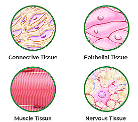
1. Epithelial Tissue
This tissue covers both internal and external body surfaces, with tightly packed cells and minimal intercellular matrix. It comes in two forms: simple, which is a single layer of cells serving as protective linings, and compound, which consists of multiple layers and serves secretion and absorption functions.
This tissue has a free surface, which faces either a body fluid or the outside environment and thus provides a covering or a lining for some part of the body. The cells are compactly packed with little intercellular matrix. There are two types of epithelial tissues namely simple epithelium and compound epithelium.
(a) Simple epithelium is composed of a single layer of cells and functions as a lining for body cavities, ducts, and tubes.
(b) The compound epithelium consists of two or more cell layers and has a protective function as it does in our skin.
On the basis of the structural modification of the cells, the simple epithelium is further divided into three types.
These are (i) Squamous, (ii) Cuboidal, (iii) Columnar, (iv) Ciliated
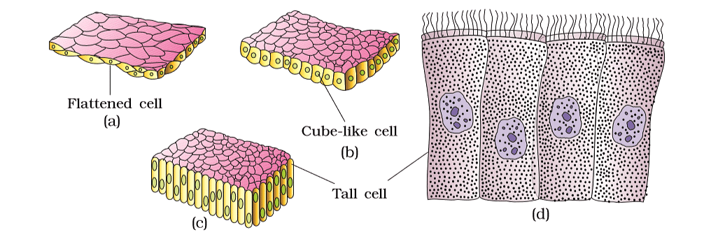
(i) The squamous epithelium is made of a single thin layer of flattened cells with irregular boundaries. They are found in the walls of blood vessels and air sacs of the lungs and are involved in functions like forming a diffusion boundary.
(ii) The cuboidal epithelium is composed of a single layer of cube-like cells. This is commonly found in ducts of glands and tubular parts of nephrons in kidneys and its main functions are secretion and absorption. The epithelium of the proximal convoluted tubule (PCT) of the nephron in the kidney has microvilli.
(iii) The columnar epithelium is composed of a single layer of tall and slender cells. Their nuclei are located at the base. The free surface may have microvilli. They are found in the lining of the stomach and intestine and help in secretion and absorption.
(iv) If the columnar or cuboidal cells bear cilia on their free surface they are called ciliated epithelium. Their function is to move particles or mucus in a specific direction over the epithelium. They are mainly present in the inner surface of hollow organs like bronchioles and fallopian tubes.
Note: Some columnar or cuboidal cells specialize in secretion and are known as glandular epithelium. There are two main types: unicellular glands like goblet cells in the digestive tract, and multicellular glands like salivary glands. Glands are categorized based on how they release their secretions. Exocrine glands, such as those producing mucus, saliva, and digestive enzymes, release their products through ducts. In contrast, endocrine glands release their hormones directly into the surrounding fluid, as they lack ducts.
(b) The compound epithelium is made of more than one layer (multi-layered) of cells and thus has a limited role in secretion and absorption. Their main function is to provide protection against chemical and mechanical stresses.
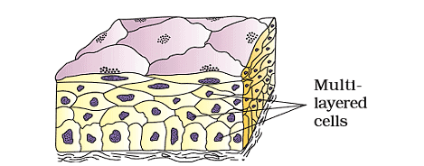
- They cover the dry surface of the skin, the moist surface of the buccal cavity, pharynx, inner lining of ducts of salivary glands, and of pancreatic ducts.
- All cells in the epithelium are held together with little intercellular material. In nearly all animal tissues, specialized junctions provide both structural and functional links between its individual cells.
- Three types of cell junctions are found in the epithelium and other tissues.
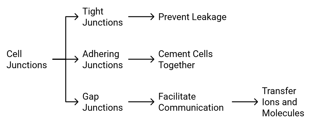 Types of Cell Junctions
 - Tight junctions help to stop substances from leaking across a tissue.
- Adhering junctions perform cementing to keep neighboring cells together.
- Gap junctions facilitate the cells to communicate with each other by connecting the cytoplasm of adjoining cells, for rapid transfer of ions, small molecules, and sometimes big molecules.
2. Connective Tissue
Connective tissues are most abundant and widely distributed in the body of complex animals. They are named connective tissues because of their special function of linking and supporting other tissues/organs of the body.
They range from soft connective tissues to specialised types, which include cartilage, bone, adipose, and blood. In all connective tissues except blood, the cells secrete fibres of structural proteins called collagen or elastin. The fibres provide strength, elasticity, and flexibility to the tissue. These cells also secrete modified polysaccharides, which accumulate between cells and fibres and act as matrix (ground substance).
Connective tissues are classified into three types:
(i) Loose connective tissue
Loose connective tissue has cells and fibres loosely arranged in a semi-fluid ground substance, for example, areolar tissue present beneath the skin. Often it serves as a support framework for epithelium. It contains fibroblasts (cells that produce and secrete fibres), macrophages, and mast cells.
 (a) Aerolar Tissue (b) Adipose Tissue
Adipose tissue is another type of loose connective tissue located mainly beneath the skin. The cells of this tissue are specialised to store fats. The excess of nutrients that are not used immediately are converted into fats and are stored in this tissue.
(ii) Dense connective tissue
Fibres and fibroblasts are compactly packed in the dense connective tissues. The orientation of fibres show a regular or irregular pattern and are called dense regular and dense irregular tissues.
- In the dense regular connective tissues, the collagen fibres are present in rows between many parallel bundles of fibres. Tendons, which attach skeletal muscles to bones and ligaments which attach one bone to another are examples of this tissue.
- Dense irregular connective tissue has fibroblasts and many fibres (mostly collagen) that are oriented differently. This tissue is present in the skin.
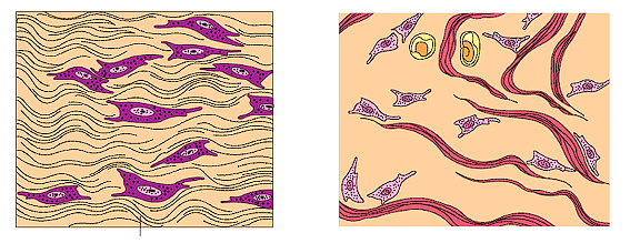 (a) Dense regular (b) Dense irregular
(iii) Specialised connective tissue
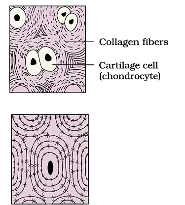
Cartilage:
- The intercellular material of cartilage is solid and pliable and resists compression.
- Cells of this tissue (chondrocytes) are enclosed in small cavities within the matrix secreted by them.
- Most of the cartilages in vertebrate embryos are replaced by bones in adults.
- Cartilage is present in the tip of nose, outer ear joints, between adjacent bones of the vertebral column, limbs and hands in adults
Bones:
- Bones have a hard and non-pliable ground substance rich in calcium salts and collagen fibres which give bone its strength.
- It is the main tissue that provides structural frame to the body and support and protect softer tissues and organs.
- The bone cells (osteocytes) are present in the spaces called lacunae.
- Limb bones, such as the long bones of the legs, serve weight-bearing functions.
- They also interact with skeletal muscles attached to them to bring about movements.
- The bone marrow in some bones is the site of the production of blood cells.
Blood:
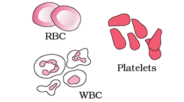
Blood is a fluid connective tissue containing plasma, red blood cells (RBC), white blood cells (WBC) and platelets. It is the main circulating fluid that helps in the transport of various substances.
3. Muscle Tissue
Each muscle is made of many long, cylindrical fibres arranged in parallel arrays. These fibres are composed of numerous fine fibrils, called myofibrils. Muscle fibres contract (shorten) in response to stimulation, then relax (lengthen) and return to their uncontracted state in a coordinated fashion. Their action moves the body to adjust to the changes in the environment and to maintain the positions of the various parts of the body.
Muscles are of three types: skeletal, smooth, and cardiac.
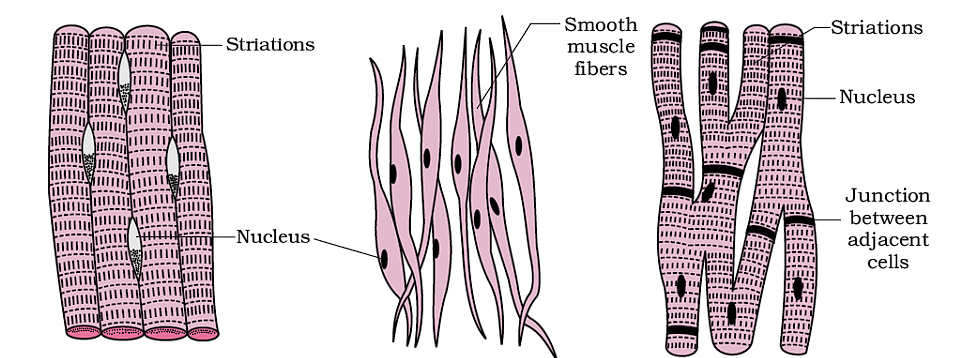
Skeletal muscle tissue
- Skeletal muscle tissue is closely attached to skeletal bones.
- In a typical muscle such as the biceps, striated (striped) skeletal muscle fibres are bundled together in a parallel fashion.
- A sheath of tough connective tissue encloses several bundles of muscle fibres.
Smooth muscle fibres
- Smooth muscle fibres taper at both ends (fusiform) and do not show striations.
- Cell junctions hold them together and they are bundled together in a connective tissue sheath.
- The wall of internal organs such as the blood vessels, stomach, and intestine contains this type of muscle tissue.
- Smooth muscles are 'involuntary' as their functioning cannot be directly controlled.
- We usually are not able to make it contract merely by thinking about it as we can do with skeletal muscles.
Cardiac muscle tissue
- Cardiac muscle tissue is a contractile tissue present only in the heart.
- Cell junctions fuse the plasma membranes of cardiac muscle cells and make them stick together.
- Communication junctions (intercalated discs) at some fusion points allow the cells to contract as a unit, i.e., when one cell receives a signal to contract, its neighbors are also stimulated to contract.
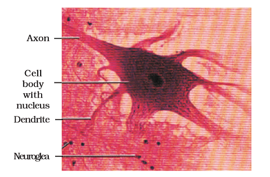
4. Neural Tissue
Neural tissue exerts the greatest control over the body’s responsiveness to changing conditions. Neurons, the unit of neural system are excitable cells.The neuroglial cell which constitute the rest of the neural system protect and support neurons.
- Neuroglia make up more than one-half the volume of neural tissue in our body.
- When a neuron is suitably stimulated, an electrical disturbance is generated which swiftly travels along its plasma membrane.
- Arrival of the disturbance at the neuron’s endings, or output zone, triggers events that may cause stimulation or inhibition of adjacent neurons and other cells.
Organ & Organ System
In multicellular organisms, basic tissues organize themselves to form organs, which then come together to create organ systems.
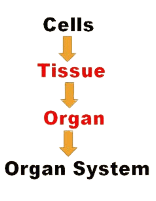
- This organisation is necessary for the efficient and coordinated activities of the millions of cells that make up an organism.
- Each organ in our body consists of one or more types of tissues, such as the heart, which contains all four tissue types: epithelial, connective, muscular, and neural.
- There is a noticeable evolutionary trend in the complexity of organs and organ systems, which will be studied in more detail in later classes.
Now, you will learn about the morphology and anatomy of a Cockroach and frog, which represents vertebrates.
Morphology refers to the study of the form or external appearance of organisms, while anatomy refers to the study of the internal structure of animal organs.
Morphology of Cockroach
The cockroach, despite being widely disliked, has been around for over 300 million years and remains a prevalent insect. Let's delve into its outer appearance and inner structure.
 Morphology of Cockroach
Morphology:
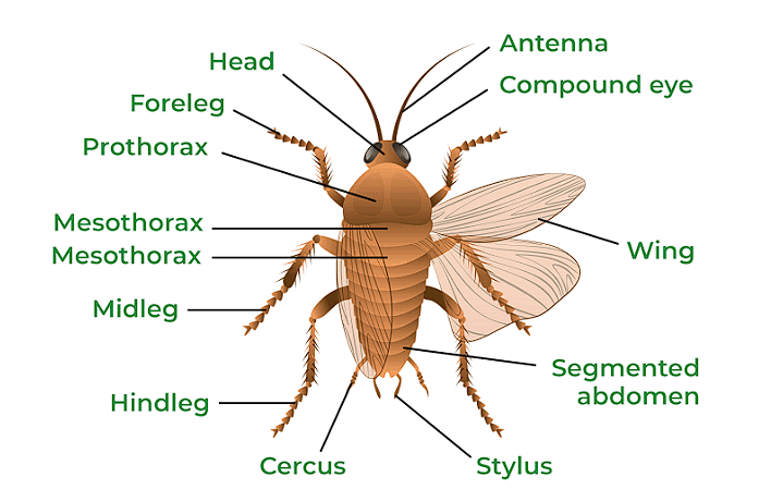
- Head: It has compound eyes and long antennae. The mouth, equipped with mandibles and other mouthparts, is located at the front, along with a flexible tongue-like structure.
- Thorax: Divided into three parts - prothorax, mesothorax, and metathorax. Legs and wings emerge from these segments.
- Abdomen: Consists of 10 segments, with reproductive organs in segments 7 to 9 and filamentous structures called cerci at the end.
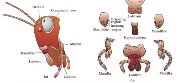 Head region of cockroach : (a) parts of head region (b) mouth parts
Cockroach Appearance:
The cockroach's body is divided into three parts: head, thorax, and abdomen, protected by a hard, brown exoskeleton made of chitin. It has distinct male and female genders, with males being longer. The head is triangular with compound eyes and antennae. The chest has three segments, each with legs and wings. The abdomen has 10 segments, with reproductive organs in segments 7 to 9. Both male and female cockroaches have cerci at the end of the abdomen.
Anatomy of Cockroach
- Alimentary Canal: The digestive tract is divided into three parts: foregut, midgut, and hindgut. Food enters through the mouth, passes through the pharynx and esophagus, and is stored in the crop. Then it moves to the gizzard for grinding before entering the midgut, where digestion occurs. Waste is eliminated through the hindgut.
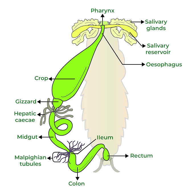 Alimentary Canal of Cockroach
- Blood Vascular System: Cockroaches have an open circulatory system with a fluid called hemolymph. Organs are bathed in hemolymph within the hemocoel. The heart, a tube with chambers, pumps hemolymph throughout the body.
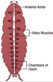 Open Circulatory System of Cockroach
- Respiratory System: Cockroaches breathe through a system of tubes called tracheae, which open to the outside through spiracles. Oxygen is transported to tissues through these tubes.
- Nervous System: The nervous system consists of ganglia arranged segmentally throughout the body. Ganglia in the chest and abdomen control various functions. Receptors such as antennae, eyes, and cerci detect stimuli.
- Excretory System: Waste is removed by Malpighian tubules, which absorb nitrogenous waste and convert it to uric acid. This acid is expelled through the hindgut, making cockroaches uricotelic.
- Reproductive System:
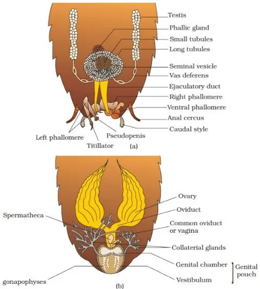 Reproductive system of cockroach : (a) male (b) female
Dioecious Species: Cockroaches have distinct male and female reproductive systems.
Male Reproductive Anatomy:
Testes: Located in the 4th to 6th abdominal segments, Vas Deferens and Ejaculatory Ducts: Lead from the testes to the male gonopore near the anus, Accessory Gland: Mushroom-shaped, located in the 6th-7th segments, aids in reproduction, Phallomeres: Chitinous structures surrounding the gonopore, part of the external genitalia, Spermatophores: Sperm bundles formed for copulation.
Female Reproductive Anatomy:
Ovaries: Situated in the 2nd to 6th abdominal segments, Oviducts: Merge into a single vagina that opens into the genital chamber, Spermatheca: Located in the 6th segment, used for storing sperm, Oothecae: Reddish-brown egg capsules, each containing 14-16 eggs, usually placed in humid areas near food, Egg Production: Females typically produce 9-10 oothecae.
Development:
Paurometabolous: Development involves a nymphal stage that closely resembles adults, Moulting: Nymphs undergo about 13 moults before reaching adult form, Wings: Only adult cockroaches develop wings.
Frog
- Frogs, which are classified as Amphibia in the phylum Chordata, can live both on land and in freshwater.
- The most common species of frog found in India is Rana tigrina.
- Frogs are cold-blooded, which means their body temperature varies with the temperature of their environment.
- Frogs can change the color of their skin to blend in with their surroundings, which is called camouflage. This ability helps them hide from predators.
- Frogs are also known to take shelter in deep burrows during extreme weather conditions such as peak summer and winter, which is called summer sleep (aestivation) and winter sleep (hibernation) respectively. This protects them from the harsh effects of extreme heat and cold.
Morphology of a Frog
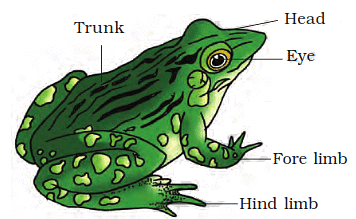 External Features of a Frog
- Frog's skin is smooth, slippery, and always moist due to the presence of mucus.
- The dorsal side of the frog's body is generally olive green with dark irregular spots, while the ventral side is uniformly pale yellow.
- Frogs absorb water through their skin and do not drink water.
- Frog's body is divided into a head and trunk, with no neck or tail.
- Frog has a pair of nostrils above its mouth and bulged eyes covered by a protective membrane called nictitating membrane.
- Frog's ears, called tympanum, are membranous structures on either side of its eyes that receive sound signals.
- Forelimbs and hind limbs of the frog help in swimming, walking, leaping, and burrowing.
- Hind limbs have five digits and are larger and more muscular than forelimbs, which have four digits.
- Frog's feet have webbed digits that aid in swimming.
- Male frogs have vocal sacs for sound production and a copulatory pad on the first digit of forelimbs, which are absent in female frogs.
- This difference in characteristics between male and female frogs is called sexual dimorphism.
Anatomy of a Frog
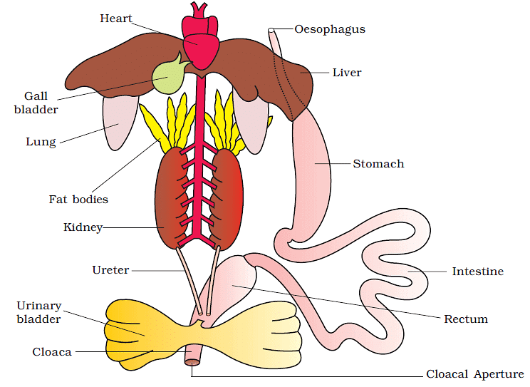 Anatomy of a Frog
(i) Digestive System of Frog
- Frog's body cavity contains different organ systems including digestive, circulatory, respiratory, nervous, excretory, and reproductive systems.
- Frog's digestive system consists of an alimentary canal and digestive glands.
- The alimentary canal is short because frogs are carnivores, so their intestine is reduced in length.
- The mouth opens into the buccal cavity, which leads to the oesophagus through the pharynx.
- The oesophagus is a short tube that opens into the stomach, which then continues as the intestine, rectum, and finally opens outside through the cloaca.
- The liver secretes bile that is stored in the gall bladder.
- Pancreas, a digestive gland, produces pancreatic juice for digestion.
- Frog captures food with its bilobed tongue.
- Digestion of food occurs in the stomach through the action of HCl and gastric juices.
- Partially digested food called chyme is passed from the stomach to the duodenum, the first part of the small intestine.
- The duodenum receives bile from the gall bladder and pancreatic juices from the pancreas through a common bile duct.
- Bile emulsifies fat and pancreatic juices digest carbohydrates and proteins.
- Final digestion takes place in the intestine.
- Digested food is absorbed through finger-like folds called villi and microvilli in the inner wall of the intestine.
- Undigested solid waste moves into the rectum and passes out through the cloaca.
(ii) Respiration in Frogs
- Frogs have two methods of respiration: cutaneous respiration in water and pulmonary respiration on land.
- In water, the skin acts as an aquatic respiratory organ, where dissolved oxygen is exchanged through the skin by diffusion.
- On land, the buccal cavity, skin, and lungs act as respiratory organs.
- The lungs are a pair of elongated, pink-colored sac-like structures located in the upper part of the trunk region (thorax).
- Air enters through the nostrils into the buccal cavity and then to the lungs for pulmonary respiration.
- During aestivation and hibernation, gaseous exchange takes place through the skin.
(iii) Vascular System of a Frog
- Frogs have a well-developed closed vascular system, which includes the heart, blood vessels, and blood.
- They also have a lymphatic system, consisting of lymph, lymph channels, and lymph nodes.
- The heart is a muscular structure with three chambers: two atria and one ventricle, covered by a membrane called pericardium.
- Blood enters the heart through the sinus venosus, a triangular structure that joins the right atrium, and receives blood from major veins called vena cava.
- The ventricle opens into a sac-like structure called conus arteriosus on the ventral side of the heart.
- Arteries carry blood from the heart to all parts of the body, while veins collect blood from different parts of the body and return it to the heart.
- Frogs have special venous connections between the liver and intestine, and the kidney and lower parts of the body, called hepatic portal system and renal portal system respectively.
- Blood is composed of plasma and cells, including nucleated red blood cells (RBCs or erythrocytes), white blood cells (WBCs or leucocytes), and platelets.
- Blood carries nutrients, gases, and water to different parts of the body during circulation, which is achieved by the pumping action of the muscular heart.
(iv) Excretory System of a Frog
- Frogs have a well-developed excretory system for eliminating nitrogenous wastes.
- The excretory system includes a pair of kidneys, ureters, cloaca, and urinary bladder.
- The kidneys are compact, dark red, bean-like structures located posteriorly in the body cavity on both sides of the vertebral column.
- Each kidney is made up of uriniferous tubules or nephrons, which are structural and functional units responsible for filtering and excreting wastes.
- In male frogs, two ureters emerge from the kidneys and act as urinogenital ducts that open into the cloaca.
- In females, the ureters and oviduct open separately into the cloaca.
- The thin-walled urinary bladder is located ventral to the rectum and also opens into the cloaca.
- Frogs excrete urea, making them ureotelic animals.
- Excretory wastes are carried by blood into the kidneys, where they are separated and excreted from the body.
(v) Control and Coordination in Frogs
- Frogs have a highly evolved system for control and coordination, which includes both the neural system and endocrine glands.
- Hormones secreted by endocrine glands are responsible for chemical coordination of various organs in the body.
- Prominent endocrine glands in frogs include pituitary, thyroid, parathyroid, thymus, pineal body, pancreatic islets, adrenals, and gonads.
- The nervous system in frogs is organized into a central nervous system (brain and spinal cord), a peripheral nervous system (cranial and spinal nerves), and an autonomic nervous system (sympathetic and parasympathetic).
- Frogs have ten pairs of cranial nerves arising from the brain, which is enclosed in a bony structure called the brain box or cranium.
- The brain in frogs is divided into forebrain, midbrain, and hindbrain.
- The forebrain includes olfactory lobes, paired cerebral hemispheres, and unpaired diencephalon.
- The midbrain is characterized by a pair of optic lobes.
- The hindbrain consists of cerebellum and medulla oblongata.
- The medulla oblongata continues into the spinal cord, which is enclosed in the vertebral column and passes out through the foramen magnum.
(vi) Sense Organs in Frogs
- Frogs have different types of sense organs, including organs of touch (sensory papillae), taste (taste buds), smell (nasal epithelium), vision (eyes), and hearing (tympanum with internal ears).
- Eyes and internal ears are well-organized structures in frogs, while the rest are cellular aggregations around nerve endings.
- Frog's eyes are a pair of spherical structures located in the orbit of the skull, and they are simple eyes with only one unit.
- External ears are absent in frogs, and only the tympanum can be seen externally.
- The ear in frogs serves both as an organ of hearing and balancing (equilibrium).
(vii) Reproduction and Ecological Importance of Frogs
- Frogs have well-organized male and female reproductive systems.
- Male reproductive organs in frogs consist of a pair of testes that are yellowish ovoid structures located near the kidneys and connected to the urinogenital duct via vasa efferentia.
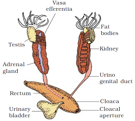 Male Reproductive System
- Female reproductive organs in frogs include a pair of ovaries located near the kidneys, with oviducts that open separately into the cloaca.
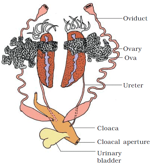 Female Reproductive System
- Fertilization in frogs is external and occurs in water, and the development involves a larval stage called tadpole, which undergoes metamorphosis to become an adult frog.
- Frogs play an important ecological role by eating insects and protecting crops, as they serve as a link in the food chain and food web in ecosystems.
- In some countries, frogs are also used as a food source for their muscular legs.
FAQs (Frequently Asked Questions)
Q. What are organs and organ systems in multicellular organisms?
Organs are formed by basic tissues organizing themselves, and organ systems are created when organs come together. This organization is necessary for the efficient and coordinated activities of cells in an organism.
Q. What are some external features of a frog?
Frog's skin is smooth, slippery, and always moist due to the presence of mucus. The dorsal side of the frog's body is generally olive green with dark irregular spots, while the ventral side is uniformly pale yellow. Frogs absorb water through their skin and do not drink water. Frog's body is divided into a head and trunk, with no neck or tail.
Q. What are the differences in characteristics between male and female frogs?
Male frogs have vocal sacs for sound production and a copulatory pad on the first digit of forelimbs, which are absent in female frogs. This difference in characteristics between male and female frogs is called sexual dimorphism.
Q. What are the organs and organ systems in the digestive system of a frog?
Frog's digestive system consists of an alimentary canal and digestive glands. The alimentary canal is short because frogs are carnivores, so their intestine is reduced in length. The mouth opens into the buccal cavity, which leads to the esophagus through the pharynx. The esophagus is a short tube that opens into the stomach, which then continues as the intestine, rectum, and finally opens outside through the cloaca. The liver secretes bile that is stored in the gall bladder. Pancreas, a digestive gland, produces pancreatic juice for digestion.
Q. How do frogs respire in water and on land?
Frogs have two methods of respiration: cutaneous respiration in water and pulmonary respiration on land. In water, the skin acts as an aquatic respiratory organ, where dissolved oxygen is exchanged through the skin by diffusion. On land, the buccal cavity, skin, and lungs act as respiratory organs. The lungs are a pair of elongated, pink colored sac-like structures located in the upper part of the trunk region (thorax). Air enters through the nostrils into the buccal cavity and then to the lungs for pulmonary respiration. During aestivation and hibernation, gaseous exchange takes place through the skin.
Q. What is the vascular system of a frog?
Frogs have a well-developed closed vascular system, which includes the heart, blood vessels, and blood. They also have a lymphatic system, consisting of lymph, lymph channels, and lymph nodes. The heart is a muscular structure with three chambers: two atria and one ventricle, covered by a membrane called pericardium. Blood enters the heart through the sinus venosus, a triangular structure that joins the right atrium, and receives blood from major veins called vena cava. The ventricle opens into a sac-like structure called conus arteriosus on the ventral side of the heart. Arterial blood is carried to various parts of the body by arteries, and venous blood returns to the heart through veins.
|
169 videos|400 docs|136 tests
|


