UPSC Exam > UPSC Notes > Zoology Optional Notes for UPSC > Development of Eye and Heart
Development of Eye and Heart | Zoology Optional Notes for UPSC PDF Download
Development of Eye
- Neural tube interacts with cranial ectodermal placodes.
- Interaction leads to formation of principal sensory organs of the skull.
- Cranial ectodermal placodes are series of epidermal thickenings.
The mechanism of eye development:
- Head ectoderm interacts with involuting endoderm and mesoderm during gastrulation.
- Interaction predisposes head ectoderm towards lens formation.
- Optic vesicle, emerging from diencephalon, places lens in relation to retina.
- Optic vesicle contact triggers development of lens placode, later forming lens.
- Optic vesicle transforms into two-walled optic cup with differentiating layers.
- Outer layer differentiates into pigmented retina.
- Inner layer undergoes fast cell division, producing various retinal cells.
- Retinal ganglion cells carry electrical signals to the brain via optic nerve.
- Anterior tip of neural plate expresses transcription factors Six3, Pax6, and Rx1.
- Optic vesicles arise from this domain.
- Pax6 crucial for lens and retina growth, shared feature in photoreceptive cells.
- Sonic hedgehog secretion divides single eye field into bilateral fields.
- Alterations in sonic hedgehog gene or protein processing can lead to cyclopia.
- Sonic hedgehog protein from prechordal plate suppresses Pax6 expression, dividing the eye field.
Neural differentiation of the retina:
- Neural retina develops into layered array of various neuronal types.
- Similarity to cerebral and cerebellar cortices observed.
- Layers include rod and cone photoreceptor cells, ganglion cell bodies, and bipolar interneurons.

- Horizontal neurons conduct electrical impulses within the retina plane.
- Amacrine neurons lack long axons.
- Müller glial cells maintain retina integrity.
- Striated, laminar pattern formed during early retinal development.
- Pattern created through cell division, migration, and differential cell death.
- Single neuroblast precursor cell from retinal germinal layer can give rise to at least three different types of neurons or two different types of neurons and a glial cell.
Differentiation of lens and cornea:
- Lens placode folds and touches fresh ectoderm during lens development.
- Lens vesicle induces ectoderm to form transparent cornea.
- Proper curvature of cornea crucial for focusing light onto retina.
- Intraocular fluid pressure essential for maintaining cornea curvature.
- Ring of scleral bones, likely from neural crest, maintains intraocular pressure.
- Changes in cell structure, shape, and production of crystallins needed for lens tissue differentiation.
- Crystallins, lens-specific proteins, crucial for lens transparency and light direction onto retina.
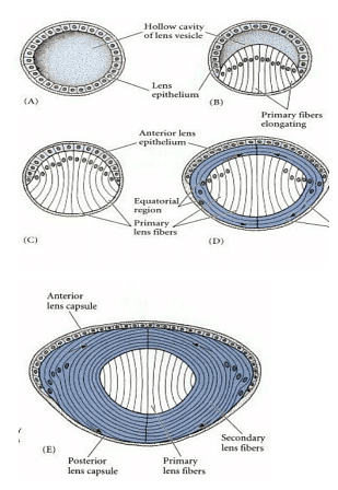
- Inner cells of lens vesicle expand and transform into lens fibers under neural retina influence.
- Lens fibers produce crystallins, filling cells and leading to nucleus extrusion.
- Crystallin-synthesizing fibers cover gap between two layers of lens vesicle.
- Anterior lens vesicle cells form ongoing germinal epithelium.
- Dividing cells move towards vesicle's equator, lengthen, and transform.

- Lens consists of three distinct zones:
- Anterior zone: Contains proliferating epithelial cells.
- Equatorial zone: Characterized by cellular elongation.
- Posterior and middle zone: Comprised of fiber cells containing crystalline.
- Ongoing laying down of fibers maintains this arrangement throughout animal's lifetime.
- Transition from epithelial cell to lens fiber in adult chicken takes two years.
- Iris, a pigmented and skeletal tissue, located in front of lens.
- Iris muscles regulate pupil's size.
- Part of iris derived from ectodermal layer, unlike other body muscles from mesoderm.
- Optic cup section forming iris continuous with neural retina but does not produce photoreceptors.
Development of Heart
- Circulatory system is a significant achievement of lateral plate mesoderm.
- It comprises heart, blood cells, and intricate network of blood vessels.
- Nourishes growing vertebrate embryo.
- Heart is the first functional organ and circulatory system is the first functional component of embryo.
- Splanchnic mesoderm sections on both sides of body specialize for heart development.
- Interaction with surrounding tissue leads to specialization and formation of vertebrate heart.
Specification of heart tissue and fusion of heart rudiments:
- Amniote vertebrate embryos are flattened discs.
- Lateral plate mesoderm doesn't fully cover yolk sac.
- Early primitive streak, slightly posterior to Hensen's node, is where cardiogenic mesoderm originates.
- Cardiogenic mesoderm cells divide through the streak.
- Division generates two clusters of mesodermal cells lateral to Hensen's node.
- These clusters are the source of cells forming endothelium lining of heart, cushion cells of valves, Purkinje conducting fibers, and atrial/ventricular muscles.

- Presumptive heart cells migrate anteriorly between ectoderm and endoderm in chick embryo at 18-20 hours old.
- Migration occurs while maintaining close contact with endodermal surface.
- Migration halts at lateral walls of anterior gut tube.
- Direction of movement guided by foregut endoderm.
- Cardiac mesoderm moves opposite to rotation of heart region endoderm about embryo.
- Anterior-to-posterior concentration gradient of fibronectin in endoderm directs migration.
- Some cardiogenic cells designated by endoderm and primitive streak develop into heart muscles.
- Cerberus and unidentified substance (presumably BMP2) induce cardiogenic cells to produce Nkx2-5 transcription factor in anterior endoderm.
- Nkx2-5 directs mesoderm to develop into cardiac tissue, triggering production of other transcription factors (GATA, MEF2 families).
- Transcription factors stimulate expression of genes coding for proteins specific to heart muscle.
- Ventricular cells become defined before atrial cells.
- Advancing heart-forming primordia experience separate cell differentiation.
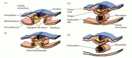
- Cells display N-cadherin on apices and unite to form epithelium as they move.
- Endocardium formed when small population of cells detach from epithelium and downregulate N-cadherin.
- Myocardium comprised of epithelial cells develops into heart's pumping muscles.

- Endocardial cells produce numerous heart valves and regulate neural tissue positioning in heart.
- They secrete proteins controlling myocardial growth.
- Splanchnic mesoderm folds inward during neurulation to form foregut.
- Myocardium becomes one tube as movement pulls two cardiac tubes together.
- "Cardio Bifida" results in development of distinct hearts on each side of body.
- Previously paired coelomic chambers combine to form body cavity housing heart.
- Fusion of coelomic chambers occurs around 29 hours in chick development.
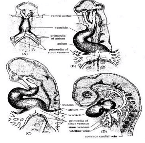
- Apertures of vitelline veins in heart formed by unfused posterior sections of endocardium.
- Veins transport nutrients from yolk sac to sinus venosus.
- Blood enters atrial portion of heart through flap resembling valve.
- Truncus arteriosus contractions accelerate blood flow into aorta.
- Heart begins beating while primordia are still joining.
- Sinus venosus acts as pacemaker for contractions.
- Wave of muscular contraction propagates from where contractions start in tubular heart.
- Heart pumps blood before intricate valve system fully develops.
- Heart muscle cells contract naturally.
- By fourth day, chick embryo's ECG similar to adult's.
- Contractions in embryo controlled by electrical inputs from medulla oblongata via vagus nerve.

Looping and formation of heart chambers:
- Three-day-old chick embryo has two-chambered heart with atrium and ventricle.
- Blood cycle observed with blood entering lower chamber and pumped out through aorta.
- Heart's looping changes original anterior-posterior polarity to right-left polarity.
- Right ventricle area located ahead of left ventricle area due to looping.
- Left-right patterning proteins necessary for looping process.
- Nkx2-5 controls Hand1 and Hand2 transcription factors in heart primordium.
- Hand1 limited to left ventricle and Hand2 to right once looping begins.
- Disruption of Hand proteins leads to improper ventricle development.
- Pitx-2 transcription factor crucial for correct cardiac looping, activated on left side of lateral plate mesoderm.
- Pitx-2 may control expression of proteins like lectin to manage physical strain of heart components.
- Xin gene mediates cytoskeletal alterations required for heart looping, activated by transcription factors Nkx2-5 and MEF2C.
- Various transcription factors limited to specific regions of heart tube define atrium-ventricle separation.
- Myocardium cells release substance (likely transforming growth factor-β3) triggering endocardium cells to separate and enter hyaluronate-rich cardiac jelly, partitioning tube into distinct atrium and ventricle.
The document Development of Eye and Heart | Zoology Optional Notes for UPSC is a part of the UPSC Course Zoology Optional Notes for UPSC.
All you need of UPSC at this link: UPSC
|
198 videos|351 docs
|
FAQs on Development of Eye and Heart - Zoology Optional Notes for UPSC
| 1. How does the eye develop in the human body? |  |
Ans. The eye develops in the human body through a complex process known as embryogenesis, where specialized cells differentiate and organize to form the various structures of the eye, including the lens, retina, and optic nerve.
| 2. What role does the heart play in the development of the circulatory system? |  |
Ans. The heart plays a crucial role in the development of the circulatory system by pumping oxygen-rich blood to all parts of the body, providing nutrients and removing waste products. The heart begins to form early in embryonic development and undergoes various stages of growth to become fully functional.
| 3. How do genetic factors influence the development of the eye and heart? |  |
Ans. Genetic factors play a significant role in the development of the eye and heart, as specific genes control the formation and function of these organs. Mutations in these genes can lead to congenital abnormalities or diseases affecting the eye and heart.
| 4. What are some common developmental disorders associated with the eye and heart? |  |
Ans. Some common developmental disorders associated with the eye include cataracts, glaucoma, and retinoblastoma, while common heart disorders include congenital heart defects, arrhythmias, and coronary artery disease.
| 5. How can environmental factors impact the development of the eye and heart? |  |
Ans. Environmental factors such as exposure to toxins, infections, and maternal health can impact the development of the eye and heart during pregnancy. It is crucial to maintain a healthy lifestyle and seek prenatal care to minimize the risk of developmental issues in these organs.
Related Searches


