UPSC Exam  >  UPSC Notes  >  Medical Science Optional Notes for UPSC  >  Electron Microscopic Appearance of a Skeletal Muscle

Electron Microscopic Appearance of a Skeletal Muscle | Medical Science Optional Notes for UPSC PDF Download

Overview

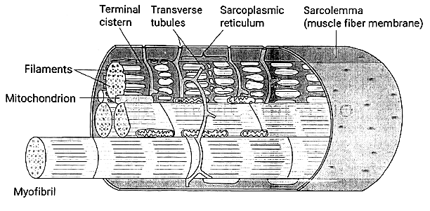
  • Muscle fibers are composed of myofibrils, which are further divided into individual filaments.

  • Myofilaments within the muscle contain multiple proteins, collectively forming the contractile machinery of the skeletal muscle.

  • The contractile mechanism in skeletal muscle is heavily reliant on proteins such as myosin-II, actin, tropomyosin, and troponin.

  • Troponin is comprised of three subunits: troponin I, troponin T, and troponin C.

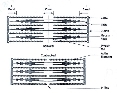
  • Each myofibril consists of interdigitating thick and thin filaments arranged longitudinally within sarcomeres.

  • Sarcomeres, the repeating units within myofibrils, contribute to the distinctive banding pattern observed in striated muscle.

  • A sarcomere extends from the Z line to another Z line, defining the structural and functional unit of muscle contraction.

    Electron Microscopic Appearance of a Skeletal Muscle | Medical Science Optional Notes for UPSC

Thick Filaments

  • Found in the A band at the center of the sarcomere and are composed of myosin.

  • Myosin consists of six polypeptide chains, comprising one pair of heavy chains and two pairs of light chains.

  • Each myosin molecule possesses two "heads" attached to a single "tail."

  • The myosin heads play a crucial role in the process of muscle contraction, as they bind ATP and actin, participating in the formation of cross-bridges.

    Thick Filament
    Thick Filament

Thin Filaments

  • Anchored at the Z lines and located in the I bands of the sarcomere.

  • Interdigitate with the thick filaments within a portion of the A band.

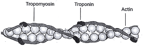
  • Composed of actin, tropomyosin, and troponin.

  • Troponin acts as the regulatory protein, allowing cross-bridge formation when it binds to Ca2+.

  • Troponin is a complex of three globular proteins:

    1. Troponin T ("T" for tropomyosin): Attaches the troponin complex to tropomyosin.
    2. Troponin I ("I" for inhibition): Inhibits the interaction of actin and myosin.
    3. Troponin C ("C" for Ca2+): The Ca2+-binding protein that, when bound to Ca2+, facilitates the interaction of actin and myosin.
Electron Microscopic Appearance of a Skeletal Muscle | Medical Science Optional Notes for UPSC

T Tubules

  • Form an extensive tubular network open to the extracellular space, facilitating the transmission of depolarization from the sarcolemmal membrane to the cell interior.

  • Positioned at the junctions of A bands and I bands.

  • Contain a voltage-sensitive protein known as the dihydropyridine receptor (DHPR).

  • Depolarization induces a conformational change in the dihydropyridine receptor.

Question for Electron Microscopic Appearance of a Skeletal Muscle
Try yourself:
Which protein helps in the formation of cross-bridges during muscle contraction?
View Solution

Sarcoplasmic Reticulum

  • Internal tubular structure crucial for Ca2+ storage and release in excitation-contraction coupling.

  • Features terminal cisternae in intimate contact with T tubules, forming a triad arrangement.

  • Membrane includes the Ca2+-ATPase (Ca2+ pump) responsible for transporting Ca2+ from the intracellular fluid into the SR interior, maintaining low intracellular [Ca2+].

  • Contains a Ca2+ release channel called the ryanodine receptor.

    Electron Microscopic Appearance of a Skeletal Muscle | Medical Science Optional Notes for UPSC

Latch Phenomenon in Smooth Muscle

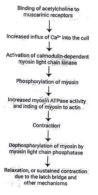
  • Dephosphorylation of myosin light chain kinase: Does not always result in smooth muscle relaxation.

  • Latch Bridge Mechanism: Myosin cross-bridges stay attached to actin even after the cytoplasmic Ca2+ concentration decreases.

  • Outcome: Sustained contraction with minimal energy expenditure, particularly crucial in vascular smooth muscle.

  • Relaxation: Presumably happens when the Ca2+-calmodulin complex eventually dissociates.

    Electron Microscopic Appearance of a Skeletal Muscle | Medical Science Optional Notes for UPSC

There is no troponin; instead, Ca2+ regulates myosin on the thick filaments.

Question for Electron Microscopic Appearance of a Skeletal Muscle
Try yourself:
What is the role of the sarcoplasmic reticulum in excitation-contraction coupling?
View Solution

Nerve muscle physiology-Repeats

Muscle physiology
Q1: Differentiate between isometric and isotonic contraction in skeletal muscle. Give one example of each type. (2015)
Q2: What are modes of contraction in a skeletal muscle? Give suitable examples. Explain length-tension relationship in skeletal muscle. (2018)
Q3: Name the contractile proteins in skeletal muscle. What is the electron microscopic appearance of muscle? Write sequence of events in muscular contraction. (2011)
Q4: Compare with the help of a diagram the length-tension relationship for skeletal, cardiac and smooth muscles. What is the latch phenomenon in smooth muscle (2001)?

The document Electron Microscopic Appearance of a Skeletal Muscle | Medical Science Optional Notes for UPSC is a part of the UPSC Course Medical Science Optional Notes for UPSC.
All you need of UPSC at this link: UPSC
7 videos|219 docs

Top Courses for UPSC

FAQs on Electron Microscopic Appearance of a Skeletal Muscle - Medical Science Optional Notes for UPSC

1. What is the role of the sarcoplasmic reticulum in muscle contraction?
Ans. The sarcoplasmic reticulum is a specialized structure in muscle cells that plays a crucial role in muscle contraction. It stores and releases calcium ions, which are essential for the contraction process. When a muscle is stimulated, an action potential travels along the muscle cell membrane and reaches the sarcoplasmic reticulum. This triggers the release of calcium ions into the muscle cell, leading to the contraction of the muscle fibers.
2. What is the latch phenomenon in smooth muscle?
Ans. The latch phenomenon is a characteristic feature of smooth muscle contraction. It refers to the ability of smooth muscle to maintain a sustained contraction for an extended period of time with minimal energy expenditure. Unlike skeletal muscle, which requires continuous energy input to maintain contraction, smooth muscle can maintain a contracted state without using significant amounts of ATP. This prolonged contraction in smooth muscle is important for functions such as maintaining blood pressure and controlling the movement of substances through organs like the intestines.
3. How does the electron microscopic appearance of skeletal muscle differ from smooth muscle?
Ans. The electron microscopic appearance of skeletal muscle and smooth muscle differs in several aspects. In skeletal muscle, the fibers are multinucleated and have a well-organized arrangement of sarcomeres, giving them a striated appearance. The sarcomeres contain thick and thin filaments arranged in a regular pattern. In contrast, smooth muscle fibers are uninucleated and lack the striations seen in skeletal muscle. They have a more irregular arrangement of thick and thin filaments, giving them a non-striated appearance. Smooth muscle cells also have dense bodies, which serve as anchor points for the contractile filaments.
4. How does the sarcoplasmic reticulum regulate muscle contraction?
Ans. The sarcoplasmic reticulum plays a crucial role in regulating muscle contraction. It stores and releases calcium ions, which are necessary for the interaction between the thick and thin filaments in the muscle fibers. When a muscle is at rest, the sarcoplasmic reticulum actively pumps calcium ions into its lumen, maintaining a low concentration of calcium in the cytoplasm. Upon stimulation, an action potential triggers the release of calcium ions from the sarcoplasmic reticulum into the muscle cell. This increase in calcium concentration allows the thick and thin filaments to interact, leading to muscle contraction. After contraction, the sarcoplasmic reticulum reabsorbs the released calcium ions, returning the muscle to its relaxed state.
5. What are the implications of the latch phenomenon in smooth muscle?
Ans. The latch phenomenon in smooth muscle has important implications for various physiological processes. It allows smooth muscle to maintain a sustained contraction without exhausting its energy reserves. This is particularly important in organs such as the uterus, where prolonged contractions are required during childbirth. The latch mechanism also enables smooth muscle to maintain a constant tension for extended periods, contributing to functions like regulating blood pressure and controlling the movement of substances through organs like the digestive tract. Understanding the latch phenomenon in smooth muscle is crucial for developing treatments for conditions involving smooth muscle dysfunction, such as hypertension and gastrointestinal disorders.
Explore Courses for UPSC exam

Top Courses for UPSC

Signup for Free!
Signup to see your scores go up within 7 days! Learn & Practice with 1000+ FREE Notes, Videos & Tests.
10M+ students study on EduRev
Related Searches

MCQs

,

Summary

,

Semester Notes

,

Electron Microscopic Appearance of a Skeletal Muscle | Medical Science Optional Notes for UPSC

,

Extra Questions

,

mock tests for examination

,

Important questions

,

past year papers

,

Sample Paper

,

Electron Microscopic Appearance of a Skeletal Muscle | Medical Science Optional Notes for UPSC

,

Exam

,

video lectures

,

study material

,

practice quizzes

,

ppt

,

Free

,

shortcuts and tricks

,

Viva Questions

,

Previous Year Questions with Solutions

,

Electron Microscopic Appearance of a Skeletal Muscle | Medical Science Optional Notes for UPSC

,

Objective type Questions

,

pdf

;