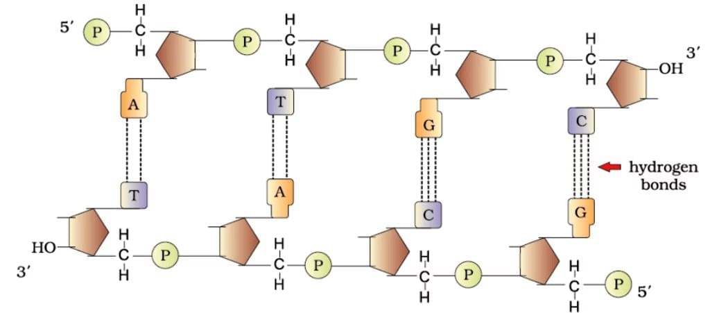
Structure of Polynucleotide Chain: Single Strand
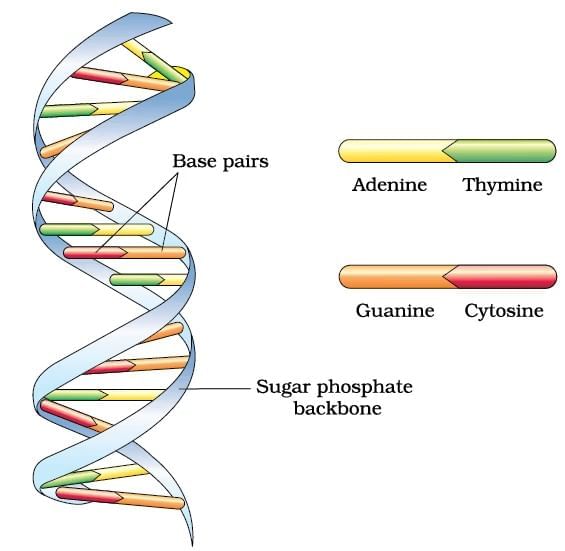
Structure of Polynucleotide Chain : Double Stranded
DNA Double Helix
DNA in both prokaryotic and eukaryotic cells is compactly organized despite its large length, which allows it to fit within the microscopic confines of a cell nucleus or nucleoid.
Prokaryotic DNA Packaging:
Eukaryotic DNA Packaging:
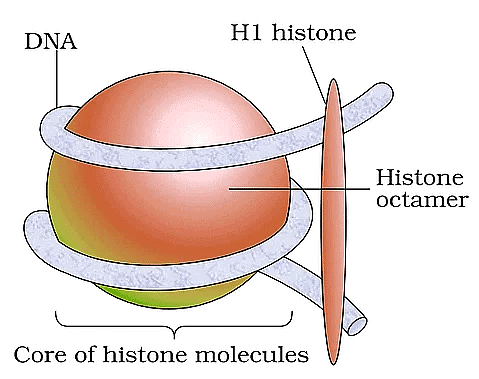
Nucleosome
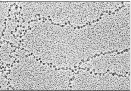
EM Picture: Beads on String
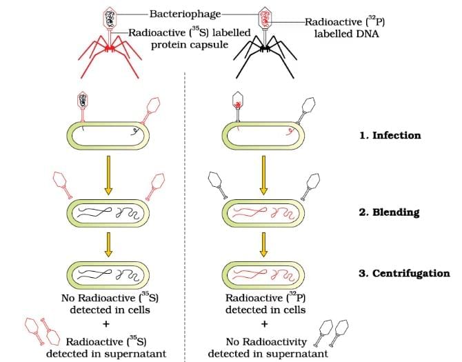
Hershey and Chase Experiment
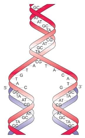
Watson-Crick model for semiconservative DNA replication
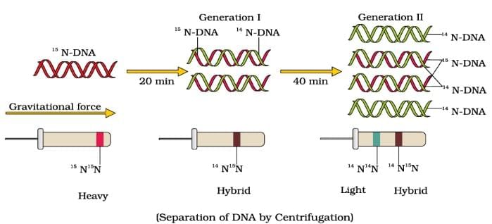
Meselson and Stahl's experiment in 1958 provided definitive evidence for the semiconservative model of DNA replication. Here's a summary of their method and findings:
Isotopic Labeling: They grew Escherichia coli in a medium containing the heavy nitrogen isotope N15 as the sole nitrogen source over many generations. This allowed N15 to be incorporated into the newly synthesized DNA, making it denser than normal N14 DNA.
Transfer to Normal Nitrogen: The bacteria were then transferred to a medium containing the normal ^14N isotope. DNA samples were taken at specific time intervals as the bacteria continued to reproduce.
Density Gradient Centrifugation: The extracted DNA was centrifuged using a cesium chloride (CsCl) density gradient. This method separates DNA based on its density, allowing differentiation between heavy (N15), light (N14 ), and hybrid (N15/N14 ) DNA.
Results and Analysis:
Conclusion: These findings supported the idea that DNA replication is semiconservative, where each new DNA molecule consists of one old (parental) and one new (daughter) strand. Subsequent experiments in higher organisms confirmed that semiconservative replication is a universal mechanism.
Meselson and Stahl’s Experiment
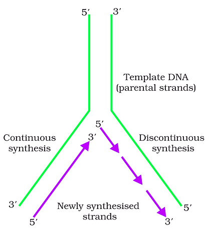
During DNA replication in long DNA molecules, the two strands are not fully separated along their entire length due to the high energy requirement for such an unwinding. Instead, replication occurs at specific sections known as replication forks where the DNA helix opens up.
Direction of Polymerization: DNA-dependent DNA polymerases, the enzymes responsible for adding nucleotides to the growing DNA strand, can only synthesize DNA in the 5' to 3' direction. This directional synthesis introduces complexities at the replication fork.
Continuous vs. Discontinuous Synthesis:
Fragment Joining: The discontinuously synthesized Okazaki fragments on the lagging strand are later connected to form a continuous strand through the action of the enzyme DNA ligase, which links the sugar-phosphate backbones of adjacent fragments.
This method of replication ensures that the DNA is accurately copied despite the directional limitations of the DNA polymerases and the complex structure of the DNA molecule.
Replication Fork
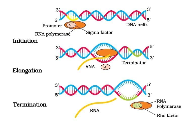
A transcription unit in DNA comprises three main elements:
The template strand (3'→5') acts as the mold for RNA synthesis, while the coding strand (5'→3') mirrors the RNA sequence (except for thymine in place of uracil) and serves as a reference for gene regulation. The arrangement of promoter and terminator defines which strand serves as the template, and additional regulatory sequences may influence gene expression further.
Schematic Structure of Transcription Unit
Process of Transcription in Bacteria
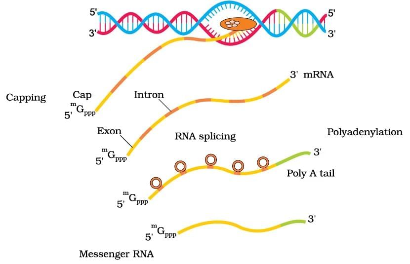
Process of Transcription in Eukaryotes
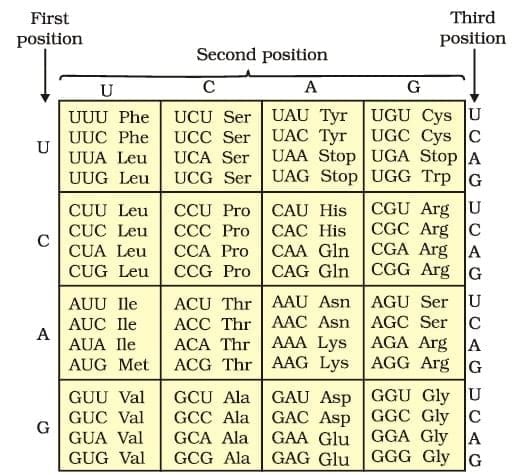
The salient features of genetic code are as follows:
(i) The codon is triplet. 61 codons code for amino acids and 3 codons do not code for any amino acids, hence they function as stop codons.
(ii) Some amino acids are coded by more than one codon, hence the code is degenerate.
(iii) The codon is read in mRNA in a contiguous fashion. There are no punctuations.
(iv) The code is nearly universal: for example, from bacteria to human UUU would code for Phenylalanine (phe). Some exceptions to this rule have been found in mitochondrial codons, and in some protozoans.
(v) AUG has dual functions. It codes for Methionine (met) , and it also act as initiator codon. (vi) UAA, UAG, UGA are stop terminator codons.
The Codons for the Various Amino Acids
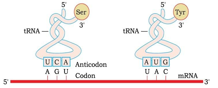
tRNA– the Adapter Molecule
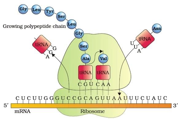
Process of Translation
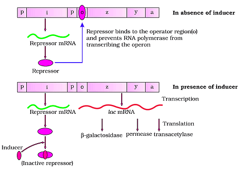
The Lac Operon
The Human Genome Project (HGP), launched in 1990 and completed in 2003, was a large-scale initiative aimed at mapping all human genes and sequencing the 3 billion base pairs in human DNA.
Key achievements include identifying 20,000-25,000 genes, revealing that less than 2% of the genome codes for proteins, and highlighting extensive repeated sequences that contribute to genomic structure and evolution.
Techniques like automated DNA sequencing and specialized computer programs were essential for managing the massive data involved. The project's insights are foundational for advancing personalized medicine, understanding genetic disorders, and exploring human evolution.
Salient Features of Human Genome
(i) The human genome contains 3164.7 million bp.
(ii) The average gene consists of 3000 bases, but sizes vary greatly, with the largest known human gene being dystrophin at 2.4 million bases.
(iii) The total number of genes is estimated at 30,000–much lower than previous estimates of 80,000 to 1,40,000 genes. Almost all
(99.9 per cent) nucleotide bases are exactly the same in all people.
(iv) The functions are unknown for over 50 per cent of the discovered genes.
(v) Less than 2 per cent of the genome codes for proteins.
(vi) Repeated sequences make up very large portion of the human genome.
(vii) Repetitive sequences are stretches of DNA sequences that are repeated many times, sometimes hundred to thousand times. They are thought to have no direct coding functions, but they shed light on chromosome structure, dynamics and evolution.
(viii) Chromosome 1 has most genes (2968), and the Y has the fewest (231).
(ix) Scientists have identified about 1.4 million locations where singlebase DNA differences (SNPs – single nucleotide polymorphism, pronounced as ‘snips’) occur in humans. This information promises to revolutionise the processes of finding chromosomal locations for disease-associated sequences and tracing human history.
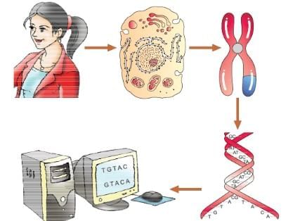
A representative diagram of human genome project
Ans: (d)
A transcription unit in DNA is critical for the process of transcription, wherein a particular segment of DNA is copied into RNA (especially mRNA) by the enzyme RNA polymerase. This unit is composed of sequences that include both coding regions, which are directly transcribed into RNA, and regulatory regions, which ensure that transcription is initiated and terminated at the correct locations on the DNA.The correct answer is: Option D: Promotor, Structural gene, Terminator
Here's a detailed explanation of each component:
Promoter: The promoter is a sequence in DNA that signals the RNA polymerase to start transcription. It is located at the upstream end (5' end) of the gene. Promoters are essential for transcription initiation and are typically found just before the genes they regulate.
Structural gene: This region of the transcription unit is actually expressed or translated into protein (or functional RNA), depending on the kind of gene. These genes contain the functional sequences that are copied during the transcription process.
Terminator: The terminator is found at the downstream end (3' end) of the transcription unit and includes sequences that signal the RNA polymerase enzyme to stop transcription. This ensures that the newly synthesized RNA contains only the necessary genetic message.
The other options contain components that do not accurately define the typical structure of a transcription unit:
Option A mixes regulatory proteins and DNA regions, which does not accurately represent the structural components of a transcription unit.
Option B includes "transposons" which are genetic elements that can move around within the genomes but are not typically part of the transcription unit.
Option C again refers to regulatory proteins (inducer and repressor) along with structural genes, confusing the functions of proteins and DNA regions.
Therefore, Option D correctly represents the standard components of a transcription unit in the context of gene transcription in DNA.
Q2: Match List - I with List - II. (NEET 2022 Phase 2)
Choose the correct answer from the options given below
(a) (a) - (iii), (b) - (i), (c) - (iv), (d) - (ii)
(b) (a) - (iii), (b) - (ii), (c) - (i), (d) - (iv)
(c) (a) - (iv), (b) - (iii), (c) - (ii), (d) - (i)
(d) (a) - (iv), (b) - (i), (c) - (iii), (d) - (ii)
Ans: (c)
In Iac operon,
78 videos|276 docs|174 tests
|
1. What is the structure of polynucleotide chains in DNA? |
2. How does DNA get packaged into a compact structure? |
3. What was the significance of the Hershey and Chase experiment? |
4. What is semi-conservative DNA replication according to the Watson and Crick model? |
5. What are the key differences in transcription between prokaryotes and eukaryotes? |
|
Explore Courses for NEET exam
|